. 2016 Apr:145:380-392.
doi: 10.1016/j.exer.2016.02.009. Epub 2016 Feb 27.

G-quartet oligonucleotide mediated delivery of proteins into photoreceptors and retinal pigment epithelium via intravitreal injection

Affiliations

G-quartet oligonucleotide mediated delivery of proteins into photoreceptors and retinal pigment epithelium via intravitreal injection

Derek Leaderer et al. Exp Eye Res. 2016 Apr.

Abstract

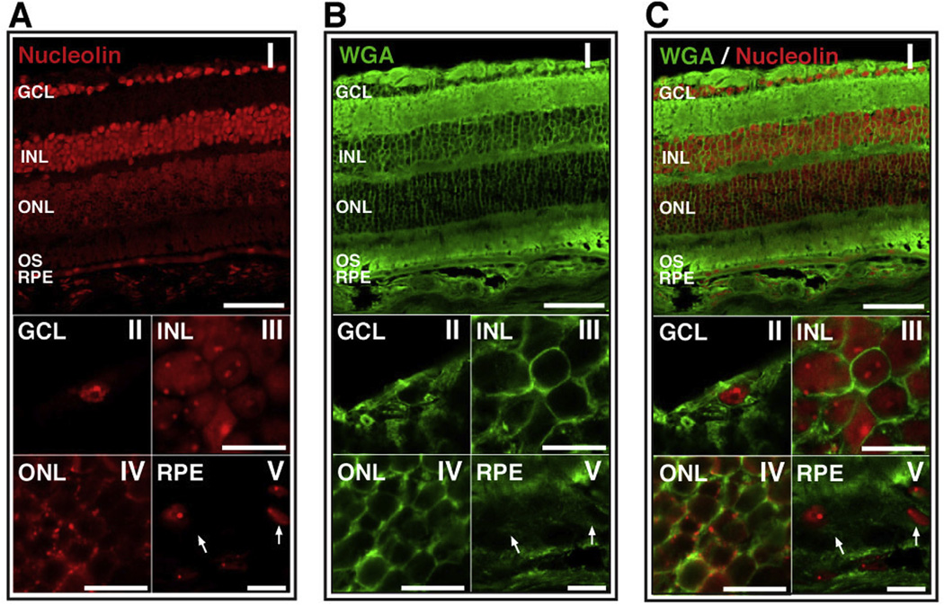
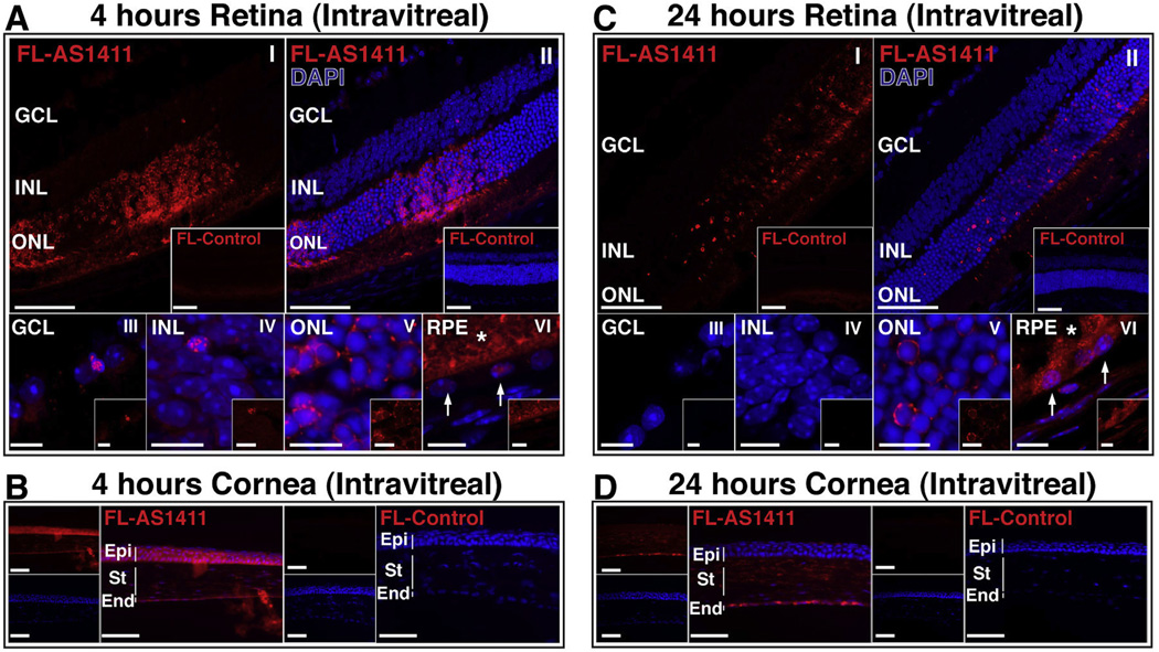
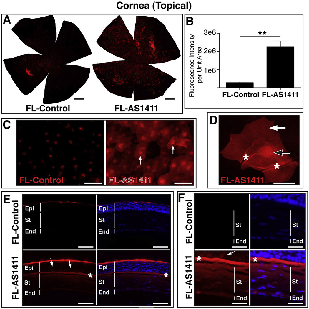
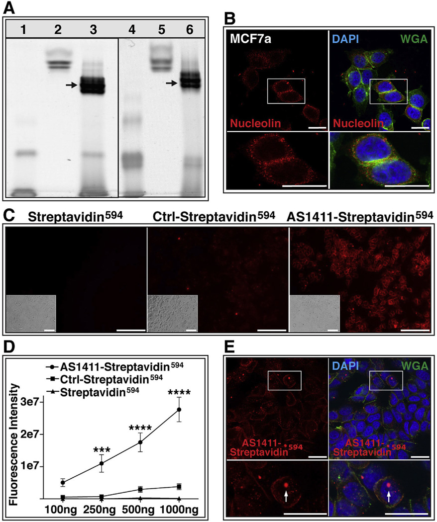
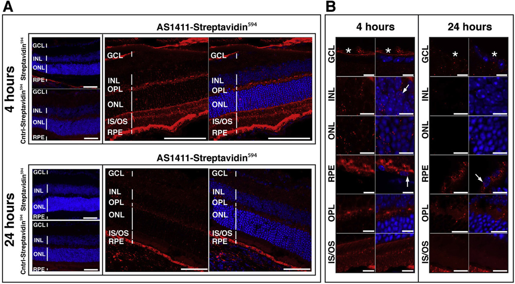
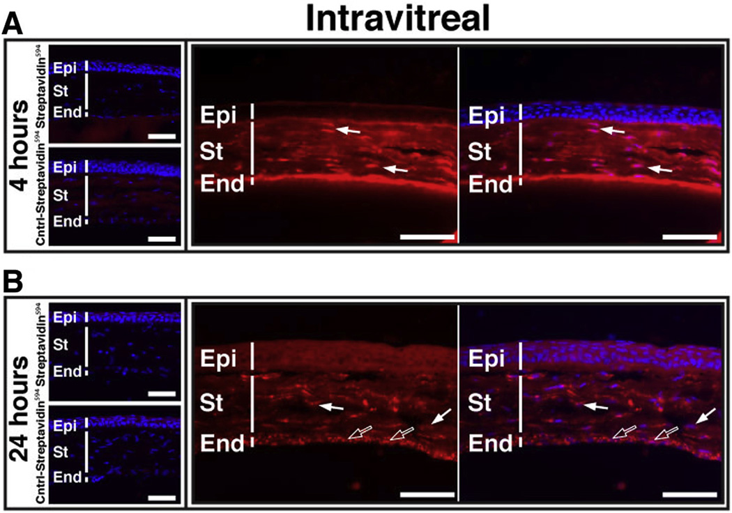
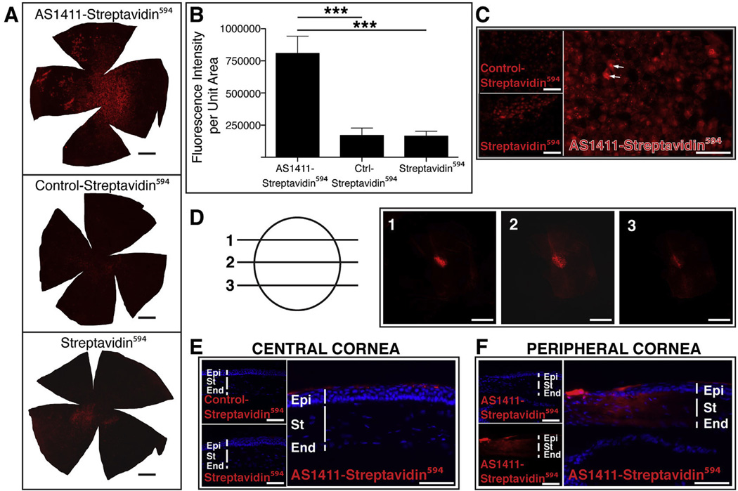
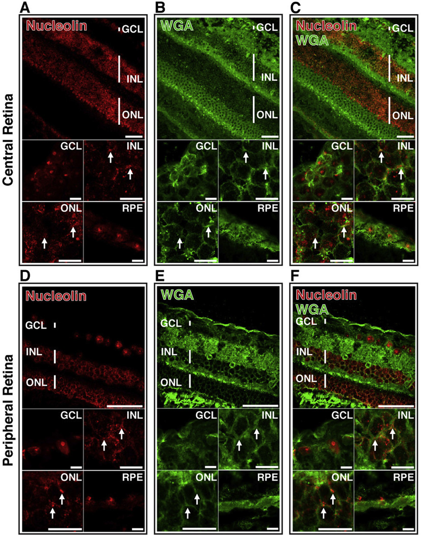
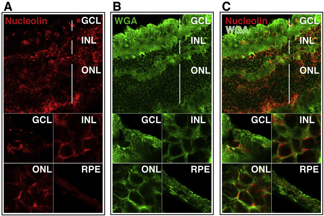
There is currently no available method to efficiently deliver proteins across the plasma membrane of photoreceptor or retinal pigment epithelium (RPE) cells in vivo. Thus, current clinical application of recombinant proteins in ophthalmology is limited to the use of proteins that perform their biological function extracellularly. The ability to traverse biological membranes would enable the mobilization of a significantly larger number of proteins with previously well characterized properties. Nucleolin is abundantly present on the surface of rapidly dividing cells including cancer cells. Surprisingly, nucleolin is also present on the surface of photoreceptor cell bodies. Here we investigated whether nucleolin can be utilized as a gateway for the delivery of proteins into retinal cells following intravitreal injection. AS1411 is a G-quartet aptamer capable of targeting nucleolin. Subsequent to intravitreal injection, fluorescently labeled AS1411 localized to various retinal cell types including the photoreceptors and RPE. AS1411 linked to streptavidin (a ∼50 kDa protein) via a biotin bridge enabled the uptake of Streptavidin into photoreceptors and RPE. AS1411-Streptavidin conjugate applied topically to the cornea allowed for uptake of the conjugate into the nucleus and cytoplasm of corneal endothelial cells. Clinical relevance of AS1411 as a delivery vehicle was strongly indicated by demonstration of the presence of cell surface nucleolin on the photoreceptors, inner neurons and ganglion cells of human retina. These data support exploration of AS1411 as a means of delivering therapeutic proteins to diseased retina.

Keywords: Aptamer; Cornea; Intravitreal; Nucleolin; Retina; Shuttle; Topical.


Figures

Fig. 1
Fig. 1
Nucleolin is present on the surface of cells of the outer nuclear layer in BALB/c retina. (A) Confocal image of BALB/c retinal section stained for nucleolin; (I) whole retina, (II)–(V) increased magnification of GCL, INL, ONL and RPE, respectively. Arrows indicate the nuclei of RPE cells. (B) Confocal image of BALB/c retinal section stained with the cell surface marker, wheat germ agglutinin (WGA); (I) whole retina, (II)–(V) increased magnification of GCL, INL, ONL and RPE, respectively. Arrows indicate the cell surface of RPE cells. (C) Merged image of nucleolin and WGA staining indicates co-localization of these markers only on the ONL (yellow). Top panels scale bar = 60 µm; lower panels scale bar = 10 µm. GCL, ganglion cell layer, INL, inner nuclear layer; ONL, outer nuclear layer; RPE, retinal pigment epithelium. (For interpretation of the references to color in this figure legend, the reader is referred to the web version of this article.)
Fig. 2
Fig. 2
AS1411 administered by intravitreal injection localizes to a variety of ocular cells in vivo. (A,C) Representative confocal images of BALB/c retina harvested 4 h and 24 h following intravitreal injection of 0.3 nmol AlexaFluro594 labeled AS1411 (FL-AS1411) or Control oligonucleotide (FL-Control; Insets). Top panels scale bar = 60 µm; lower panels scale bar = 10 µm; (I, II) whole retina, (III–VI) higher magnification of GCL, INL, ONL and RPE. Arrows indicate RPE nuclei; asterisk indicates outer segments. (B,D) Representative images of BALB/c cornea harvested 4 h and 24 h following intravitreal injection of 0.3 nmol FL-AS1411 or FL-Control. Scale bar = 60 µm. DAPI (blue) counterstains nuclei. n = 6 eyes/3 mice per condition/time point. GCL-gcmglion cell layer; INL-inner nuclear layer; ONL-outer nuclear layer; RPE-retinal pigment epithelium; Epi-epithelium; St-stroma; End-endothelium. (For interpretation of the references to color in this figure legend, the reader is referred to the web version of this article.)
Fig. 3
Fig. 3
Topical application of AS1411 results in uptake by corneal cells in vivo. (A) Representative corneal flat mounts harvested 2 h post topical application of 1 nmol FL-AS1411 or FL-Control [scale bar = 0.5 mm]. (B) Quantitation of fluorescent signal per area of topically treated corneas (p = 0.002, n = 6 for each of FL-AS1411, FL-Control). (C) Higher magnification of flat mounts from FL-AS1411 or FL-Control treated cornea. Scale bar = 60 µm. Arrows indicate nuclear localization. (D) A representative image from a series of confocal laser scanned micrographs of a corneal epithelial cell showing FL-AS1411 in the nucleus (black/white arrow) and cytoplasm (white arrow). Asterisks indicate areas of wrinkling, typical of superficial epithelial cells. Scale bar = 20 µm. (E) Transverse sections of corneas topically treated with FL-AS1411 or FL-Control shows FL-AS1411 in all layers of the cornea. Basal and polygonal cells of the epithelium are indicated by arrows. FL-Control was observed only in the superficial squamous cell layer of the epithelium. Asterisks indicate the anterior limiting lamina. Scale bar = 60 µm. (F) Higher magnification of the deeper layers of the epithelium, the stroma and the endothelium show FL-AS1411 permeation of these layers, while FL-Control is undetectable. Scale bar = 30 µm n = 4 eyes/2 mice for each of FL-AS1411, FL-Control. Epi-epithelium; St-stroma; End-endothelium.
Fig. 4
Fig. 4
AS1411 delivers protein to cells in vitro. (A) Native polyacrylamide gel confirms conjugation of streptavidin594 with biotinylated AS1411. Lane 1: biotinylated AS1411; Lanes 2 and 5: Streptavidin594; Lane 3: AS1411-Streptavidin594 conjugate; Lane 4: biotinylated control oligonucleotide; Lane 6: Control-Streptavidin594 conjugate;. Arrows indicate conjugation products. (B) MCF7a cells probed with antibody against nucleolin (red) or stained with wheat germ agglutinin (WGA; green) and DAPI (blue). Scale bar = 20 µm (C) MCF7a cells incubated for 1 h with 1000 ng of Streptavidin594, Ctrl-Streptavidin594, or AS1411-Streptavidin594. Bright-field images shown in insert Scale bar = 120 µm. (D) Quantification of raw fluorescence intensity of MCF7a cells incubated with increasing doses of Streptavidin594, Ctrl-Streptavidin594, or AS1411-Streptavidin594 (study performed twice in triplicate) (*** = p < 0.001; **** = p < 0.0001). (E) Confocal microscopy of AS1411-Streptavidin594 (red) incubated cells stained with WGA (green) and DAPI (blue). Arrow indicates nuclear localization of AS1411-Streptavidin594. Scale bar = 20 µm. Ctrl-Control. (For interpretation of the references to color in this figure legend, the reader is referred to the web version of this article.)
Fig. 5
Fig. 5
AS1411-Streptavidin594 transduces mouse retinal cells following intravitreal injection. (A) Sections of BALB/c retina harvested 4 h and 24 h following intravitreal injection of 1.5 µg of either AS1411-Streptavidin594, Control-Streptavidin594, or unconjugated Streptavidin594. Sections were counterstained with DAPI (blue; n = 6 eyes/3 mice per condition/time point). Scale bar = 60 µm. (B) Confocal microscopy of retinal sections at 4 and 24 h following intravitreal delivery of AS1411 -Streptavidin594. Scale bar = 10 µm. Asterisks indicate nerve fiber layer. Arrows indicate red fluorescence signal in the nucleus. Cntrl-Control; GCL-ganglion cell layer; INL-inner nuclear layer; ONL-outer nuclear layer; IS/OS-inner/outer segments; RPE-retinal pigment epithelium; OPL-outer plexiform layer. (For interpretation of the references to color in this figure legend, the reader is referred to the web version of this article.)
Fig. 6
Fig. 6
AS1411-Streptavidin594 transduces mouse cornea following intravitreal injection. Corneal sections of BALB/c eyes harvested (A) 4 h and (B) 24 h following intravitreal injection of 1.5 µg of either AS1411-Streptavidin594, Control-Streptavidin594 or Streptavidin594. Scale bar = 60 µm. Arrows indicate nuclear localization (solid arrows, stromal fibroblasts; hollow arrows, endothelial cells). Cntrl-control; Epi-epithelium; St-stroma; End-endothelium.
Fig. 7
Fig. 7
Topical Application of AS1411-streptavidin594 results in uptake by corneal cells and nuclear localization in vivo. (A) Corneal flat mounts harvested 2 h post topical application of 5 µg of AS1411-Streptavidin594, Control-Streptavidin594 or Streptavidin594. Scale bar = 0.5 mm. (B) Quantitation of the intensity of fluorescence of streptavidin594 conjugates per area of corneal flatmount (n = 6 per condition, *** = p < 0.001). (C) Higher magnification images of AS1411-Streptavidin594, Control-Streptavidin594 or Streptavidin594 treated corneal flat mounts. Scale bar =120 µm. Arrows indicate examples of nuclear binding. (D) Confocal series of a flatmount of AS1411-Streptavidin594 treated cornea indicating nuclear localization within a corneal epithelial cell. Scale bar = 20 µm. (E) Transverse sections of corneas treated topically with AS1411-Streptavidin594, Control-Streptavidin594 or Streptavidin594 (n = 4 eyes/2 mice per condition). Scale bar = 60 µm. (F) Transverse section of AS1411-strepatividin594 treated cornea showing transduction of stroma and endothelium at the corneal limbus. Scale bar = 60 µm. Ctrl-Control; Epi-epithelium; St-stroma; End-endothelium.
Fig. 8
Fig. 8
Cell surface nucleolin is present on neurons of non-human primate retina. Confocal microscopy of retinal sections of Cynomolgus monkey (macaca fascicularis) stained with anti-nucleolin antibody (A,D; red) and WGA (B,E; green). Images from both central and peripheral retina are shown. Overlay of images A and B (C) and D and E (F) indicates co-localization of nucleolin and WGA (yellow). Arrows indicate regions of co-localization. Top panels scale bar = 60 µm; lower panels scale bar = 10 µm. GCL-ganglion cell layer; INL-inner nuclear layer; ONL-outer nuclear layer; RPE-retinal pigment epithelium. (For interpretation of the references to color in this figure legend, the reader is referred to the web version of this article.)
Fig. 9
Fig. 9
Cell surface nucleolin is present in all neuronal layers of adult human retina. Confocal images of sections of human retina probed with anti-nucleolin antibody (A; red) and WGA (B; green). Overlay of images A and B (C) indicates co-localization of nucleolin and WGA (yellow). Top panels scale bar = 60 µm; lower panels scale bar = 10 µm. GCL-ganglion cell layer, INL-inner nuclear layer, ONL-outer nuclear layer, RPE-retinal pigment epithelium. (For interpretation of the references to color in this figure legend, the reader is referred to the web version of this article.)

References

    1. Aravind A, Jeyamohan P, Nair R, et al. AS1411 aptamer tagged PLGA-lecithin-PEG nanoparticles for tumor cell targeting and drug delivery. Biotechnol. Bioeng. 2012;109:2920–2931. - PubMed
    1. Bates PJ, Laber DA, Miller DM, Thomas SD, Trent JO. Discovery and development of the G-rich oligonucleotide AS1411 as a novel treatment for cancer. Exp. Mol. Pathol. 2009;86:151–164. - PMC - PubMed
    1. Binder C, Read SP, Cashman SM, Kumar-Singh R. Nuclear targeted delivery of macromolecules to retina and cornea. J. Gene Med. 2011;13:158–170. - PubMed
    1. Borer RA, Lehner CF, Eppenberger HM, Nigg EA. Major nucleolar proteins shuttle between nucleus and cytoplasm. Cell. 1989;56:379–390. - PubMed
    1. Carvalho LS, Vandenberghe LH. Promising and delivering gene therapies for vision loss. Vis. Res. 2015;111:124–133. - PMC - PubMed

Publication types

MeSH terms