. 2016 Jun;28(6):541-51.
doi: 10.1016/j.cellsig.2016.02.015. Epub 2016 Feb 24.

Dual role of vinculin in barrier-disruptive and barrier-enhancing endothelial cell responses

Affiliations

Dual role of vinculin in barrier-disruptive and barrier-enhancing endothelial cell responses

Anna A Birukova et al. Cell Signal. 2016 Jun.

Abstract

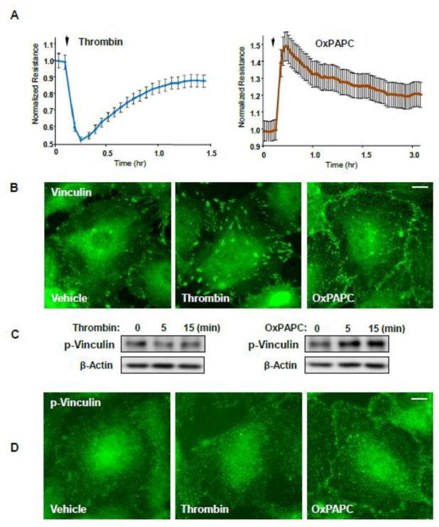
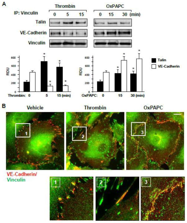
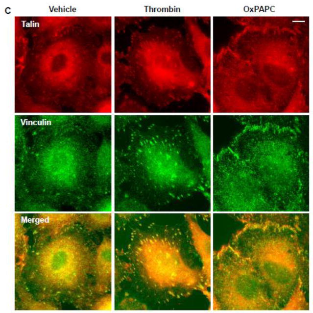
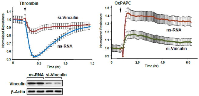
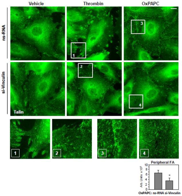
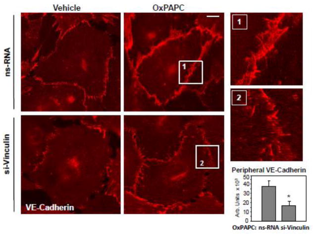
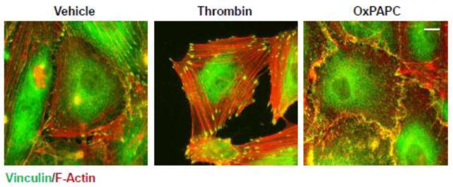
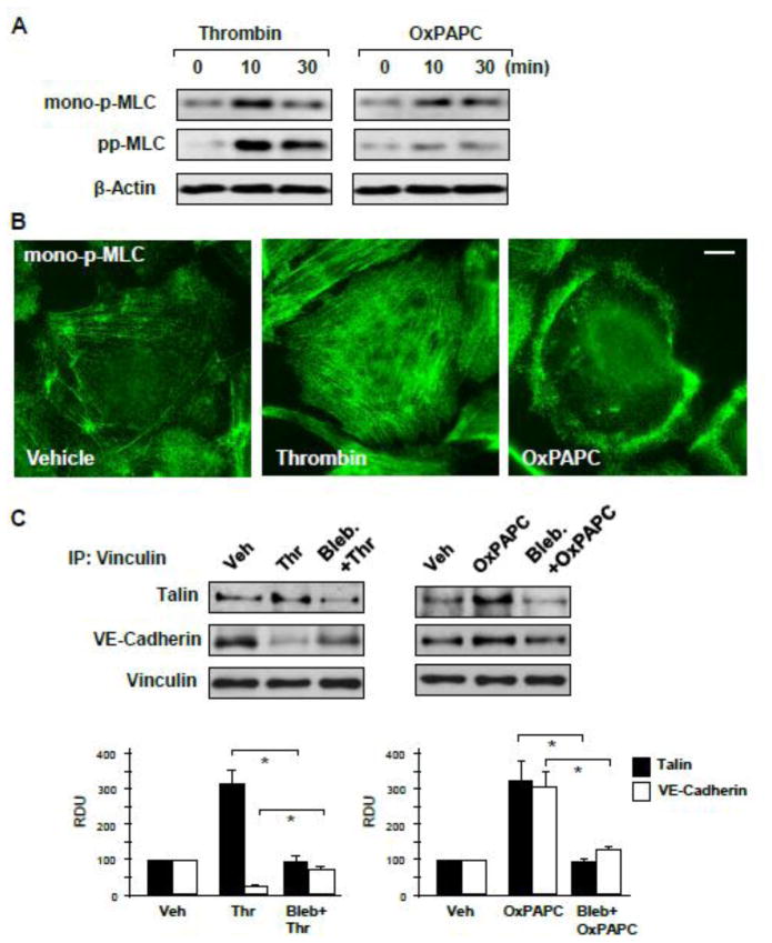
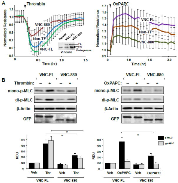
Endothelial cell (EC) barrier disruption induced by edemagenic agonists such as thrombin is a result of increased actomyosin contraction and enforcement of focal adhesions (FA) anchoring contracting stress fibers, which leads to cell retraction and force-induced disruption of cell junctions. In turn, EC barrier enhancement by oxidized phospholipids (OxPAPC) and other agonists is a result of increased tethering forces due to enforcement of the peripheral actin rim and enhancement of cell-cell adherens junction (AJ) complexes promoting EC barrier integrity. This study tested participation of the mechanosensitive adaptor, vinculin, which couples FA and AJ to actin cytoskeleton, in control of the EC permeability response to barrier disruptive (thrombin) and barrier enhancing (OxPAPC) stimulation. OxPAPC and thrombin induced different patterns of FA remodeling. Knockdown of vinculin attenuated both, OxPAPC-induced decrease and thrombin-induced increase in EC permeability. Thrombin stimulated the vinculin association with FA protein talin and suppressed the interaction with AJ protein, VE-cadherin. In contrast, OxPAPC stimulated the vinculin association with VE-cadherin. Thrombin and OxPAPC induced different levels of myosin light chain (MLC) phosphorylation and caused different patterns of intracellular phospho-MLC distribution. Thrombin-induced talin-vinculin and OxPAPC-induced VE-cadherin-vinculin association were abolished by myosin inhibitor blebbistatin. Expression of the vinculin mutant unable to interact with actin attenuated EC permeability changes and MLC phosphorylation caused by both, thrombin and OxPAPC. These data suggest that the specific vinculin interaction with FA or AJ in different contexts of agonist stimulation is defined by development of regional actyomyosin-based tension and participates in both, the barrier-disruptive and barrier-enhancing endothelial responses.

Keywords: Actin; Endothelium; Focal adhesions; Oxidized phospholipids; Rho; Thrombin; Vascular permeability.


Figures

Figure 1
Figure 1. Effects of thrombin and OxPAPC on endothelial permeability and vinculin distribution
A - HPAEC plated on microelectrodes were treated with thrombin (0.2 U/ml) or OxPAPC (15 μg/ml) followed by TER measurements. The TER curves represent pooled data from three independent experiments. B - Cells grown on glass coverslips were stimulated with thrombin or OxPAPC for 15 or 30 min, respectively. Vinculin localization was analyzed by immunofluorescence staining with corresponding antibody. Results are representative of three independent experiments. C - Western blot analysis of phospho-Y822 vinculin levels in HPAEC stimulated with thrombin (0.2 U/ml) or OxPAPC (15 μg/ml) at the indicated time points. D - Cells grown on glass coverslips were stimulated with thrombin or OxPAPC for 5 or 15 min, respectively. Phospho-Y822 vinculin localization was analyzed by immunofluorescence staining with corresponding antibody. Bar - 5 μm. Results are representative of three independent experiments.
Figure 2
Figure 2. Effects of thrombin and OxPAPC on focal adhesion and adherens junction remodeling
A – EC were stimulated with thrombin (0.2 U/ml) or OxPAPC (15 μg/ml), followed by vinculin immunoprecipitation under non-denaturing conditions. Presence of talin and VE-cadherin in immune complexes was tested by Western blot. Bar graphs depict quantitative analysis of Western blot data; n=3; *P<0.05 vs. vehicle. B and C – Thrombin- or OxPAPC-induced vinculin redistribution and its colocalization with VE-cadherin (B) and talin (C) was evaluated by immunofluorescence staining of formaldehyde-fixed EC for vinculin (green) and VE-cadherin or talin (red). Bar - 5 μm. Higher magnification insets show details of vinculin and VE-cadherin co-localization (yellow). Results are representative of three to five independent experiments.
Figure 3
Figure 3. Effects of vinculin knockdown on thrombin and OxPAPC permeability response
Human pulmonary EC were transfected with vinculin-specific or non-specific siRNA. TER was measured in EC stimulated with thrombin (0.2 U/ml) or OxPAPC (15 μg/ml). The TER curves represent pooled data from three independent experiments. siRNA-induced vinculin protein depletion was confirmed by western blot.
Figure 4
Figure 4. Effect of vinculin knockdown on agonist-induced focal adhesion remodeling
Human pulmonary EC were transfected with vinculin-specific or non-specific siRNA. Focal adhesion remodeling in response to thrombin treatment (0.2 U/ml, 10 min) or OxPAPC (15 μg/ml, 30 min) treatment was analyzed by immunofluorescence staining for talin. Bar - 5 μm. Higher magnification insets show details of focal adhesion remodeling in control and stimulated EC. Bar graph represents quantitative analysis of OxPAPC-induced peripheral accumulation of focal adhesions in control and vinculin-depleted EC. Results are representative of four independent experiments.
Figure 5
Figure 5. Effect of vinculin knockdown on OxPAPC-induced adherens junction remodeling
Human pulmonary EC were transfected with vinculin-specific or non-specific siRNA. Adherens junction remodeling in response to OxPAPC (15 μg/ml, 30 min) was analyzed by immunofluorescence staining for VE-cadherin. Bar - 5 μm. Higher magnification insets show details of adherens junction remodeling in control and stimulated EC. Bar graph represents quantitative analysis of the areas covered by adherens junctions in control and vinculin-depleted EC stimulated with OxPAPC. Results are representative of four independent experiments.
Figure 6
Figure 6. Analysis of cytoskeletal remodeling in response thrombin and OxPAPC
HPAEC grown on glass coverslips were stimulated thrombin treatment (0.2 U/ml, 10 min) or OxPAPC (15 μg/ml, 30 min) followed by double immunofluorescence staining for F-actin (red) and vinculin (green). Areas of actin and vinculin co-localization appear in yellow. Bar - 5 μm. Results are representative of three independent experiments.
Figure 7
Figure 7. Analysis of contractile signaling in response to thrombin and OxPAPC
A – MLC mono- and di-phosphorylation was analyzed by Western blotting with corresponding antibodies in control, thrombin (0.2 U/ml) or OxPAPC (15 μg/ml) treated EC. Probing for β-actin was used as a normalization control. B – Intracellular localization of mono-phosphorylated MLC in control and agonist-stimulated cells (thrombin 10 min, OxPAPC 30 min) was determined by immunofluorescence staining with specific antibody. Bar - 5 μm. C – HPAEC were pretreated with vehicle or blebbistatin (15 μM, 30 min), followed by stimulation with thrombin (0.2 U/ml, 10 min) or OxPAPC (15 μg/ml, 30 min). Co-immunoprecipitation assays using vinculin antibody were performed, and talin and VE-cadherin content in the immunoprecipitates was detected using appropriate antibody. Bar graphs depict quantitative analysis of Western blot data; n=3; *P<0.05 vs. thrombin or OxPAPC alone.
Figure 8
Figure 8. Effect of vinc880 mutant on thrombin- and OxPAPC-induced alterations of EC permeability
HPAEC were transfected with full length (VNC-FL) or vinculin-880 (VNC-880) mutant or left un-transfected followed by stimulation with thrombin (0.2 U/ml) or OxPAPC (15 μg/ml). A – TER measurements were performed over the time indicated. The TER curves represent pooled data from three independent experiments. Inset: The levels of ectopically expressed GFP-tagged wild type and vinculin-880 mutant as well as endogenous vinculin were detected by immunoblotting using anti-GFP and vinculin antibodies. B - Cells were stimulated with thrombin or OxPAPC for 10 min or 30 min, respectively. Western blot analysis of agonist-induced mono- and di-phosphorylation of MLC. Probing for β-actin was used as a normalization control. Bar graphs depict quantitative analysis of Western blot data; n=3; *P<0.05 vs. VNC-FL.

References

    1. Beckers CM, van Hinsbergh VW, van Nieuw Amerongen GP. Thromb Haemost. 2010;103(1):40–55. - PubMed
    1. Spindler V, Schlegel N, Waschke J. Cardiovasc Res. 2010;87(2):243–253. - PubMed
    1. Murthy KS. Annu Rev Physiol. 2006;68:345–374. - PubMed
    1. Bass MD, Morgan MR, Roach KA, Settleman J, Goryachev AB, Humphries MJ. J Cell Biol. 2008;181(6):1013–1026. - PMC - PubMed
    1. Guilluy C, Swaminathan V, Garcia-Mata R, O’Brien ET, Superfine R, Burridge K. Nat Cell Biol. 2011;13(6):722–727. - PMC - PubMed

Publication types

LinkOut - more resources