Review
. 2016 Apr;89(4):779-91.
doi: 10.1016/j.kint.2015.11.021. Epub 2016 Jan 21.

Autophagy in acute kidney injury

Affiliations
Review

Autophagy in acute kidney injury

Gur P Kaushal et al. Kidney Int. 2016 Apr.

Abstract

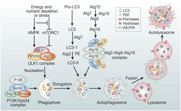
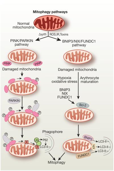
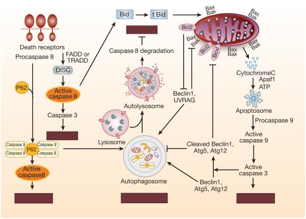
Autophagy is a conserved multistep pathway that degrades and recycles damaged organelles and macromolecules to maintain intracellular homeostasis. The autophagy pathway is upregulated under stress conditions including cell starvation, hypoxia, nutrient and growth-factor deprivation, endoplasmic reticulum stress, and oxidant injury, most of which are involved in the pathogenesis of acute kidney injury (AKI). Recent studies demonstrate that basal autophagy in the kidney is vital for the normal homeostasis of the proximal tubules. Deletion of key autophagy proteins impaired renal function and increased p62 levels and oxidative stress. In models of AKI, autophagy deletion in proximal tubules worsened tubular injury and renal function, highlighting that autophagy is renoprotective in models of AKI. In addition to nonselective sequestration of autophagic cargo, autophagy can facilitate selective degradation of damaged organelles, particularly mitochondrial degradation through the process of mitophagy. Damaged mitochondria accumulate in autophagy-deficient kidneys of mice subjected to ischemia-reperfusion injury, but the precise mechanisms of regulation of mitophagy in AKI are not yet elucidated. Recent progress in identifying the interplay of autophagy, apoptosis, and regulated necrosis has revived interest in examining shared pathways/molecules in this crosstalk during the pathogenesis of AKI. Autophagy and its associated pathways pose potentially unique targets for therapeutic interventions in AKI.

Keywords: acute kidney injury; apoptosis; autophagy; cell death; cisplatin nephrotoxicity; ischemia reperfusion; mitophagy.


Figures

Figure 1
Figure 1
Overview of the autophagy pathway and its molecular machinery: Formation of autophagosome involves a series of steps mediated by functional complexes containing Atg proteins: (i) The ULK1/2 kinase complex composed of ULK1/2, Atg13, RB1CC1/FIP200, and Atg101 is recruited to the phagophore membranes. It is required for initiation of autophagy and is regulated by mTORC1 and AMPK. (ii) PI3K/Vps34 complex composed of Vps34, Vps 15, Beclin-1, Ambra and Atg14L is required for nucleation of the phagophore membrane.(iii) Atg9 vesicles are required for membrane expansion for the autophagosome assembly. (iv) Atg12–Atg5–Atg16 multimeric complex and Atg8/LC3-II are two ubiquitin-like conjugation systems that are involved in the elongation and expansion steps in the autophagosome formation. LC3-II remains present on both the membranes of the autophagosome and can bind to selective substrates including p62. The mature autophagosome then fuses with lysosome to form autolysosome. Upon fusion with lysosome, the cytoplasmic contents sequestered in the autophagosome contents are degraded by the lysosomal hydrolases.
Figure 2
Figure 2
PINK/PARKIN and BNIP3/NIX/FUNDC1 pathways of mtophagy. In the PINK/PARKIN pathway, mitochondrial deploarization or mitochondrial fission leads to localization of PINK1 in the outer membrane of the depolarized mitochondria. PINK1 then recruits E3 ubiquitin ligase, PARKIN, to the mitochondria and activates PARKIN to facilitate ubiquitination of mitochondrial surface proteins for autophagic clearance of the damaged mitochondria. Polyubiquitinated outer mitochondrial proteins interact with p62 bound to autophagosomal LC3, thereby targeting the mitochondrion for mitophagy In the BNIP3/NIX/FUNDC1 pathway, mitochondrial outer membrane proteins BNIP3, NIX, or FUNDC1 bind to LC3-II via their LIR motifs facing the cytosol and promote selective clearance of mitochondria. NIX is involved in the process of mitochondrial clearance during reticulocytes maturation and BNIP3 is upregulated in renal IR injury and overexpression of BNIP3 in NRK-53E cells selectively induced mitophagy72.
Figure 3
Figure 3
Cross-talk between apoptosis and autophagy. Common proteins in the autophagy and apoptotic pathways are shared and intimately linked in cross-talk between apoptosis and autophagy regulating cell death in mammalian cells. Degradation of key autophagy proteins such as Atg5, Atg3, Atg4D, Beclin-1 and Atg12 by active caspases represents one mechanism that limits continuous autophagosome formation. Antiapoptic Bcl-2 family members bind to Beclin-1 and restrict availability Beclin-1 for autophagosome formation and suppress autophagy. Autophagy substrate P62 participates in recruitment of caspase-8 that facilitates self-oligomerization of caspase-8 and subsequent activation. Caspase-8 once recruited to the autophagosome can also be degraded that suppresses apoptosis.

References

    1. De Duve C. The lysosome. Sci Am. 1963;208:64–72. - PubMed
    1. Deter RL, De Duve C. Participation of lysosomes in cellular autophagy induced in rat liver by glucagon. J Cell Biol. 1967;35:C11–16. P. B. - PMC - PubMed
    1. Klionsky DJ. Autophagy revisited: a conversation with Christian de Duve. Autophagy. 2008;4:740–743. - PubMed
    1. Yang Z, Klionsky DJ. Mammalian autophagy: core molecular machinery and signaling regulation. Curr Opin Cell Biol. 2010;22:124–131. - PMC - PubMed
    1. Mizushima N, Komatsu M. Autophagy: renovation of cells and tissues. Cell. 2011;147:728–741. - PubMed

Publication types

MeSH terms

LinkOut - more resources