

Ultrasound-based gestational-age estimation in late pregnancy
- PMID: 26924421
- PMCID: PMC6680349
- DOI: 10.1002/uog.15894
Ultrasound-based gestational-age estimation in late pregnancy
Abstract
Objective: Accurate gestational-age (GA) estimation, preferably by ultrasound measurement of fetal crown-rump length before 14 weeks' gestation, is an important component of high-quality antenatal care. The objective of this study was to determine how GA can best be estimated by fetal ultrasound for women who present for the first time late in pregnancy with uncertain or unknown menstrual dates.
Methods: INTERGROWTH-21st was a large, prospective, multicenter, population-based project performed in eight geographically defined urban populations. One of its principal components, the Fetal Growth Longitudinal Study, aimed to develop international fetal growth standards. Each participant had their certain menstrual dates confirmed by first-trimester ultrasound examination. Fetal head circumference (HC), biparietal diameter (BPD), occipitofrontal diameter (OFD), abdominal circumference (AC) and femur length (FL) were measured every 5 weeks from 14 weeks' gestation until delivery. For each participant, a single, randomly selected ultrasound examination was used to explore all candidate biometric variables and permutations to build models to predict GA. Regression equations were ranked based upon minimization of the mean prediction error, goodness of fit and model complexity. An automated machine learning algorithm, the Genetic Algorithm, was adapted to evaluate > 64 000 potential polynomial equations as predictors.
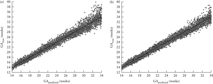
Results: Of the 4607 eligible women, 4321 (94%) had a pregnancy without major complications and delivered a live singleton without congenital malformations. After other exclusions (missing measurements in GA window and outliers), the final sample comprised 4229 women. Two skeletal measures, HC and FL, produced the best GA prediction, given by the equation loge (GA) = 0.03243 × (loge (HC))2 + 0.001644 × FL × loge (HC) + 3.813. When FL was not available, the best equation based on HC alone was loge (GA) = 0.05970 × (loge (HC))2 + 0.000000006409 × (HC)3 + 3.3258. The estimated uncertainty of GA prediction (half width 95% interval) was 6-7 days at 14 weeks' gestation, 12-14 days at 26 weeks' gestation and > 14 days in the third trimester. The addition of FL to the HC model led to improved prediction intervals compared with using HC alone, but no further improvement in prediction was afforded by adding AC, BPD or OFD. Equations that included other measurements (BPD, OFD and AC) did not perform better.
Conclusions: Among women initiating antenatal care late in pregnancy, a single set of ultrasound measurements combining HC and FL in the second trimester can be used to estimate GA with reasonable accuracy. We recommend this tool for underserved populations but considerable efforts should be implemented to improve early initiation of antenatal care worldwide. © 2016 Authors. Ultrasound in Obstetrics & Gynecology published by John Wiley & Sons Ltd on behalf of International Society of Ultrasound in Obstetrics and Gynecology.
Keywords: dating; fetal growth; gestational age.
© 2016 The Authors. Ultrasound in Obstetrics & Gynecology published by John Wiley & Sons Ltd on behalf of the International Society of Ultrasound in Obstetrics and Gynecology.
Figures

Comment in
-
Re: Ultrasound-based gestational-age estimation in late pregnancy. A. T. Papageorghiou, B. Kemp, W. Stones, E. O. Ohuma, S. H. Kennedy, M. Purwar, L. J. Salomon, D. G. Altman, J. A. Noble, E. Bertino, M. G. Gravett, R. Pang, L. Cheikh Ismail, F. C. Barros, A. Lambert, Y. A. Jaffer, C. G. Victora, Z. A. Bhutta and J. Villar, for the International Fetal and Newborn Growth Consortium for the 21st Century (INTERGROWTH-21st). Ultrasound Obstet Gynecol 2016; 48: 719-726.Ultrasound Obstet Gynecol. 2016 Dec;48(6):693. doi: 10.1002/uog.17355. Ultrasound Obstet Gynecol. 2016. PMID: 27933707 No abstract available.
References
-
- Ioannou C, Talbot K, Ohuma E, Sarris I, Villar J, Conde‐Agudelo A, Papageorghiou AT. Systematic review of methodology used in ultrasound studies aimed at creating charts of fetal size. BJOG 2012; 119 : 1425–1439. - PubMed
-
- Blencowe H, Cousens S, Oestergaard MZ, Chou D, Moller A, Narwal R, Adler A, Vera Garcia C, Rohde S, Say L, Lawn JE. National, regional, and worldwide estimates of preterm birth rates in the year 2010 with time trends since 1990 for selected countries: a systematic analysis and implications. Lancet 2012; 379 : 2162–2172. - PubMed
-
- de Onis, Blössner M , Villar J. Levels and patterns of intrauterine growth retardation in developing countries. Eur J Clin Nutr 1998; 52 Suppl 1 : S5–15. - PubMed
-
- Savitz DA, Terry JW, Dole N, Thorp JM, Siega‐Riz AM, Herring AH. Comparison of pregnancy dating by last menstrual period, ultrasound scanning, and their combination. Am J Obstet Gynecol 2002; 187 : 1660–1666. - PubMed
-
- Thorsell M, Kaijser M, Almström H, Andolf E. Expected day of delivery from ultrasound dating versus last menstrual period – obstetric outcome when date mismatch. BJOG 2008; 115 : 585–589. - PubMed
Publication types
MeSH terms
LinkOut - more resources
Full Text Sources
Other Literature Sources
Research Materials
