Dynamic capsule restructuring by the main pneumococcal autolysin LytA in response to the epithelium
- PMID: 26924467
- PMCID: PMC4773454
- DOI: 10.1038/ncomms10859
Dynamic capsule restructuring by the main pneumococcal autolysin LytA in response to the epithelium
Abstract
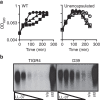
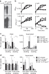
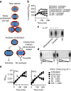
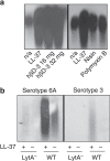
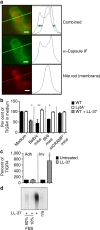
Bacterial pathogens produce complex carbohydrate capsules to protect against bactericidal immune molecules. Paradoxically, the pneumococcal capsule sensitizes the bacterium to antimicrobial peptides found on epithelial surfaces. Here we show that upon interaction with antimicrobial peptides, encapsulated pneumococci survive by removing capsule from the cell surface within minutes in a process dependent on the suicidal amidase autolysin LytA. In contrast to classical bacterial autolysis, during capsule shedding, LytA promotes bacterial survival and is dispersed circumferentially around the cell. However, both autolysis and capsule shedding depend on the cell wall hydrolytic activity of LytA. Capsule shedding drastically increases invasion of epithelial cells and is the main pathway by which pneumococci reduce surface bound capsule during early acute lung infection of mice. The previously unrecognized role of LytA in removing capsule to combat antimicrobial peptides may explain why nearly all clinical isolates of pneumococci conserve this enzyme despite the lethal selective pressure of antibiotics.
Figures

Similar articles
-
Pleiotropic effects of cell wall amidase LytA on Streptococcus pneumoniae sensitivity to the host immune response.Infect Immun. 2015 Feb;83(2):591-603. doi: 10.1128/IAI.02811-14. Epub 2014 Nov 17. Infect Immun. 2015. PMID: 25404032 Free PMC article.
-
[Value of demonstration of pneumococcal surface antigen A and autolysin genes for the identification of Streptococcus pneumoniae clinical isolates].Mikrobiyol Bul. 2009 Jan;43(1):11-7. Mikrobiyol Bul. 2009. PMID: 19334375 Turkish.
-
Tissue-specific contributions of pneumococcal virulence factors to pathogenesis.J Infect Dis. 2004 Nov 1;190(9):1661-9. doi: 10.1086/424596. Epub 2004 Sep 21. J Infect Dis. 2004. PMID: 15478073
-
Recent trends on the molecular biology of pneumococcal capsules, lytic enzymes, and bacteriophage.FEMS Microbiol Rev. 2004 Nov;28(5):553-80. doi: 10.1016/j.femsre.2004.05.002. FEMS Microbiol Rev. 2004. PMID: 15539074 Review.
-
Molecular analysis of antibiotic tolerance in pneumococci.Int J Med Microbiol. 2002 Jul;292(2):75-9. doi: 10.1078/1438-4221-00193. Int J Med Microbiol. 2002. PMID: 12195738 Review.
Cited by
-
Time-resolved dual RNA-seq reveals extensive rewiring of lung epithelial and pneumococcal transcriptomes during early infection.Genome Biol. 2016 Sep 27;17(1):198. doi: 10.1186/s13059-016-1054-5. Genome Biol. 2016. PMID: 27678244 Free PMC article.
-
A multiomics analysis of direct interkingdom dynamics between influenza A virus and Streptococcus pneumoniae uncovers host-independent changes to bacterial virulence fitness.PLoS Pathog. 2022 Dec 21;18(12):e1011020. doi: 10.1371/journal.ppat.1011020. eCollection 2022 Dec. PLoS Pathog. 2022. PMID: 36542660 Free PMC article.
-
Interactions of Bacteriophages and Bacteria at the Airway Mucosa: New Insights Into the Pathophysiology of Asthma.Front Allergy. 2021 Jan 26;1:617240. doi: 10.3389/falgy.2020.617240. eCollection 2020. Front Allergy. 2021. PMID: 35386933 Free PMC article. Review.
-
Proteomic Adaptation of Streptococcus pneumoniae to the Human Antimicrobial Peptide LL-37.Microorganisms. 2020 Mar 14;8(3):413. doi: 10.3390/microorganisms8030413. Microorganisms. 2020. PMID: 32183275 Free PMC article.
-
Resistance Mechanisms to Antimicrobial Peptides in Gram-Positive Bacteria.Front Microbiol. 2020 Oct 21;11:593215. doi: 10.3389/fmicb.2020.593215. eCollection 2020. Front Microbiol. 2020. PMID: 33193264 Free PMC article. Review.
References
-
- Sorensen U. B., Henrichsen J., Chen H. C. & Szu S. C. Covalent linkage between the capsular polysaccharide and the cell wall peptidoglycan of Streptococcus pneumoniae revealed by immunochemical methods. Microb. Pathog. 8, 325–334 (1990). - PubMed
-
- Goldman M. J. et al. Human β-defensin-1 is a salt-sensitive antibiotic in lung that is inactivated in cystic fibrosis. Cell 88, 553–560 (1997). - PubMed
-
- McCray P. B. & Bentley L. Human airway epithelia express a beta-defensin. Am. J. Respir. Cell. Mol. Biol. 16, 343–349 (1997). - PubMed
MeSH terms
Substances
LinkOut - more resources
Full Text Sources
Other Literature Sources

