. 2016 Mar 3;98(3):473-489.
doi: 10.1016/j.ajhg.2016.01.006. Epub 2016 Feb 25.

Mutations in Subunits of the Activating Signal Cointegrator 1 Complex Are Associated with Prenatal Spinal Muscular Atrophy and Congenital Bone Fractures

Affiliations

Mutations in Subunits of the Activating Signal Cointegrator 1 Complex Are Associated with Prenatal Spinal Muscular Atrophy and Congenital Bone Fractures

Ellen Knierim et al. Am J Hum Genet. .

Abstract

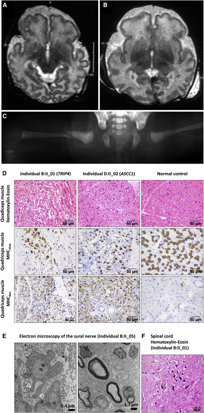
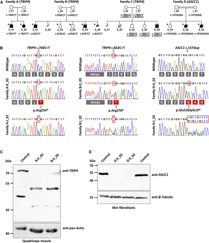
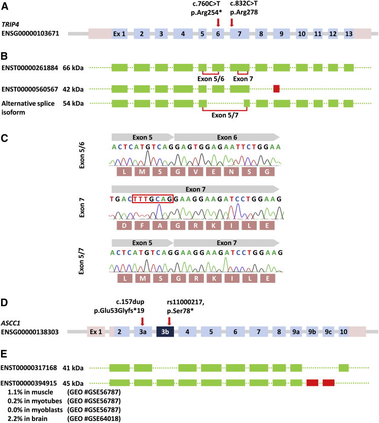
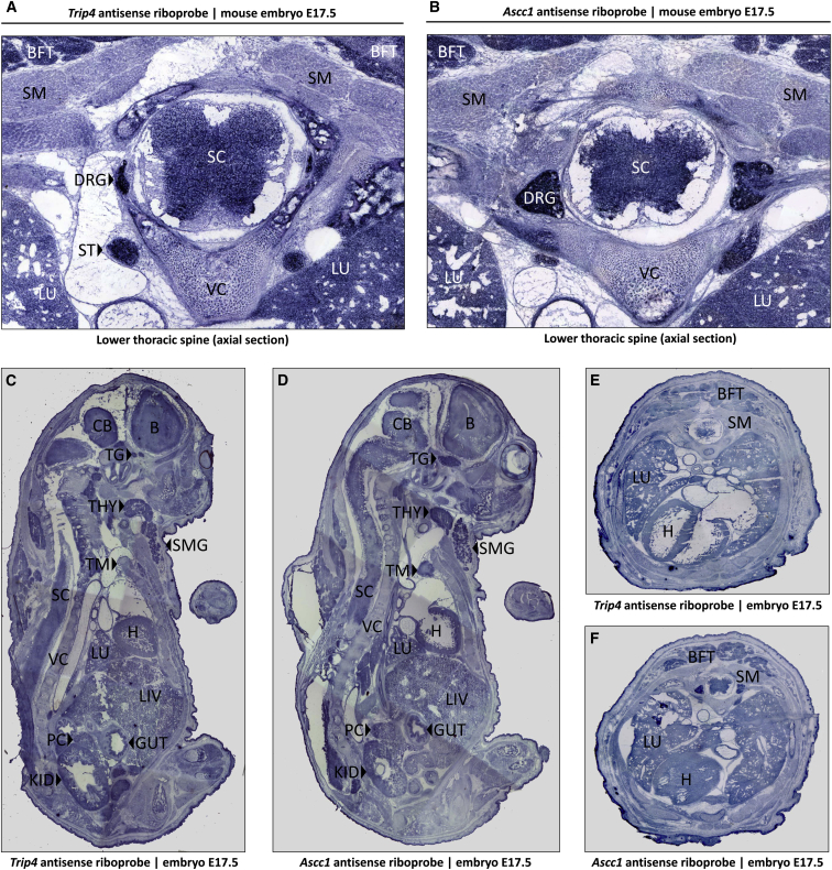
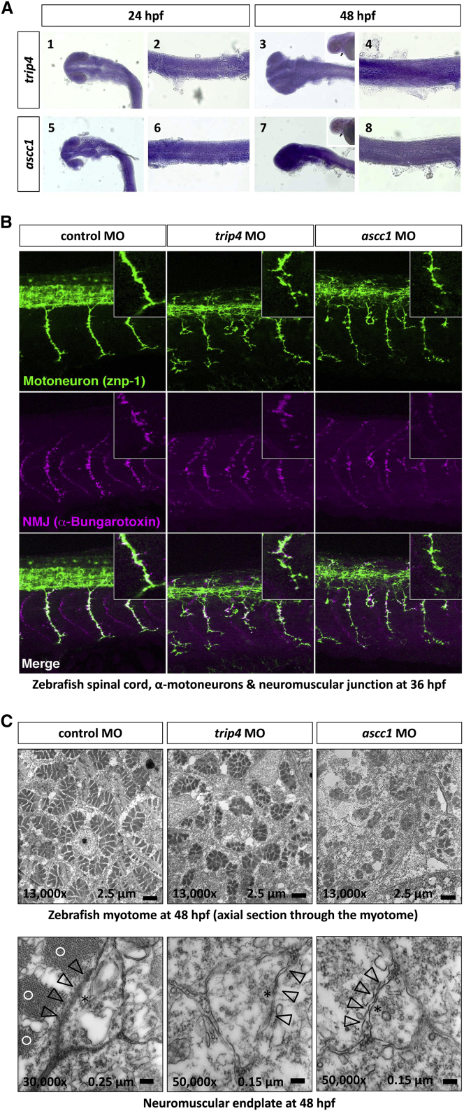
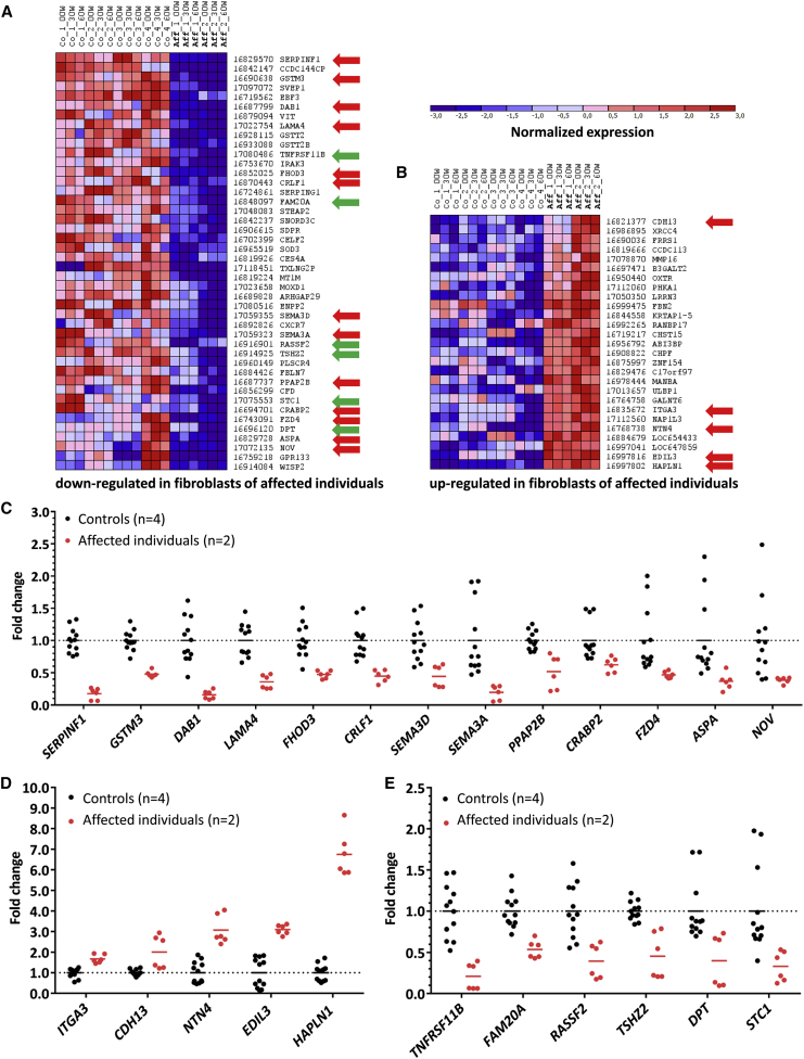
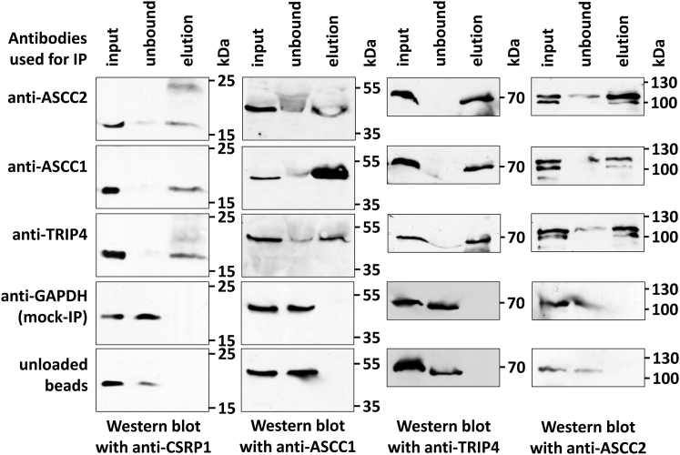
Transcriptional signal cointegrators associate with transcription factors or nuclear receptors and coregulate tissue-specific gene transcription. We report on recessive loss-of-function mutations in two genes (TRIP4 and ASCC1) that encode subunits of the nuclear activating signal cointegrator 1 (ASC-1) complex. We used autozygosity mapping and whole-exome sequencing to search for pathogenic mutations in four families. Affected individuals presented with prenatal-onset spinal muscular atrophy (SMA), multiple congenital contractures (arthrogryposis multiplex congenita), respiratory distress, and congenital bone fractures. We identified homozygous and compound-heterozygous nonsense and frameshift TRIP4 and ASCC1 mutations that led to a truncation or the entire absence of the respective proteins and cosegregated with the disease phenotype. Trip4 and Ascc1 have identical expression patterns in 17.5-day-old mouse embryos with high expression levels in the spinal cord, brain, paraspinal ganglia, thyroid, and submandibular glands. Antisense morpholino-mediated knockdown of either trip4 or ascc1 in zebrafish disrupted the highly patterned and coordinated process of α-motoneuron outgrowth and formation of myotomes and neuromuscular junctions and led to a swimming defect in the larvae. Immunoprecipitation of the ASC-1 complex consistently copurified cysteine and glycine rich protein 1 (CSRP1), a transcriptional cofactor, which is known to be involved in spinal cord regeneration upon injury in adult zebrafish. ASCC1 mutant fibroblasts downregulated genes associated with neurogenesis, neuronal migration, and pathfinding (SERPINF1, DAB1, SEMA3D, SEMA3A), as well as with bone development (TNFRSF11B, RASSF2, STC1). Our findings indicate that the dysfunction of a transcriptional coactivator complex can result in a clinical syndrome affecting the neuromuscular system.

Keywords: ASCC1; TRIP4; arthrogryposis multiplex congenita; bone fractures; exome sequencing; neuromuscular unit; respiratory distress; spinal muscular atrophy; zebrafish model.


Figures

Figure 1
Figure 1
Clinical Presentation of the Affected Children (A and B) Axial T2-weighted cranial MRI images of affected children D.II_02 (A) (at a corrected gestational age of 39 weeks) and D.II_03 (B) (at a corrected gestational age of 37 weeks) with a simplified gyral pattern of the frontal lobes and enlargement of the external CSF spaces. Myelination of the brain stem and the basal ganglia is normal. (C) X-ray of the bilateral congenital femoral fractures of individual D.II_03. (D) Muscle histology demonstrates a reduction in fiber size and an increase in fiber-size variation in two individuals with a TRIP4 and ASCC1 mutation, in contrast to fiber size and variation in an age-matched control individual. The grouping of the larger type I fibers (marked by MHCslow), in contrast to a normal checkerboard pattern in the control individual, is characteristic for prenatal SMA. The intense staining of the muscles of the affected children for MHCdev highlights their immaturity. (E) Ultrastructure of a sural nerve biopsy specimen with normal myelinization but with loss of unmyelinated axons as documented by “empty” pouches (open triangles). (F) Presence of multiple intensely stained apoptotic α-motoneurons in the anterior horn of the spinal cord in a postmortem sample.
Figure 2
Figure 2
Pedigrees of the Families and Molecular Genetic Findings (A) The pedigrees of all investigated families with their respective genotypes are depicted below the symbols. Individuals marked with an asterisk were used for autozygosity mapping. (B) Variants identified by WES were verified by Sanger sequencing for segregation in all family members. Below the electropherograms, the reading frame of the respective amino acids is provided in the three letter code. Both TRIP4 mutations resulted in a premature termination codon, whereas the ASCC1 mutation led to a frameshift with a termination codon after insertion of 19 non-original amino acids. (C) Western blot of a muscle protein extract from two individuals with a TRIP4 mutation and from a control individual. The premature termination codons in both affected children lead to upregulation of an alternative splice isoform at ∼53 kDa that excludes both mutant positions. Anti-pan-Actin band density was used as a loading control. (D) Western blot of a protein extract from cultured fibroblasts of both children from family D and of two control individuals. The blot demonstrates the complete absence of the ASCC1 band in the affected children. β-tubulin band density was used as a loading control.
Figure 3
Figure 3
Splice Isoforms of TRIP4 and ASCC1 (A) Genomic structure of TRIP4 (not drawn to scale). The introns are indicated by gray lines. (B) The exons included into the various mRNA splice isoforms are depicted in green, and the red box indicates a frameshift. (C) Sequence traces of the exon splice junctions. Their respective localization is marked on (B). The alternative splice acceptor site in exon 7 is highlighted by a red box. (D) Genomic structure of ASCC1 (not drawn to scale). (E) The transcript encoding the 41 kDa ASCC1 is the most abundant. Exon 3b, which contains a missense mutation in 5.4% of the ExAC alleles and leads to the truncation of the protein (p.Ser78), is only present in a rare ASCC1 45 kDa splice variant found in 0%–2% of the indicated GEO RNA-seq datasets.
Figure 4
Figure 4
Gene Expression Study in E17.5 Mouse Embryos In situ hybridizations of cryosections from E17.5 mouse embryos demonstrate the nearly identical expression patterns of Trip4 (A, C, and E) and of Ascc1 (B, D, and F) mRNA. At the lower thoracic level (A and B), the highest expression levels were seen in the spinal cord, dorsal root ganglia, paraspinal sympathetic ganglia, muscle, lung, and brown fat tissue. In the parasagittal sections, the highest expression levels were seen on the thyroid and submandibular salivary gland and the trigeminal ganglion. Abbreviations are as follows: B, brain; BFT, brown fat tissue; CB, cerebellum; DRG, dorsal root ganglion; GUT, gut; H, heart; KID, kidney; LIV, liver; LU, lung; PC, pancreas; SC, spinal cord; SM, skeletal muscle; SMG, submandibular salivary gland; ST, sympathetic tract; TG, trigeminal ganglion; TG, thyroid gland; TM; thymus; VC, vertebral column.
Figure 5
Figure 5
Studies on Zebrafish Embryos Expression of trip4 and ascc1 mRNA. (A) trip4 is expressed ubiquitously in the head (A1) and trunk (A2) at 24 hpf and at 48 hpf (A3 and A4). ascc1 is also expressed ubiquitously in the head (A5) and trunk (A6) at 24 hpf and 48 hpf (A7 and A8). The arrowheads indicate the heart, verifying that both trip4 and ascc1 are expressed in cardiac muscles. (B) MO-mediated trip4 and ascc1 knockdown in zebrafish larvae led to a severe derangement of α-motoneuron axons and the myotome. In the morphants, we found a perturbed outgrowth of α-motoneuron axons projecting to the trunk muscle in every somite segment at 36 hpf. The α-motoneurons were short, thin, and fragile with abnormal branches in trip4 morphants and ascc1 morphants. In these morphants, we additionally see ectopic outgrowth of motoneurons from the spinal cord. The α-motoneurons are labeled with the znp-1 antibody (green). Labeling with α-bungarotoxin (purple) displays the formation of neuromuscular junctions that form along with the α-motoneurons. The neuromuscular junctions were thin, reduced in number, and disorganized in the trip4 and ascc1 morphants. (C) Electron-microscopic images of axial sections through the zebrafish myotome at 48 hpf. The rosette-like formation of myofibrils is greatly disturbed with reduced size and numbers. The lower panels show a neuromuscular endplate of the control morphants (left) with a normal thickened basal lamina, which is directly adjacent to the contractile elements of the myofibril. This is in contrast to the endplates from the trip4 and ascc1 morphants, which are smaller and have a disrupted basal lamina and no adjacent contractile elements. An asterisk denotes clusters of neurotransmitter vesicles (vesicle diameter 30–40 nm); open arrowheads denote the synaptic cleft and basal lamina of the neuromuscular endplate; open circles denote sarcomers (contractile elements) in the vicinity of the neuromuscular endplate. Note the higher magnification of the morphant endplate.
Figure 6
Figure 6
Gene Expression Analysis of Wild-Type and ASCC1 Mutant Fibroblasts (A) Clustergram of genes downregulated in fibroblasts of affected children with FDR < 0.01. Blue denotes normalized downregulation; red denotes upregulation (see attached scale above). The numbers on the right identify the Affymetrix Human GeneChip 2.0 probe set. The gene names are given on the far right column. The red arrows depict genes involved in neurogenesis; green arrows depict genes involved in bone metabolism. Abbreviations are as follows: Co, control; Aff, affected individual with ASCC1 mutation; 00W, serum starved for 12 hr; 30W, serum challenge for 30 min; 60W, serum challenge for 60 min; FDR, false discovery rate. (B) Genes upregulated in fibroblasts of affected children with FDR < 0.01. (C and D) Genes involved in neurogenesis that are downregulated (C) or upregulated (D) in ASCC1 mutant fibroblasts (red dots). (E) Downregulated genes involved in bone metabolism and development (red dots). Each dot represents a separate hybridization experiment. Black dots denote control individuals; red dots denote affected individuals. The horizontal lines represent the arithmetic mean.
Figure 7
Figure 7
Coimmunoprecipitation of CSRP1 with Three Subunits of the ASC-1 Complex Immunoprecipitation was done with anti-TRIP4, anti-ASCC1, and anti-ASCC2 antibodies. For mock immunoprecipitation, we used GAPDH antibodies, and for empty controls, we used unloaded Protein-G-Sepharose beads. Western blots from input, flow-through (unbound), and bound protein (elution) were serially incubated with anti-TRIP4, anti-ASCC1, anti-ASCC2, as well as with anti-CSRP1 antibodies.

References

    1. Hoff J.M., Loane M., Gilhus N.E., Rasmussen S., Daltveit A.K. Arthrogryposis multiplexa congenita: an epidemiologic study of nearly 9 million births in 24 EUROCAT registers. Eur. J. Obstet. Gynecol. Reprod. Biol. 2011;159:347–350. - PubMed
    1. Hall J.G. Arthrogryposis (multiple congenital contractures): diagnostic approach to etiology, classification, genetics, and general principles. Eur. J. Med. Genet. 2014;57:464–472. - PubMed
    1. Bamshad M., Van Heest A.E., Pleasure D. Arthrogryposis: a review and update. J. Bone Joint Surg. Am. 2009;91(Suppl 4):40–46. - PMC - PubMed
    1. Beck A.E., McMillin M.J., Gildersleeve H.I.S., Kezele P.R., Shively K.M., Carey J.C., Regnier M., Bamshad M.J. Spectrum of mutations that cause distal arthrogryposis types 1 and 2B. Am. J. Med. Genet. A. 2013;161A:550–555. - PMC - PubMed
    1. McMillin M.J., Below J.E., Shively K.M., Beck A.E., Gildersleeve H.I., Pinner J., Gogola G.R., Hecht J.T., Grange D.K., Harris D.J., University of Washington Center for Mendelian Genomics Mutations in ECEL1 cause distal arthrogryposis type 5D. Am. J. Hum. Genet. 2013;92:150–156. - PMC - PubMed

Publication types

MeSH terms