. 2016:2016:6595610.
doi: 10.1155/2016/6595610. Epub 2016 Jan 27.

Imaging Findings of Plasmacytoma of Both Breasts as a Preceding Manifestation of Multiple Myeloma

Affiliations

Imaging Findings of Plasmacytoma of Both Breasts as a Preceding Manifestation of Multiple Myeloma

Young Mi Park. Case Rep Med. 2016.

Abstract

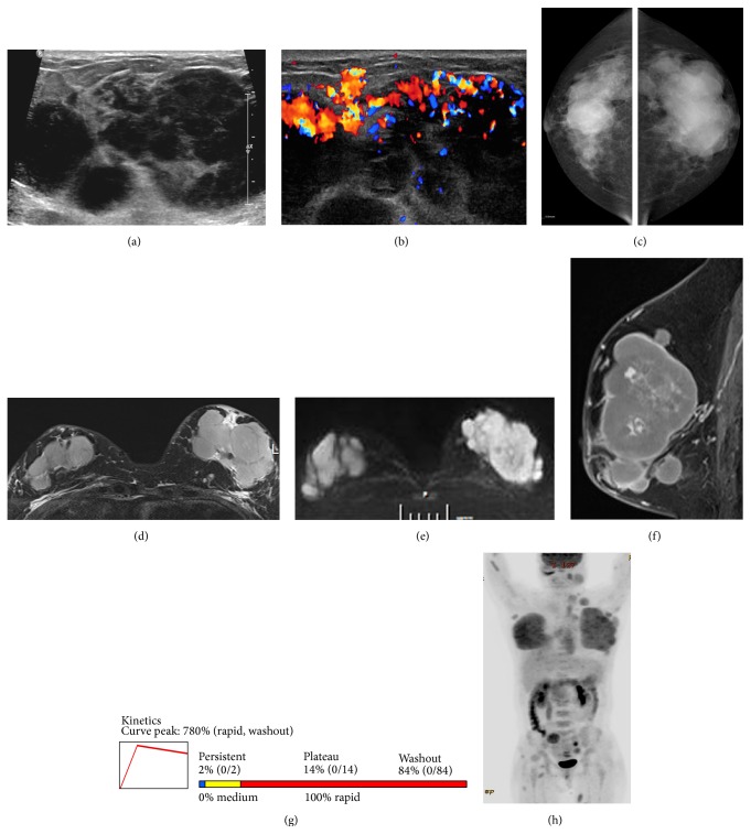
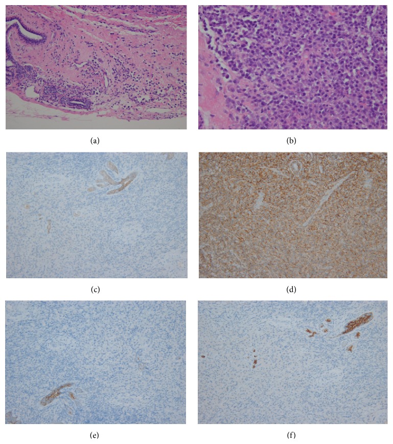
Breast plasmacytoma is an extremely rare tumor. It can occur as a primary isolated tumor or as an extramedullary manifestation in multiple myeloma. This report describes the unusual case of a primary extramedullary plasmacytoma that progressed to multiple myeloma within 15 months in a 35-year-old woman. The patient had been initially diagnosed with a primary extramedullary plasmacytoma of the epidural soft tissue at the cervical 6-thoracic 1 spine level and the stomach. The patient had received chemotherapy and the disease had been in remission. One year later, the disease recurred, affecting both breasts, right clavicle, and orbit. Three months later, the disease had progressed to multiple myeloma. I report this case, focusing on the findings of mammography, ultrasonography, magnetic resonance imaging, and positron emission tomography of bilateral breast plasmacytoma, and provide a review of the literature.


Figures

Figure 1
Figure 1
Gray scale (a) and color Doppler (b) sonograms of the breast show multiple, circumscribed, markedly hypoechoic, round or oval masses with highly increased vascularity. Mammograms (c) show circumscribed irregular conglomerated hyperdense masses invading both entire breasts. T2-weighted image of breast magnetic resonance imaging (MRI) (d) shows multiple, circumscribed, oval or round masses of high signal intensity (SI) in both breasts. The masses reveal high SI on diffusion-weighted image (e) with a low apparent diffuse coefficient value (not shown) suspicious for malignancy. Early phase of dynamic contrast-enhanced sagittal T1-weighted MRI (f) shows multiple circumscribed masses with strong homogeneous enhancement. Time-intensity curve of dynamic contrast-enhanced MRI (g) shows early strong (780% of the baseline value) and fast/delayed washout enhancement kinetics, suggestive of malignancy. Follow-up positron emission tomography-computed tomography scan (h) reveals massive breast plasmacytomas (SUVmax 6.3), invading both entire breasts. Metastatic lymphadenopathies throughout the whole body and metastatic masses in the abdomen, left cheek, both shoulders, chest wall, right buttock, and both thighs are shown.
Figure 2
Figure 2
Histopathological analysis ((a) HE ×200) of left breast mass shows infiltration of plasmacytoid cells around the ductal breast tissue. The plasmacytoid cells are eccentrically located with slightly enlarged nuclei compared to mature plasma cells ((b) HE ×400). Photomicrographs show a positive reaction for CD 138 ((c) ×200), indicating plasma cell origin, and positive reactions for E-cadherin ((d) ×200), CK5/6 ((e) ×200), and CK7 ((f) ×200) in the entrapped breast ductal tissue, but negative in infiltrated cells, suggesting a nonepithelial origin.

References

    1. Kyle R. A., Gertz M. A., Witzig T. E., et al. Review of 1027 patients with newly diagnosed multiple myeloma. Mayo Clinic Proceedings. 2003;78(1):21–33. doi: 10.4065/78.1.21. - DOI - PubMed
    1. Surov A., Holzhausen H.-J., Ruschke K., Arnold D., Spielmann R.-P. Breast plasmacytoma. Acta Radiologica. 2010;51(5):498–504. doi: 10.3109/02841851003712924. - DOI - PubMed
    1. The International Myeloma Working Group. Criteria for the classification of monoclonal gammopathies, multiple myeloma and related disorders: a report of the International Myeloma Working Group. British Journal of Haematology. 2003;121(5):749–757. doi: 10.1046/j.1365-2141.2003.04355.x. - DOI - PubMed
    1. Soutar R., Lucraft H., Jackson G., et al. Guidelines on the diagnosis and management of solitary plasmacytoma of bone and solitary extramedullary plasmacytoma. British Journal of Haematology. 2004;124(6):717–726. doi: 10.1111/j.1365-2141.2004.04834.x. - DOI - PubMed
    1. Alexiou C., Kau R. J., Dietzfelbinger H., et al. Extramedullary plasmacytoma: tumor occurrence and therapeutic concepts. Cancer. 1999;85(11):2305–2314. doi: 10.1002/(sici)1097-0142(19990601)85:11<2305::aid-cncr2>3.0.co;2-3. - DOI - PubMed

LinkOut - more resources