. 2016:2016:7469549.
doi: 10.1155/2016/7469549. Epub 2016 Jan 27.

Assessment of the Protective Role of Prenatal Zinc versus Insulin Supplementation on Fetal Cardiac Damage Induced by Maternal Diabetes in Rat Using Caspase-3 and KI67 Immunohistochemical Stains

Affiliations

Assessment of the Protective Role of Prenatal Zinc versus Insulin Supplementation on Fetal Cardiac Damage Induced by Maternal Diabetes in Rat Using Caspase-3 and KI67 Immunohistochemical Stains

Ahmed S Shams et al. Cardiol Res Pract. 2016.

Abstract

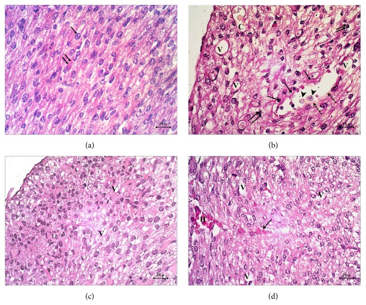
Maternal diabetes mellitus (DM) affects early organogenesis. Metabolic disorders of DM are associated with a depleted zinc status. This study evaluated the effect of maternal DM on cardiac development of rat fetuses and protective roles of prenatal zinc versus insulin supplementation. Pregnant rats were divided into 4 groups ((I) control, (II) STZ-induced DM, (III) STZ-induced DM treated with Zn, and (IV) STZ induced DM treated with insulin), all sacrificed on GD 20. Fetal heart weight of diabetic rats showed significant decrease compared to controls (P < 0.05). H&E stained section of controls had normal appearance of the myocardium, compared to diabetics that showed myocardial disarray with characteristic degenerative changes. Sections of zinc treated group showed restored architecture of normal myofibrils with minimal degenerative changes, while those of insulin treated group show partial restoration of the normal architecture of cardiomyocytes with focal improvement of cardiac tissue. Caspase-3 immunostained slides showed positive cytoplasmic immunoreactivity in diabetic group. But KI67 immunostained slides revealed negative nuclear immunoreaction in diabetics. We observed that gestational diabetes was associated with increased risk of fetal myocardial damage that might be caused by increased apoptotic level. Treating diabetic pregnant subjects with zinc and insulin was associated with improvement in myocardial integrity.


Figures

Figure 1
Figure 1
Photomicrograph of H&E (×400) sections in the fetal myocardium. (a) Control group showing normal striated pattern of the cardiomyocytes with centrally located nuclei (arrow) and eosinophilic cytoplasm (double arrow). (b) STZ-induced DM group showing myocardial disarray, marked vacuolar degeneration of the cardiomyocytes (V), fragmentation of the nuclei of cardiomyocytes (arrow) and pyknotic changes of the nuclei (arrow head), congested blood vessels (C), and discontinuation of the vessel wall (dashed arrow) with the presence of subendocardial thickening (double arrow). (c) STZ-induced DM treated with zinc group showing restored architecture of cardiomyocytes with the presence of mild vacuolar degeneration (V). (d) STZ-induced DM treated with insulin group showing focal improvement of the myocardium with the presence of vacuolar degeneration (V) and myocardial hemorrhage (H) with extravasated red blood cells within the myocardial tissue (arrow).
Figure 2
Figure 2
Photomicrograph of caspase-3 immunohistochemical staining (×400) sections in the fetal myocardium. (a) control group. (b) STZ-induced DM group. (c) STZ-induced DM treated with zinc group. (d) STZ-induced DM treated with insulin group. Shown is positive brownish cytoplasmic immunohistochemical staining with caspase-3 in group (b) which seems to be downregulated in both group (c) and group (d); this brownish cytoplasmic immunohistochemical staining is almost absent in group (a).
Figure 3
Figure 3
A photomicrograph of KI67 immunohistochemical staining (×400) sections in the fetal myocardium. (a) Control group showing dense positive brown nuclear immunohistochemical staining with KI67. (b) STZ-induced DM group showing negative nuclear immunohistochemical staining with KI67. (c) STZ-induced DM treated with zinc group showing moderate positive nuclear immunohistochemical staining with KI67. (d) STZ-induced DM treated with insulin group showing minimal positive nuclear immunohistochemical staining with KI67.

References

    1. Shaw J. E., Sicree R. A., Zimmet P. Z. Global estimates of the prevalence of diabetes for 2010 and 2030. Diabetes Research and Clinical Practice. 2010;87(1):4–14. doi: 10.1016/j.diabres.2009.10.007. - DOI - PubMed
    1. Aneja A., Tang W. H. W., Bansilal S., Garcia M. J., Farkouh M. E. Diabetic cardiomyopathy: insights into pathogenesis, diagnostic challenges, and therapeutic options. The American Journal of Medicine. 2008;121(9):748–757. doi: 10.1016/j.amjmed.2008.03.046. - DOI - PubMed
    1. Kumar S. D., Dheen T. S., Tay S. S. W. Maternal diabetes induces congenital heart defects in mice by altering the expression of genes involved in cardiovascular development. Cardiovascular Diabetology. 2007;6, article 34 doi: 10.1186/1475-2840-6-34. - DOI - PMC - PubMed
    1. Kumar S. D., Yong S.-K., Dheen S. T., Bay B.-H., Tay S. S.-W. Cardiac malformations are associated with altered expression of vascular endothelial growth factor and endothelial nitric oxide synthase genes in embryos of diabetic mice. Experimental Biology and Medicine. 2008;233(11):1421–1432. doi: 10.3181/0806-RM-186. - DOI - PubMed
    1. Devereux R. B., Roman M. J., Paranicas M., et al. Impact of diabetes on cardiac structure and function: the Strong Heart study. Circulation. 2000;101(19):2271–2276. doi: 10.1161/01.cir.101.19.2271. - DOI - PubMed

LinkOut - more resources