

Osteochondritis dissecans of the capitellum in adolescents
- PMID: 26925381
- PMCID: PMC4757654
- DOI: 10.5312/wjo.v7.i2.102
Osteochondritis dissecans of the capitellum in adolescents
Abstract
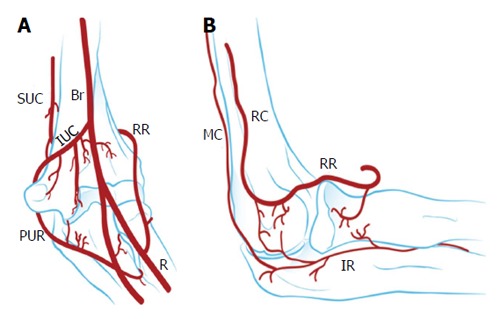
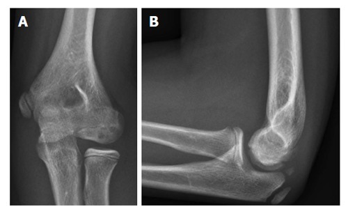
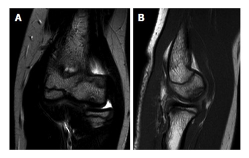
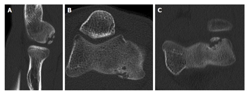
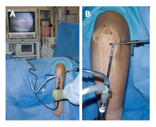
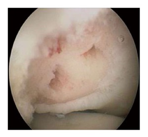
Osteochondritis dissecans (OCD) is a disorder of articular cartilage and subchondral bone. In the elbow, an OCD is localized most commonly at the humeral capitellum. Teenagers engaged in sports that involve repetitive stress on the elbow are at risk. A high index of suspicion is warranted to prevent delay in the diagnosis. Plain radiographs may disclose the lesion but computed tomography and magnetic resonance imaging are more accurate in the detection of OCD. To determine the best treatment option it is important to differentiate between stable and unstable OCD lesions. Stable lesions can be initially treated nonoperatively with elbow rest or activity modification and physical therapy. Unstable lesions and stable lesions not responding to conservative therapy require a surgical approach. Arthroscopic debridement and microfracturing has become the standard initial procedure for treatment of capitellar OCD. Numerous other surgical options have been reported, including internal fixation of large fragments and osteochondral autograft transfer. The aim of this article is to provide a current concepts review of the etiology, clinical presentation, diagnosis, treatment, and outcomes of elbow OCD.
Keywords: Adolescent; Arthroscopy; Athletes; Bone marrow stimulation; Capitellum; Cartilage; Elbow; Osteoarthritis; Osteochondritis dissecans; Overhead sports.
Figures
References
-
- Bruns J. [Osteochondrosis dissecans] Orthopade. 1997;26:573–584. - PubMed
-
- Kida Y, Morihara T, Kotoura Y, Hojo T, Tachiiri H, Sukenari T, Iwata Y, Furukawa R, Oda R, Arai Y, et al. Prevalence and Clinical Characteristics of Osteochondritis Dissecans of the Humeral Capitellum Among Adolescent Baseball Players. Am J Sports Med. 2014;42:1963–1971. - PubMed
-
- Baratz M, Yi SJ. Osteochondritis dissecans of the elbow. In: Eygendaal D, editor. The elbow. The treatment of basic elbow pathology. Nieuwegein: Arko Sports Media; 2009. pp. 139–148.
-
- Kenniston JA, Beredjiklian PK, Bozentka DJ. Osteochondritis dissecans of the capitellum in fraternal twins: case report. J Hand Surg Am. 2008;33:1380–1383. - PubMed
-
- Douglas G, Rang M. The role of trauma in the pathogenesis of the osteochondroses. Clin Orthop Relat Res. 1981;(158):28–32. - PubMed
Publication types
LinkOut - more resources
Full Text Sources
Other Literature Sources
