Review
. 2016 May 23:11:343-64.
doi: 10.1146/annurev-pathol-012615-044351. Epub 2016 Feb 25.

Pathogenesis of Staphylococcus aureus Bloodstream Infections

Affiliations
Review

Pathogenesis of Staphylococcus aureus Bloodstream Infections

Lena Thomer et al. Annu Rev Pathol. .

Abstract

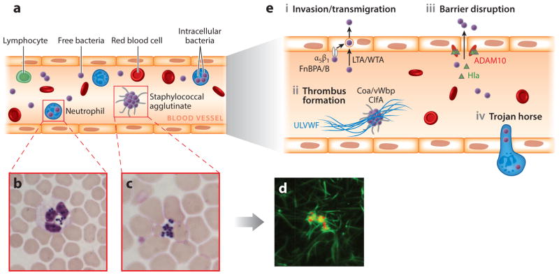
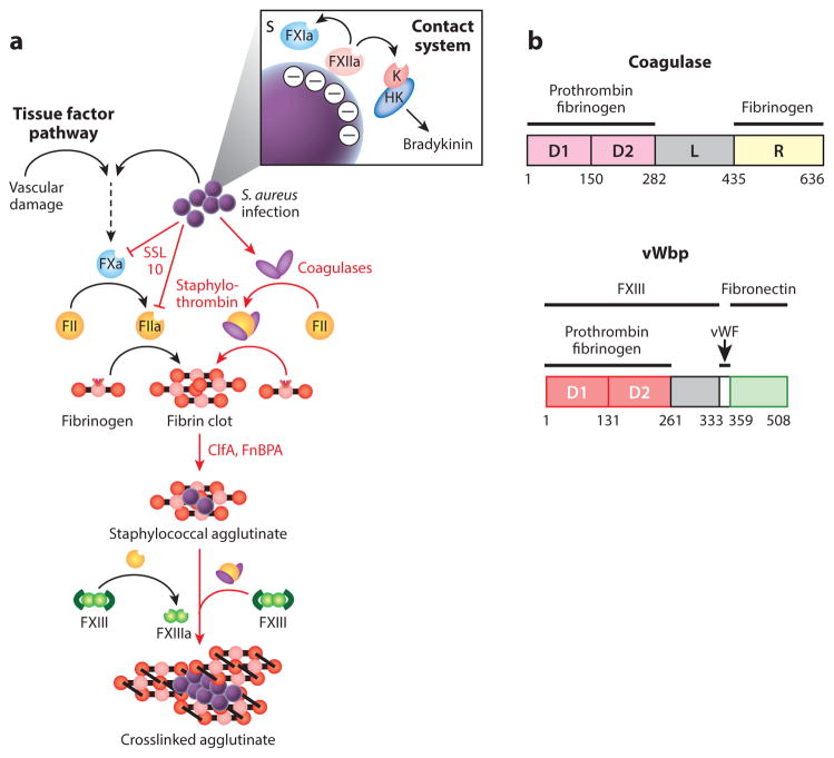
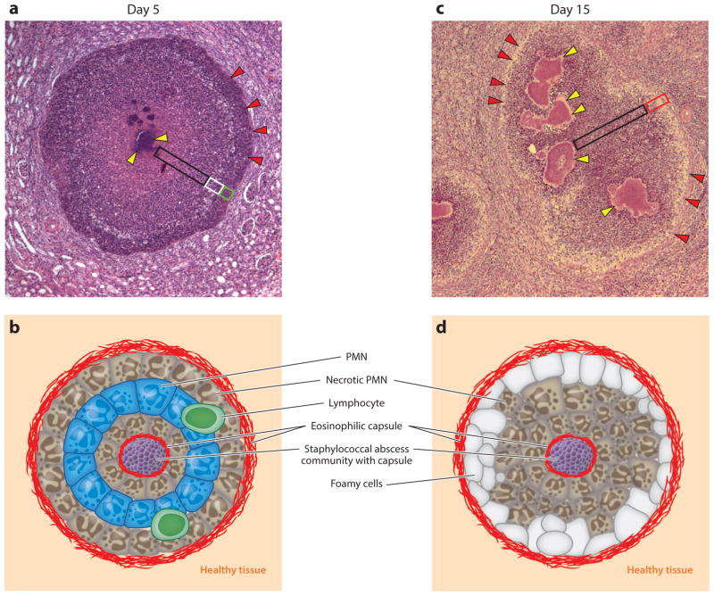
Staphylococcus aureus, a Gram-positive bacterium colonizing nares, skin, and the gastrointestinal tract, frequently invades the skin, soft tissues, and bloodstreams of humans. Even with surgical and antibiotic therapy, bloodstream infections are associated with significant mortality. The secretion of coagulases, proteins that associate with and activate the host hemostatic factor prothrombin, and the bacterial surface display of agglutinins, proteins that bind polymerized fibrin, are key virulence strategies for the pathogenesis of S. aureus bloodstream infections, which culminate in the establishment of abscess lesions. Pathogen-controlled processes, involving a wide spectrum of secreted factors, are responsible for the recruitment and destruction of immune cells, transforming abscess lesions into purulent exudate, with which staphylococci disseminate to produce new infectious lesions or to infect new hosts. Research on S. aureus bloodstream infections is a frontier for the characterization of protective vaccine antigens and the development of immune therapeutics aiming to prevent disease or improve outcomes.

Keywords: abscess formation; agglutination; coagulation; immune evasion; vaccine.


Figures

Figure 1
Figure 1
Survival of Staphylococcus aureus in the bloodstream and dissemination into host tissues. (a) Following intravenous inoculation of mice with ~107 colony-forming units (CFU), S. aureus survives in the bloodstream and disseminates from the vasculature into organ tissues. Bacteria from the initial inoculum cannot be detected in the bloodstream within 24 h. (bc) Microscopy of Giemsa-stained blood samples from mice infected for 2 h reveals staphylococci that are (b) associated with neutrophils or (c) agglutinated outside of immune cells. Staphylococci phagocytosed by neutrophils deploy several virulence strategies that ensure their survival (see text for details). (d ) Extracellular S. aureus cocci expressing mCherry (red fluorescence) interact with fibrin ( green fluorescence) to generate large bacterial agglutinates covered with a shield of fibrin cables, thereby preventing phagocytosis. The image shows S. aureus mixed for 5 min with anticoagulated human plasma that has been supplemented with 5% Alexa488-conjugated human fibrinogen. Staphylococcal agglutination requires Coa/vWbp (coagulases) and prothrombin as well as ClfA (agglutinin) and fibrinogen. Agglutinins are S. aureus surface proteins that bind coagulase-derived fibrin cables to produce large aggregates of bacteria covered with fibrin. (e) Models for S. aureus exit from the bloodstream. (i ) FnBPA and FnBPB (staphylococcal surface proteins) bind fibronectin and interact with integrin α5β1 on the surface of the vascular endothelium, thereby triggering invasion of cells and transmigration. Wall teichoic acid (WTA) and lipoteichoic acid (LTA) of S. aureus, polymers in the bacterial envelope, have also been proposed to promote staphylococcal invasion of host cells. (ii ) Staphylococci induce formation of fibrin thrombi via Coa/vWbp- and ClfA-mediated agglutination and bind to von Willebrand factor (vWF) on endothelial surfaces, generating Ultra Large vWF (ULVWF) polymers. (iii ) Staphylococci secrete Hla, a toxin that binds to the ADAM10 receptor to disrupt the physiological barrier functions of the vascular endothelium. (iv) The Trojan horse model, whereby neutrophils with intracellular S. aureus extravasate to deliver bacteria into host tissues.
Figure 2
Figure 2
Agglutination and Staphylococcus aureus interference with host hemostasis. (a) Vascular damage and tissue factor or contact activation and bradykinin are pathways for hemostasis—a cascade of proteolytic activation for the zymogens of clotting factors culminating in the conversion of prothrombin (FII) to thrombin (FIIa), which cleaves fibrinopeptides A and B from fibrinogen, thereby triggering self-assembly of fibrin cables either to stem blood loss from injured vasculature or to immobilize pathogens that have activated the contact system. S. aureus bypasses the host hemostasis pathways to activate FII/prothrombin via the secretion of coagulases (Coa and vWbp), thus yielding staphylothrombin and, via nonproteolytic activation of fibrin-stabilizing factor (FXIII), the transglutaminase responsible for crosslinking fibrin cables. ClfA, an agglutinin of the surface of S. aureus, captures staphylothrombin-derived fibrin cables, thereby agglutinating staphylococci into large aggregates of bacteria enclosed by a fibrin shield. (b) A diagram illustrating the primary translational products of Coa and vWbp from S. aureus strain Newman; the catalytic D1 and D2 domains are shown in light and dark red boxes, and the R domain of coagulase with repeats is shown in yellow. The function of the linker domain (L) is not known. The number of repeats in the R region of coagulase varies between strains. The binding sites for prothrombin, fibrinogen, FXIII, and fibronectin are delineated. Abbreviation: vWF, von Willebrand factor.
Figure 3
Figure 3
Staphylococcus aureus abscess lesions in murine renal tissue. (a) Microscopy image of hematoxylin-eosin-stained, thin-sectioned renal tissue 5 days following intravenous inoculation with S. aureus. Bacteria are organized into staphylococcal abscess communities (SACs) ( yellow arrowheads) at the center of the lesion. Depending on sectioning, SACs may appear fragmented; however, bacterial communities remain connected and are enclosed by eosinophilic deposits of fibrin and surrounded by zones of necrotic neutrophils (black box), healthy-appearing neutrophils (white box), and necrotic immune cells ( green box). Abscess lesions are demarcated from renal tissues by another layer of eosinophilic deposits (red arrowheads), through which immune cells enter the lesion. (b) Schematic illustrating the histopathology features of a 5-day-old S. aureus abscess lesion. (c) Microscopy image of hematoxylin-eosin-stained, thin-sectioned renal tissue 15 days following intravenous inoculation with S. aureus. The 15-day-old lesion differs from the 5-day-old lesion in that SACs have expanded and the layering of healthy and necrotic immune cells has been supplanted by diffuse infiltrates of immune cells, cellular detritus, liquefaction (black box), and foam cells positioned at the periphery of the lesion (red box). SACs remain enclosed by fibrin deposits ( yellow arrowheads). An outer eosinophilic layer (red arrowheads) demarcates the lesion from large numbers of immune cells and from the cellular detritus that has replaced renal tissue architecture in the vicinity of the abscess lesion. (d ) Schematic illustrating the histopathology features of a 15-day-old S. aureus abscess lesion. Abbreviation: PMN, polymorphonuclear leukocyte.

References

    1. van Belkum A, Melles DC, Nouwen J, van Leeuwen WB, van Wamel W, et al. Co-evolutionary aspects of human colonisation and infection by Staphylococcus aureus. Infect Genet Evol. 2009;9:32–47. - PubMed
    1. David MZ, Daum RS. Community-associated methicillin-resistant Staphylococcus aureus: epidemiology and clinical consequences of an emerging epidemic. Clin Microbiol Rev. 2010;23:616–87. - PMC - PubMed
    1. Liu C, Bayer AS, Cosgrove SE, Daum RS, Fridkin SK, et al. Clinical practice guidelines by the Infectious Diseases Society of America for the treatment of methicillin-resistant Staphylococcus aureus infections in adults and children: executive summary. Clin Infect Dis. 2011;52:285–92. - PubMed
    1. Bagnoli F, Bertholet S, Grandi G. Inferring reasons for the failure of Staphylococcus aureus vaccines in clinical trials. Front Cell Infect Microbiol. 2012;2:16. - PMC - PubMed
    1. Ogston A. Report upon micro-organisms in surgical diseases. Br Med J. 1881;1:369–75. - PMC - PubMed

MeSH terms

LinkOut - more resources