Review
. 2016 Apr;13(4):193-204.
doi: 10.1038/nrurol.2016.13. Epub 2016 Mar 1.

Receptors, channels, and signalling in the urothelial sensory system in the bladder

Affiliations
Review

Receptors, channels, and signalling in the urothelial sensory system in the bladder

Liana Merrill et al. Nat Rev Urol. 2016 Apr.

Abstract

The storage and periodic elimination of urine, termed micturition, requires a complex neural control system to coordinate the activities of the urinary bladder, urethra, and urethral sphincters. At the level of the lumbosacral spinal cord, lower urinary tract reflex mechanisms are modulated by supraspinal controls with mechanosensory input from the urothelium, resulting in regulation of bladder contractile activity. The specific identity of the mechanical sensor is not yet known, but considerable interest exists in the contribution of transient receptor potential (TRP) channels to the mechanosensory functions of the urothelium. The sensory, transduction, and signalling properties of the urothelium can influence adjacent urinary bladder tissues including the suburothelial nerve plexus, interstitial cells of Cajal, and detrusor smooth muscle cells. Diverse stimuli, including those that activate TRP channels expressed by the urothelium, can influence urothelial release of chemical mediators (such as ATP). Changes to the urothelium are associated with a number of bladder pathologies that underlie urinary bladder dysfunction. Urothelial receptor and/or ion channel expression and the release of signalling molecules (such as ATP and nitric oxide) can be altered with bladder disease, neural injury, target organ inflammation, or psychogenic stress. Urothelial receptors and channels represent novel targets for potential therapies that are intended to modulate micturition function or bladder sensation.


Conflict of interest statement

statement M.A.V. is funded by the National Institutes of Health National Institute of Diabetes and Digestive and Kidney Diseases. The other authors declare no competing interests.

Figures

Figure 1
Figure 1. An overview of micturition reflex control and cell layers of the wall of the urinary bladder
a. Storage and elimination (voiding) of urine. The neural pathways that control lower urinary tract function maintain a reciprocal relationship between the urinary bladder and the urethral outlet. Storage reflexes are activated during bladder filling and are organized primarily in the spinal cord, whereas voiding is mediated by reflex mechanisms that are organized in the brain. During bladder filling and storage, the parasympathetic innervation of the detrusor is inhibited and the smooth and striated parts of the urethral sphincter are activated, preventing involuntary bladder emptying. During bladder filling the parasympathetic efferent pathway to the bladder, including a population of CNS (for example pontine micturition centre) neurons, is turned off. As bladder filling continues and a critical level of bladder distension is achieved, the afferent activity from mechanoreceptors in the bladder switches the pathway to the elimination mode. During elimination (voiding), parasympathetic activity is activated resulting in urinary bladder contraction, whereas sympathetic activity and somatomotor activity is withdrawn. b. Anatomical components of the urinary bladder wall. Numerous receptors (including purinergic, adrenergic, cholinergic, neurotrophin, and neuropeptide) and ion channels (transient receptor potential channels) are expressed by the anatomical components of the urinary bladder wall including the urothelium, bladder sensory nerves, interstitial cells of Cajal (ICC), and detrusor smooth muscle.
Figure 2
Figure 2. Illustration of low-level afferent nerve discharge that is present throughout bladder filling
In humans, the sensation of bladder fullness is experienced at intravesical pressures of 5 15 mmHg. On passing this tension threshold, bladder afferent discharge increases to stimulate micturition reflexes. Urinary urgency occurs in humans when intravesical pressures reach 20 25 mmHg and if not relieved, pain and/or discomfort occur when pressures exceed 30 mmHg.
Figure 3
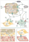
Figure 3. Hypothetical model of purinergic mechanosensory transduction and the involvement of TRP channels in the LUT focusing on potential interactions among bladder sensory nerves, urothelial cells, detrusor smooth muscle cells and ICC
Distension activates transient receptor potential (TRP) channels expressed by the urothelium leading to release of ATP, which then acts on purinergic receptors (P2X2 and/or P2X3) on suburothelial sensory nerves to convey sensory and/or nociceptive signals to the central nervous system (CNS). Several TRP channels are expressed by the urothelium and bladder sensory nerve fibres and are activated by diverse stimuli. Roles in lower urinary tract (LUT) physiology have been proposed for several TRP channels; however, the exact localization and function of TRP channels in the LUT in health and disease are still being determined. Release of ATP can be mediated by exocytosis (such as vesicular release), pannexin (Panx) channels and connexin (Cx) hemichannels. Expression of both P2X and P2Y receptors in interstitial cells of Cajal (ICC) also suggests that ATP release from the urothelium can influence the function of ICC as well as bladder sensory nerves. The signal resulting from ATP release depends on many factors including the purinergic receptors subtypes expressed, activation of G-protein-mediated and Ca2+ -mediated signalling and by the expression of ATPases and ectonucleotidases that degrade ATP. The ATP signal might influence sites beyond the urothelium and suburothelium. For example, gap junction proteins (such as connexins) are expressed by ICC and detrusor smooth muscle cells (Det) suggesting that gap junction-mediated intercellular communication could be an underlying mechanism for long-distance spread of signals from the urotheial cells to Det. Stimulation of urothelial receptors and channels can release mediators that target bladder sensory nerves and other cell types; urothelial cells can also be targets for neurotransmitters released from nerves or other cell types and can be activated by either autocrine (autoregulation) or paracrine (release from nearby nerve fibres or other cells) mechanisms. P1, purinergic 1 receptor; TRPA1, transient receptor potential channel ankyrin 1; TRPV, transient receptor potential channel vanilloid family; TRPM8, transient receptor potential channel melastatin 8; UC, urothelial cell.

Comment in

References

    1. Tank PW. In: Grant’s dissector. Stacey SL, editor. Lippincott Williams & Wilkins; 2009.
    1. Birder L, Andersson KE. Urothelial signaling. Physiol Rev. 2013;93:653–680. - PMC - PubMed
    1. Andersson KE, McCloskey KD. Lamina propria: the functional center of the bladder? Neurourol Urodyn. 2013;33:9–16. - PubMed
    1. Davidson RA, McCloskey KD. Morphology and localization of interstitial cells in the guinea pig bladder: structural relationships with smooth muscle and neurons. J Urol. 2005;173:1385–1390. - PubMed
    1. Andersson KE, Arner A. Urinary bladder contraction and relaxation: physiology and pathophysiology. Physiol Rev. 2004;84:935–986. - PubMed

Publication types

MeSH terms

Substances