. 2016 Apr;23(4):278-285.
doi: 10.1038/nsmb.3182. Epub 2016 Feb 29.

A structural ensemble of a ribosome-nascent chain complex during cotranslational protein folding

Affiliations

A structural ensemble of a ribosome-nascent chain complex during cotranslational protein folding

Lisa D Cabrita et al. Nat Struct Mol Biol. 2016 Apr.

Abstract

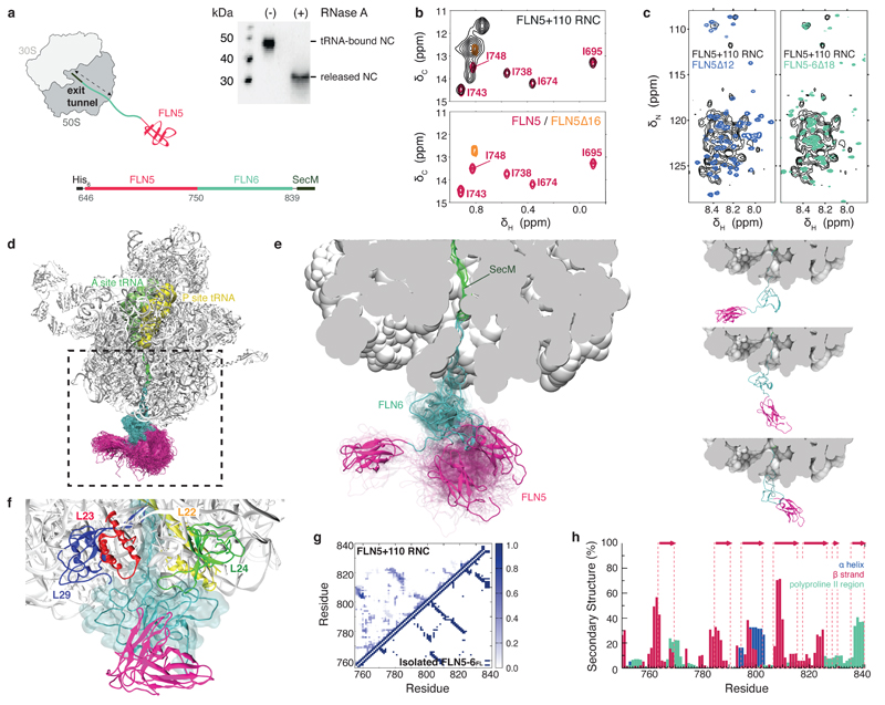
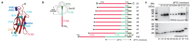
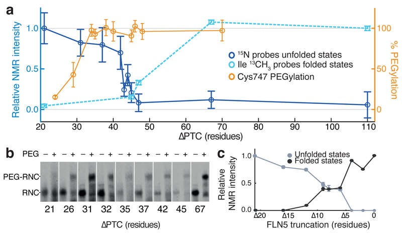
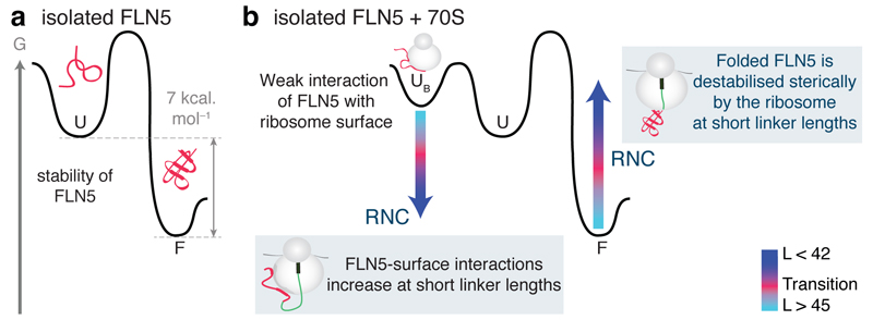
Although detailed pictures of ribosome structures are emerging, little is known about the structural and cotranslational folding properties of nascent polypeptide chains at the atomic level. Here we used solution-state NMR spectroscopy to define a structural ensemble of a ribosome-nascent chain complex (RNC) formed during protein biosynthesis in Escherichia coli, in which a pair of immunoglobulin-like domains adopts a folded N-terminal domain (FLN5) and a disordered but compact C-terminal domain (FLN6). To study how FLN5 acquires its native structure cotranslationally, we progressively shortened the RNC constructs. We found that the ribosome modulates the folding process, because the complete sequence of FLN5 emerged well beyond the tunnel before acquiring native structure, whereas FLN5 in isolation folded spontaneously, even when truncated. This finding suggests that regulating structure acquisition during biosynthesis can reduce the probability of misfolding, particularly of homologous domains.


Conflict of interest statement

Author Information

The authors declare no competing financial interests.

Figures

Figure 1
Figure 1. Structural ensemble of a ribosome-bound nascent chain.
(a) Schematic of the FLN5+110 RNC used for the ensemble calculations. The FLN5 sequence is tethered to the ribosome by a C-terminally truncated FLN6751-839 sequence and stalled using the SecM translational-arrest motif,. Anti-His western blots of purified FLN5+110 RNC, ribosome-attached with bound prolyl P-site tRNA, and also in its released form. (b) Overlay of 1H-13C correlation spectra of [U-2H; Ileδ1-13CH3 labeled] FLN5+110 RNC (black) with isolated, natively folded FLN5 (pink) and isolated unfolded FLN5∆16 (orange). (c) Overlay of 1H-15N correlation spectra of U-15N-labelled FLN5+110 RNC with isolated FLN5∆12 (blue), and unfolded FLN5-6∆18 (green). (d) NMR chemical shift restrained structural ensemble of FLN5+110 RNC, showing the disordered FLN6 linker (cyan) and the native fold acquired by FLN5 (pink). (Accession codes: PDB ID 2N62; BMRB ID 25748). (e) Close-up view of the ribosomal exit tunnel, highlighting three representative conformations of the nascent chain ensemble (left); the three representative conformations are also shown separately (right). (f) Transient interactions made between the disordered FLN6 linker in close proximity with the ribosomal proteins at the surface. (g) Probability of the formation of inter-residue contacts in the FLN5+110 RNC (shown above diagonal) and in the native state of full length (FL), isolated FLN5-6 (below diagonal) (h) Secondary structure populations of the RNC depicting β-strands (red), α-helices (blue) and polyproline II regions (green); native β-strands are indicated (red arrows).
Figure 2
Figure 2. Design and in vivo production of FLN5 ribosome-nascent chain complexes in E. coli.
(a) Structure of isolated, natively folded FLN5 (PDB: 1QFH). Mapped onto the FLN5 structure are the five isoleucines (∂ 1 methyl groups) of FLN5 (Ile674, 695, 738, 743, 748, cyan) used as probes of native structure acquisition, and the amide groups of three residues (Val682, Ala683 & Ala694, blue) selected for analysis of unfolded conformations (see text). (b) Design of the translationally-arrested RNCs to monitor nascent chain emergence and folding, in which the FLN5 sequence is tethered to the PTC via increasing lengths of the FLN6 sequence and the SecM translational arrest motif, (c) Anti-His western blots of the library of purified FLN5 RNCs shown in ribosome-bound (upper panel, see also Supplementary Fig. 1) and released (lower panel) forms.
Figure 3
Figure 3. Nascent chains of FLN5 emerging from the ribosome monitored by NMR spectroscopy.
(a) 1H-13C correlation spectra of [U-2H; Ileδ1-13CH3 labeled] FLN5 RNCs (black), isolated, natively folded FLN5 (cyan) and isolated unfolded FLN5∆16 (orange). Resonances marked “R” arise from background labeling of 70S ribosomal proteins. (b) 1H-15N correlation spectra of U-15N-labelled FLN5 RNCs, isolated FLN5∆12 (blue), and unfolded FLN5-6∆18 (green). Resonances used for the analysis of unfolded conformations are labeled in the FLN5+21 RNC spectrum.
Figure 4
Figure 4. Folding of FLN5 on the ribosome monitored by NMR spectroscopy and PEGylation.
(a) FLN5 nascent chain folding as measured by intensity changes of 15N amide resonances (blue) arising from the unfolded FLN5 domain (mean ± s.d. for n=4 (n=3 for +45 and n=2 +47); nascent chain concentration from western blot replicates), and intensity changes in Ile 13CH3 resonances (cyan) arising from native FLN5 structure (mean ± s.d. of spectral noise, n=1). Intensities are normalized and scaled relative to L = 21 (unfolded) or L = 110 (folded). The solvent accessibility of the FLN5 domain from the ribosomal exit tunnel was probed using PEGylation (orange) (mean ± s.d) of folding-incompetent FLN5 Y719E RNCs, where the native Cys747 is close to the FLN5 and FLN6 boundary. (b) Cys747 PEGylation of FLN5 Y719E RNCs results in a band shift (PEG-RNC). (c) C-terminal truncations of isolated FLN5 as measured by NMR. Averaged cross-peak intensities of folded (black) and unfolded (grey) states of FLN5 (see Supplementary Fig. 5) are mapped against truncation length.
Figure 5
Figure 5. FLN5 folding is offset on the ribosome.
A comparison of FLN5 folding on the ribosome and in isolation as depicted by the conformational states observed for FLN5 RNCs with different linker lengths, L, from the PTC, and those of C-terminal FLN5 truncations: unfolded (blue), folded (cyan) and folding transition (pink). The sequence of the FLN5 nascent chains is solvent exposed, as monitored by PEGylation, at L ≥ 31 residues where Cys747 is 34 residues from the PTC yet the domain only acquires native-like structure upon addition of a further 11-14 residues, at 42 ≤ L ≤ 45 as shown by NMR spectroscopy. Isolated FLN5 truncations are shown alongside the RNC lengths with FLN5∆4 a reference for L = 31, at which point complete PEGylation is observed. A folding offset (pink dotted arrow) is the difference observed between the initiation of FLN5 structure acquisition on the ribosome compared to that of the protein in isolation.
Figure 6
Figure 6. Residue-specific mapping of RNC interactions.
(a) An overlay of 1H-15N correlation spectra (recorded at a 1H frequency of 950 MHz) of FLN5+31 RNC (black) and unfolded, isolated FLN5∆12 (red) highlighting resonances that are significantly broadened in the RNC. (b) Relative intensities of 1H-15N resonances of folded FLN5 (5 µM) and unfolded FLN5 Y719E (8 µM) in the presence of 1 molar equivalent of 70S ribosomes. (c) Relative intensities of FLN5+21, FLN5+31, FLN5+42, FLN5+67 and FLN5+110 RNCs as compared to a reference, made up of a composite consisting of FLN5∆12 and FLN5 Y719E, to monitor unfolded FLN5 (red), and FLN5-6∆18 to monitor unfolded FLN6 (green), and folded FLN5 from 1H-13C correlation spectra (cyan). A 5-point moving average is plotted as a guide; errors derived from spectral noise, n=1. The grey shaded area denotes occluded residues inaccessible to PEGylation.
Figure 7
Figure 7. The ribosome modulates the folding landscape of FLN5 nascent chains.
(a) Schematic of a free energy diagram for isolated FLN5, showing the difference in free energy of 7 kcal mol-1 between the folded state, F, and the unfolded state, U. (b) Schematic free energy diagram for isolated FLN5 in the presence of ribosomes shows a ribosome-bound state, UB, accessible from the unfolded state. A model for how the ribosome could alter this landscape and inhibit nascent chain folding is indicated (arrows): At short linker lengths, the tethered nascent chain is subject to high effective ribosome concentrations, favoring a ribosome-bound state UB. The native state, F, is also likely to be energetically unfavorable, due to steric interactions with the ribosome. As the nascent chain increases in length, the steric effects and ribosome- associated interactions experienced by the tethered nascent chain are overcome by the stability of the folded FLN5 domain.

References

    1. Dobson CM. Protein folding and misfolding. Nature. 2003;426:884–90. - PubMed
    1. Onuchic JN, Luthey-Schulten Z, Wolynes PG. Theory of protein folding: the energy landscape perspective. Annu Rev Phys Chem. 1997;48:545–600. - PubMed
    1. Dobson CM, Sali A, Karplus M. Protein Folding: A perspective from theory and experiment. Angewandte Chemie-International Edition. 1998;37:868–93. - PubMed
    1. Cabrita LD, Dobson CM, Christodoulou J. Protein folding on the ribosome. Curr Opin Struct Biol. 2010;20:33–45. - PubMed
    1. Nicola AV, Chen W, Helenius A. Co-translational folding of an alphavirus capsid protein in the cytosol of living cells. Nat Cell Biol. 1999;1:341–5. - PubMed

Publication types

MeSH terms

Substances

LinkOut - more resources