. 2016 Apr;22(2):379-86.
doi: 10.1017/S1431927616000507. Epub 2016 Mar 1.

Foveolar Müller Cells of the Pied Flycatcher: Morphology and Distribution of Intermediate Filaments Regarding Cell Transparency

Affiliations

Foveolar Müller Cells of the Pied Flycatcher: Morphology and Distribution of Intermediate Filaments Regarding Cell Transparency

Lidia Zueva et al. Microsc Microanal. 2016 Apr.

Abstract

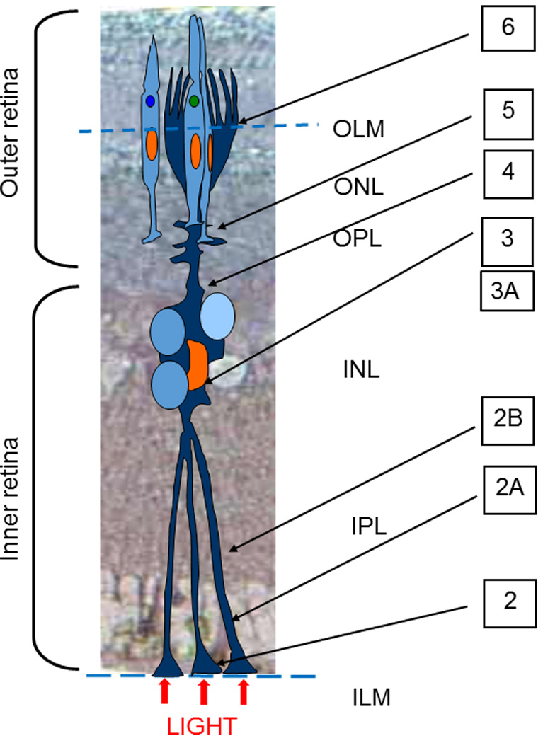
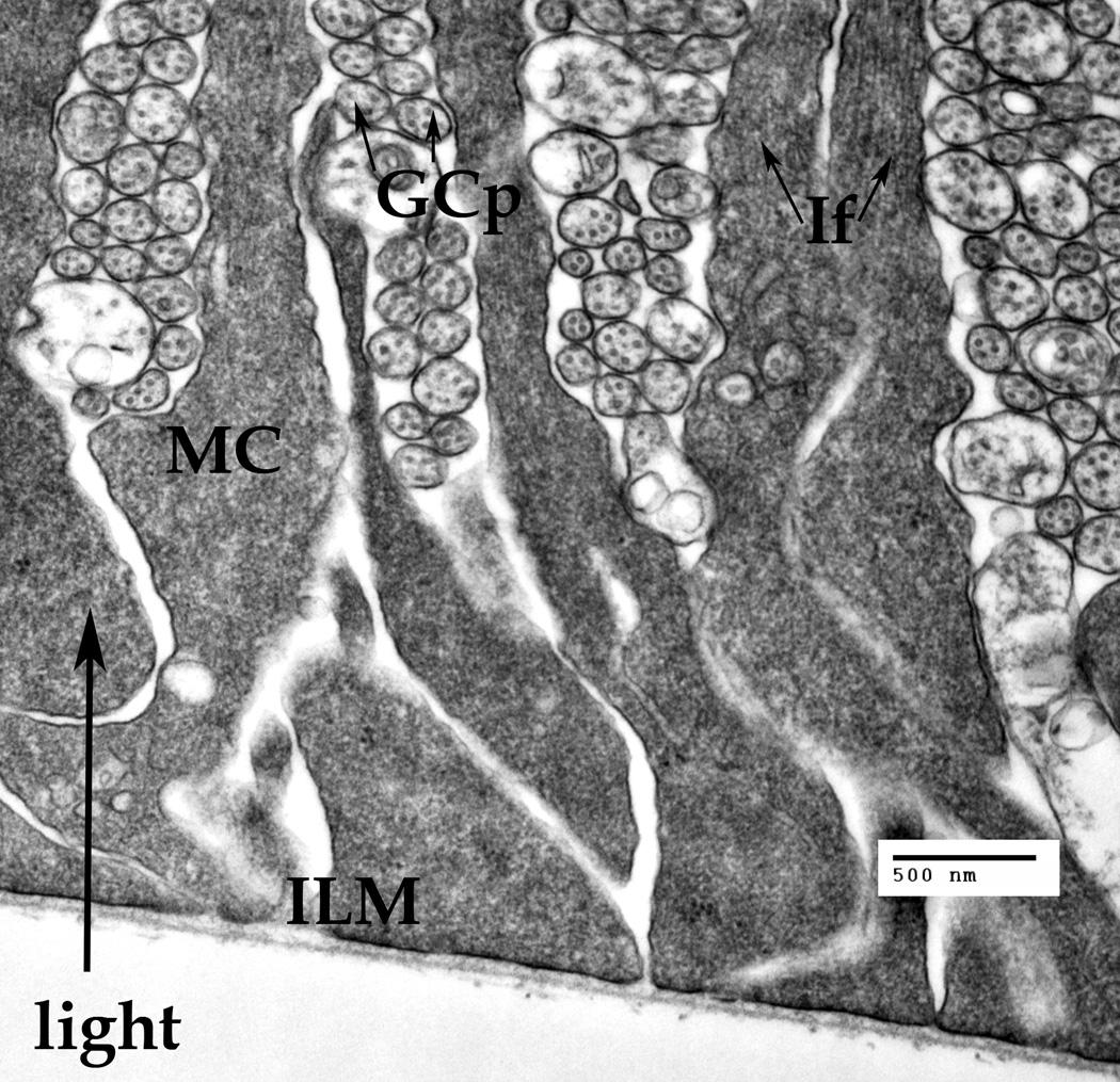
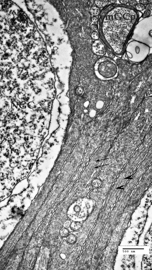
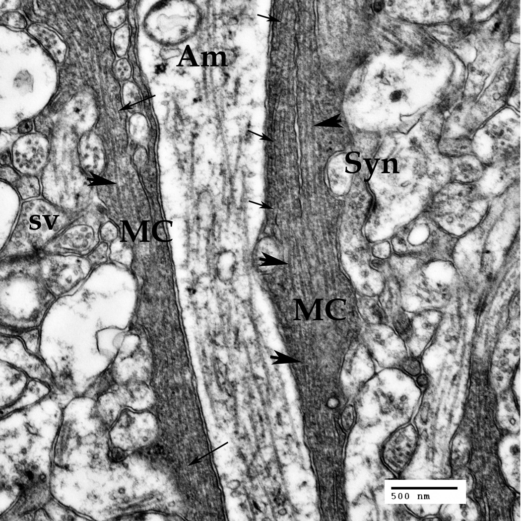
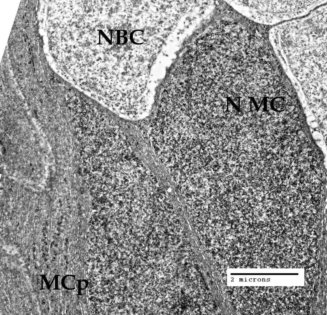
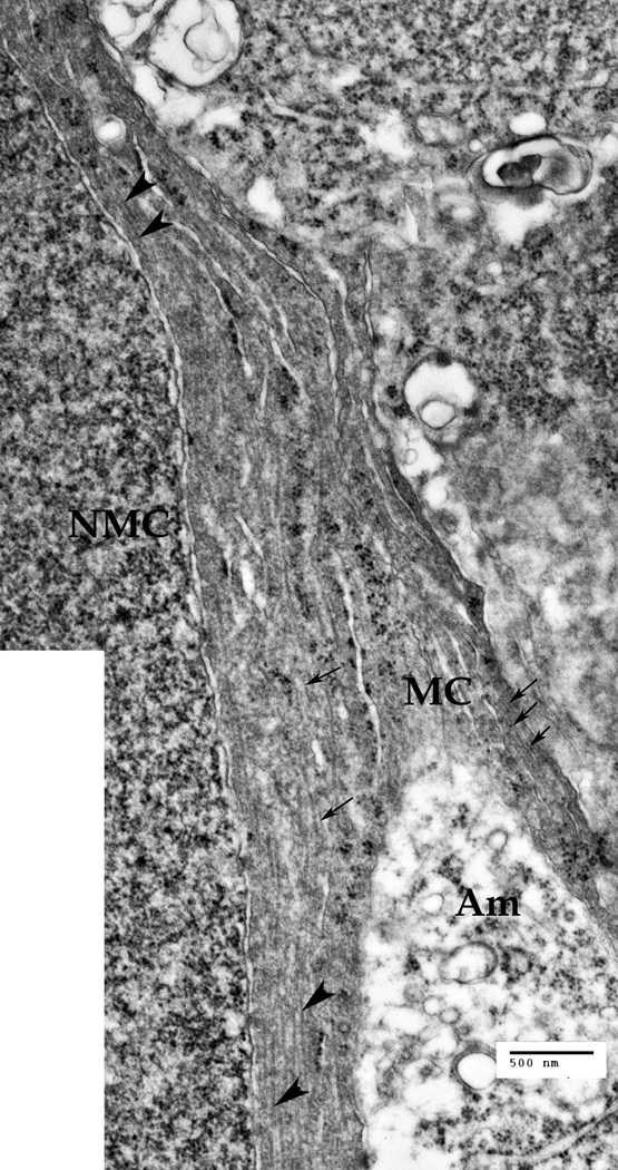
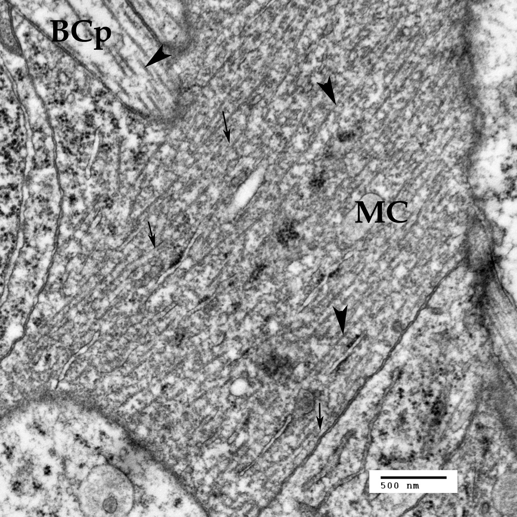
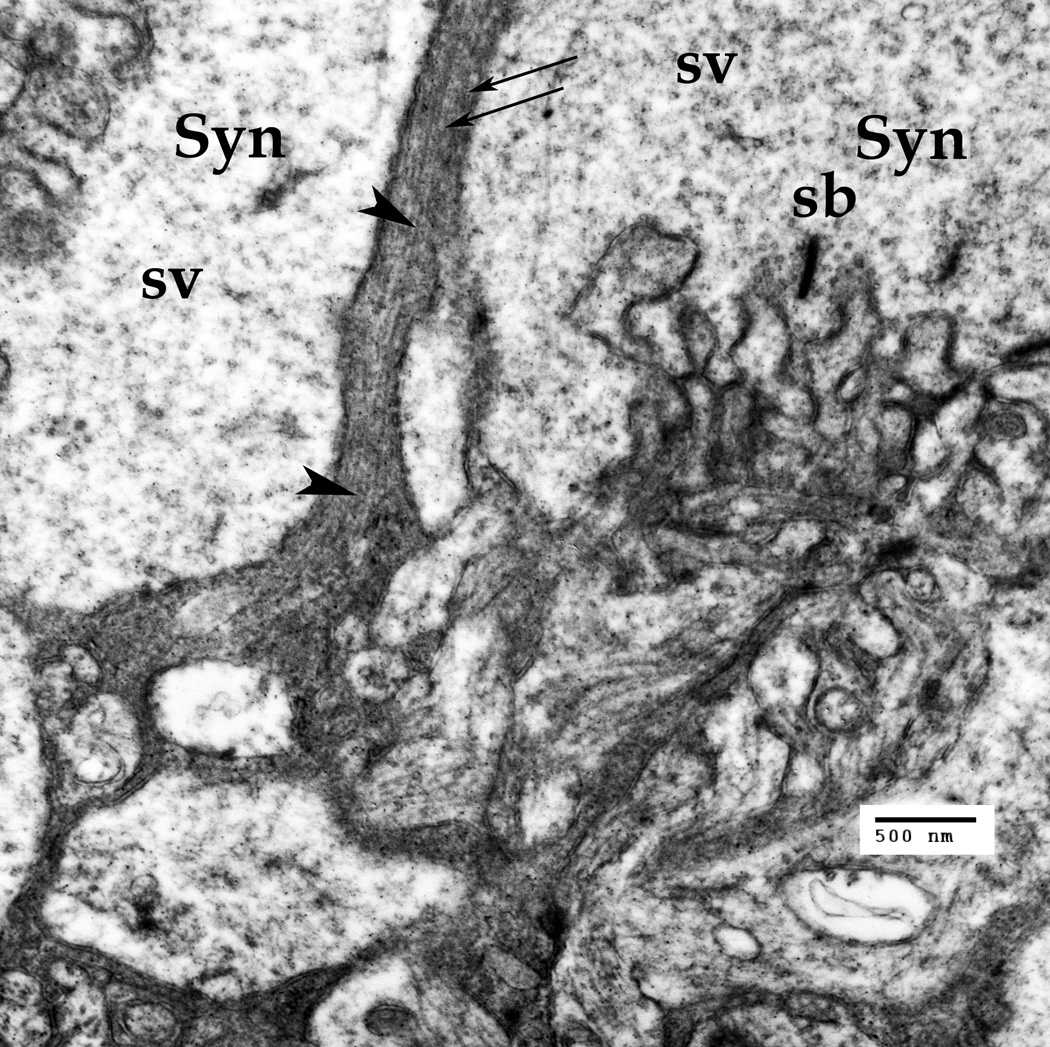
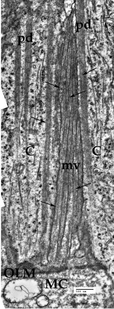
Specialized intermediate filaments (IFs) have critical importance for the clearness and uncommon transparency of vertebrate lens fiber cells, although the physical mechanisms involved are poorly understood. Recently, an unusual low-scattering light transport was also described in retinal Müller cells. Exploring the function of IFs in Müller cells, we have studied the morphology and distribution pattern of IFs and other cytoskeletal filaments inside the Müller cell main processes in the foveolar part of the avian (pied flycatcher) retina. We found that some IFs surrounded by globular nanoparticles (that we suggest are crystallines) are present in almost every part of the Müller cells that span the retina, including the microvilli. Unlike IFs implicated in the mechanical architecture of the cell, these IFs are not connected to any specific cellular membranes. Instead, they are organized into bundles, passing inside the cell from the endfeet to the photoreceptor, following the geometry of the processes, and repeatedly circumventing numerous obstacles. We believe that the presently reported data effectively confirm that the model of nanooptical channels built of the IFs may provide a viable explanation of Müller cell transparency.

Keywords: IFs; Müller cells; avian retina; transparency.


Figures

Fig. 1
Fig. 1
Sketch of a Müller cell alignment in the layers of Pied Flycatcher retina; OLM – Outer Limiting Membrane, ONL- outer nuclear layer, OPL- outer plexiform layer, INL- inner nuclear layer, IPL-inner plexiform layer, ILM-inner limiting membrane. Note the locations of the cross-sections of the electron microphotographs presented in the other Figures, marked by the figure numbers.
Fig. 2
Fig. 2
A cluster of the Müller cells' endfeet attached to the basal membrane thus forming the inner limiting membrane (ILM). The ganglion cell processes (GCp) are passing between the Müller cell endfeet. Cross-cut neuronal microtubules (arrows) are visible inside the GCps. The intermediate filaments (IF, arrows) are visible only in the narrow part of the Müller endfeet. A. A cluster of the Müller cell processes at the ganglion cells' nuclear layer of the retina. NGC – the nucleus of a ganglion cell, mGCP – the myelinated process of a ganglion cell. The Müller cells processes are forced to go around other cells and their processes. Thin arrows point to the intermediate filaments in the MCs (10–18 nm), thick arrowheads point to microtubules (22–26 nm). B. Müller cells' (MC) processes at the IPL level. A multitude of synapses (Syn) of ganglion, bipolar and amacrine (Am) neuronal cells are seen in this area. Synaptic vesicles (sv) are visible inside the synapses. Thin arrows point to the intermediate filaments (10–18 nm), thick arrowheads point to microtubules (22–26 nm). Bars: 500 nm
Fig. 2
Fig. 2
A cluster of the Müller cells' endfeet attached to the basal membrane thus forming the inner limiting membrane (ILM). The ganglion cell processes (GCp) are passing between the Müller cell endfeet. Cross-cut neuronal microtubules (arrows) are visible inside the GCps. The intermediate filaments (IF, arrows) are visible only in the narrow part of the Müller endfeet. A. A cluster of the Müller cell processes at the ganglion cells' nuclear layer of the retina. NGC – the nucleus of a ganglion cell, mGCP – the myelinated process of a ganglion cell. The Müller cells processes are forced to go around other cells and their processes. Thin arrows point to the intermediate filaments in the MCs (10–18 nm), thick arrowheads point to microtubules (22–26 nm). B. Müller cells' (MC) processes at the IPL level. A multitude of synapses (Syn) of ganglion, bipolar and amacrine (Am) neuronal cells are seen in this area. Synaptic vesicles (sv) are visible inside the synapses. Thin arrows point to the intermediate filaments (10–18 nm), thick arrowheads point to microtubules (22–26 nm). Bars: 500 nm
Fig. 3
Fig. 3
The inner nuclear layer (INL), where the nuclei of the Müller cells (NMC) and also the nuclei of the bipolar cells (NBC) are located, at low magnification (Bar: 2 µm). The cytoplasm nearby the MC nucleus, and the processes of other Müller cells, contain cytoskeletal bundles (intermediate filaments and microtubules) that go around the nuclei of both cell types. A. The INL zone at a larger magnification. Microtrubules (large arrowheads) and intermediate filaments (thin arrows) are present inside the Müller cell (MC) processes and in the vicinity of the Müller cell nucleus (NMC). Cytoskeletal bundles are going around the NMC and Amacrine cell (Am) process, as well as around the nuclei of the other cells present. Bar: 500 nm
Fig. 3
Fig. 3
The inner nuclear layer (INL), where the nuclei of the Müller cells (NMC) and also the nuclei of the bipolar cells (NBC) are located, at low magnification (Bar: 2 µm). The cytoplasm nearby the MC nucleus, and the processes of other Müller cells, contain cytoskeletal bundles (intermediate filaments and microtubules) that go around the nuclei of both cell types. A. The INL zone at a larger magnification. Microtrubules (large arrowheads) and intermediate filaments (thin arrows) are present inside the Müller cell (MC) processes and in the vicinity of the Müller cell nucleus (NMC). Cytoskeletal bundles are going around the NMC and Amacrine cell (Am) process, as well as around the nuclei of the other cells present. Bar: 500 nm
Fig. 4
Fig. 4
The Müller cell processes (MC) under the outer plexiform layer are crossing with the bipolar cell processes (BCp). Thin arrows point to the intermediate filaments in the MC (10–18 nm), thick arrowheads point to the microtubules (22–26 nm). Note that the microtubules and the intermediate filaments are much denser in the Müller cells, as compared to the neuron. Also, there are some smaller nanoparticles around both the microtubules and the IFs.
Fig. 5
Fig. 5
The Müller cell processes in the outer plexiform layer (OPL) in the synaptic zone. The cone peduncles in this zone form synapses (Syn) to both horizontal and bipolar cells. The presynaptic region has specialized synaptic bands (SB), surrounded by abundant synaptic vesicles (sv). The Müller cell processes passing through this zone carry both the intermediate filaments (fine arrows) and the microtubules. Scale bar: 500 nm
Fig. 6
Fig. 6
The Müller cell (MC) microvillus above the outer limiting membrane (OLM). The cylindrical microvillus (mv) stretches along the inner segments of the cones (C). The Cytoskeletal elements, mainly the IFs (arrows) are present inside each microvillus. Bar: 500 nm

References

    1. Agte S, Junek S, Matthias S, Ulbricht E, Erdmann I, Wurm A, Schild D, Käs JA, Reichenbach A. Müller glial cell-provided cellular light guidance through the vital guinea-pig retina. Biophys J. 2011 Dec 7;101(11):2611–2619. - PMC - PubMed
    1. Alizadeh A, Clark JI, Seeberger T, Hess J, Blankenship T, Spicer A, FitzGerald PG. Targeted genomic deletion of the lens-specific intermediate filament protein CP49. Invest Ophthalmol Vis Sci. 2002 Dec;43(12):3722–3727. - PubMed
    1. Alizadeh A, Clark J, Seeberger T, Hess J, Blankenship T, FitzGerald PG. Targeted deletion of the lens fiber cell-specific intermediate filament protein filensin. Invest Ophthalmol Vis Sci. 2003;44:5252–5258. - PubMed
    1. Alizadeh A, Clark J, Seeberger T, Hess J, Blankenship T, FitzGerald PG. Characterization of a mutation in the lens-specific CP49 in the 129 strain of mouse. Invest Ophthalmol Vis Sci. 2004 Mar;45(3):884–891. - PubMed
    1. Benedek GB. Theory of transparency of the eye. Appl Optics. 1971;10:459–471. - PubMed

Publication types

MeSH terms