. 2016 Apr;19(4):587-95.
doi: 10.1038/nn.4263. Epub 2016 Feb 29.

Circadian rhythms in neuronal activity propagate through output circuits

Affiliations

Circadian rhythms in neuronal activity propagate through output circuits

Matthieu Cavey et al. Nat Neurosci. 2016 Apr.

Abstract

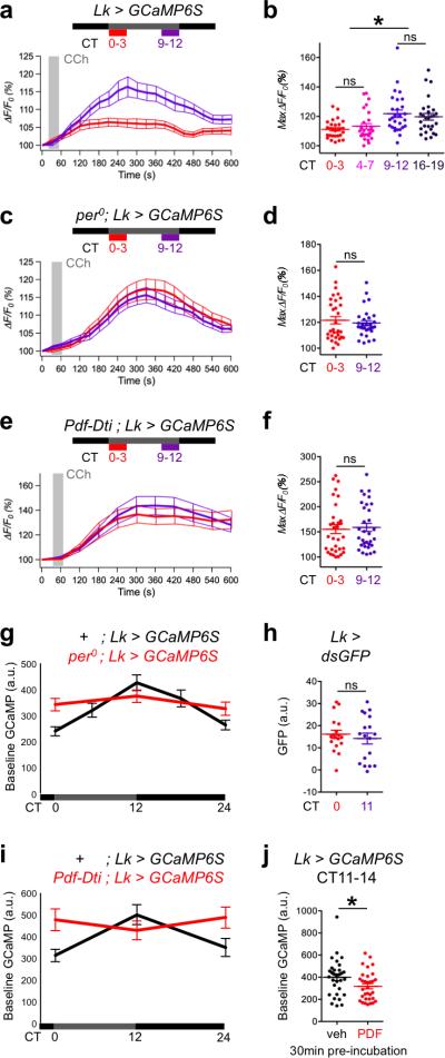
Twenty-four hour rhythms in behavior are organized by a network of circadian pacemaker neurons. Rhythmic activity in this network is generated by intrinsic rhythms in clock neuron physiology and communication between clock neurons. However, it is poorly understood how the activity of a small number of pacemaker neurons is translated into rhythmic behavior of the whole animal. To understand this, we screened for signals that could identify circadian output circuits in Drosophila melanogaster. We found that leucokinin neuropeptide (LK) and its receptor (LK-R) were required for normal behavioral rhythms. This LK/LK-R circuit connects pacemaker neurons to brain areas that regulate locomotor activity and sleep. Our experiments revealed that pacemaker neurons impose rhythmic activity and excitability on LK- and LK-R-expressing neurons. We also found pacemaker neuron-dependent activity rhythms in a second circadian output pathway controlled by DH44 neuropeptide-expressing neurons. We conclude that rhythmic clock neuron activity propagates to multiple downstream circuits to orchestrate behavioral rhythms.


Figures

Figure 1
Figure 1. Lk and Lkr mutant circadian phenotypes and location of LK and LK-R expressing neurons
(a) Average rhythm strength (power) ± SEM of UAS-RNAi lines targeting neuropeptides driven pan-neuronally with elav-Gal4. Control: elav > Dcr-2 + TRiP injection stock. Genes previously implicated in circadian rhythms or sleep are highlighted in pink, Lk in red. Dashed lines: control average ± 1 standard deviation, asterisks: power > 1 standard deviation different from control. (b-d) Representative actograms correspond to the average power of Lk and Lkr mutant flies for 10 days in DD after 3 days entrainment to 12:12 LD cycles (grey/black bars: subjective day/night). (b) RNAi knockdown of Lk and Lkr. (c-d) Lkc275 and Lkrc003 classical alleles compared to heterozygous controls. See Supplementary Tables 1 and 2 for details and sample sizes. (e) LK is expressed in sub-oesophageal LK (SELKs) and Lateral Horn LK neurons (LHLKs), which arborize very close to s-LNv dorsal projections (PDF staining). Dorsal up, ventral down here and in subsequent figures. (f-g) LHLK neurons are not clock neurons since they do not express TIM (f) or tim-Gal4 > nlsGFP (g). (h) LK-R neurons (Lkr-Gal4 > nlsGFP) are widely distributed in the central brain and (i) are not clock neurons: no TIM expression in the LNv region, DN1, DN2 (arrowheads), LNd or DN3. Scale bars: 20μm, except 100μm in e and h. >8 brains examined for each anatomical observation. (See also Supplementary Fig. 1-2).
Figure 2
Figure 2. The anatomy of LHLK/LK-R neurons suggests they lie downstream of clock neurons
(a-e) White arrowheads indicate LHLK cell bodies. (a-c) Upper panels are z-projections, lower panels are single confocal sections of the regions indicated by white dashed rectangles in upper panels. LHLKs project close to (a) s-LNvs (PDF staining), (b) DN1ps (Clk4.1-Gal4 > CD8::GFP) and (c) LNds (Mai179-Gal4; Pdf-Gal80 > CD8::GFP). (d) LHLK dendrites (Lk > DenMark) and s-LNv projections (PDF) are found in similar planes (anterior brain sections, left), whereas LHLK axon terminals (Lk > Syt::GFP) are enriched in more posterior sections (right). (e) Single confocal section of dashed rectangle in d shows that LHLK dendrites intermingle with s-LNv dorsal projections. (f) Lkr-Gal4 > CD8::GFP labels neurons in the Lateral Horn (LH), Ellipsoid Body (EB) and Pars Intercerebrallis (PI). Inset: Lkr-Gal4 is not expressed in LHLK neurons (single confocal section). (g) Lkr > FLEXAMP clone labels LK-R neurons in the LH projecting to the Fan-Shaped Body (FSB). (h) Single confocal section of the dashed square in g showing overlap of LK-R projections and LHLK arborizations and several potential contacts. (i) LHLK processes are close to LK-R dendrites (Lkr > DenMark, single confocal section). (j-k) LK-R outputs (Syt::GFP) are found predominantly in the EB (j) and FSB (k). Scale bars: 20μm, except 100μm in f and g. >8 brains examined for each anatomical observation. (See also Supplementary Fig. 3).
Figure 3
Figure 3. LHLK neurons are downstream of LNvs
Mean GCaMP6S fluorescence (thick lines) normalized to initial level (vertical lines: SEM). Grey rectangles: timing of drug perfusion. Dot plots: distribution of Maximum ΔF/F0, horizontal line shows average ± SEM. Asterisks: significant difference by Kolmogorov-Smirnov (KS) test (p<0.05), ns: no significant difference. Sample sizes: n=x;y, where x is the number of neurons in y brains, from ≥2 independent experiments. (a, left) LHLK neuron responses to 10μM Carbachol (CCh + veh, n=28;8) are reduced by simultaneously activating LNvs with 2.5 mM ATP (CCh + ATP, n=28;7, p=0.0003, D=0.536), but are still different from no CCh (veh, n=16;4, p=0.032, D=0.428). (a, right) Inhibition is lost in the absence of Pdf-LexA > P2X2 expression (p=0.425, D=0.278, n=18;9 each). ATP significantly decreases LHLK responses in brains with P2X2 vs without P2X2 (p<0.0001, D=0.786). (b) LNv activation also inhibits LHLK response to 35 mM KCl (KCl + ATP) compared to control (KCl + veh, p=0.008, D=0.5, n=20,10 each). (c, left) 20min pre-incubation with 100 μM PDF inhibits LHLK response to 10 μM CCh (p=0.0004, D=0.5, n=32;16 each). (c, middle) PDF inhibition of LHLKs is largely eliminated by 2 μM Tetrodotoxin (TTX) pre-incubation (p=0.232, D=0.25, n=32;16 each). (d) PDF inhibition of LHLKs (left, p=0.0082, D=0.458) is lost in Pdf-Dti brains (right, p=0.621, D=0.208, n=24,12 each). (e) LK-R neuron response to 100 μM CCh (n=33;4) is inhibited by 10min 100 μM LK pre-incubation (n=33;5, p<0.0001, D=0.675). (f) s-LNv responses to 100 μM CCh (n=18;6) are not changed by pre-incubation with 100 μM LK (n=17;7, p=0.262, D=0.323). (See also Supplementary Fig. 4).
Figure 4
Figure 4. LHLK neuronal activity is rhythmically regulated by clock neurons
(a-f) LHLK responses to 10 μM CCh on day 1 in DD during 3hr time windows indicated above the line graphs by colored bars; dark grey and black bars show DD cycles. n=28;14 for each sample in a-d, n=32;16 for e-f, taken from ≥2 independent experiments. (a) LHLK excitability is rhythmic in wild type brains (p=0.0029, D=0.464, KS test). (b) Quantification of a and additional statistics: CT0-3 vs CT4-7 p=0.917, D=0.143; CT9-12 vs CT16-19 p=0.917, D=0.143; CT4-7 vs CT9-12 p=0.0029, D=0.464; CT4-7 vs CT16-19 p=0.0077, D=0.428; CT0-3 vs CT16-19 p=0.0029, D=0.464. LHLK excitability is not rhythmic in per0 mutants (c-d, p=0.987, D=0.125) or Pdf-Dti brains (e-f, p=0.383, D=0.219). (g) Baseline GCaMP6S intensity per LHLK cell body in brains from 1hr time windows on day 1 in DD (n=32;16 for each data point, grey/black bars show DD cycle, error bar: SEM). Baseline GCaMP6S levels are rhythmic in wild type brains (p<0.0001, H=28.69, 4d.f. by Kruskal-Wallis one-way ANOVA) but not in per0 mutants (p=0.2746, H=2.585, 2d.f.). (h) No rhythms are observed with Lk-Gal4 expressing destabilized GFP (p=0.425, D=0.2778 by KS test, n=16;8 each). (i) The LHLK GCaMP6S rhythm (p=0.0005, H=15.41, 2d.f., Kruskal-Wallis ANOVA) is lost in brains lacking LNvs (Pdf-Dti, p=0.6386, H=0.897, 2d.f., n=32;16 for each data point). (j) PDF treatment (100μM 30min pre-incubation before imaging) reduces baseline LHLK GCaMP6S levels during their peak phase (CT11-14; p=0.0343, D=0.344 by KS test, n=32;16 each).
Figure 5
Figure 5. LHLK activity rhythms propagate to LK-R neurons
Only LK-R neurons with cell bodies in the lateral horn were imaged. Excitability (a-d) and neuronal activity (e-f) measurements and statistics as in Fig. 4. Data are from ≥2 independent experiments. (a) Lateral horn LK-R neuron excitability (response to 100 μM CCh) is rhythmic (n=80;10 for each sample, p<0.0001, D=0.375). (b) Quantification of a,c-d. (c) The rhythm is lost in per0 mutants (n=80;10 each, p=0.798, D=0.1) and (d) dampened in Lkrc003 hypomorphs, although still significant (n=80;10 each, p=0.0036, D=0.275). LK-R excitability is significantly higher in Lkrc003 mutants compared to wild type in both time windows (CT0-3 p=0.0001, D=0.337, CT9-12 p<0.0001, D=0.512) (e) Lateral horn LK-R baseline GCaMP6S levels are rhythmic in wild type brains (p<0.0001, H=200, 4d.f.; CT0 n=290;12, CT5 n=290;12, CT11 n=255;12, CT17 n=308;12, CT23 n=289;12). This rhythm is lost in per0 mutants (p=0.2918, H=2.463, 2d.f.; CT0 n=315;12, CT11 n=294;12, CT23 n=316;12) and in (f) brains lacking LNvs (Pdf-Dti, p=0.6759, H=0.7834, 2d.f.; CT0 n=277;10, CT11 n=267;10, CT23 n=273;10) compared to controls (p<0.0001, H=33.72, 2d.f.; CT0 n=234;10, CT11 n=273;10, CT23 n=267;10).
Figure 6
Figure 6. LK and LK-R neuron signaling controls locomotor activity levels
(a) Representative actograms of flies maintained 6 days in DD at 19°C and then 5 days at 28°C (red shaded area). Grey and black bars: subjective day and night. Rhythms of control flies (Lk-Gal4 / + and UAS-dTrpA1 / +) become stronger at 28°C compared to 19°C but become weaker when activating LK neurons with dTrpA1 at 28°C (Lk > dTrpA1). See Supplementary Table 2 for details and for LK-R and DH44 neuron activation data. (b-c) Acute (24hr, red shaded area) activation (b) and inhibition (c) of LK and LK-R neurons. Graphs show the population average locomotor activity over 3 days in DD, with the first 12hr of activation magnified in insets below (error bars: SEM). Asterisks: significant differences between experimental flies and both parental controls (p<0.05 by KS test). (b, left) LK neuron activation decreased locomotor activity while (b, right) LK-RR65C07 neuron activation increased locomotor activity during the first 6hr. Locomotor activity recovered to normal levels on day 3 in both experiments. Sample sizes: Lk-Gal4/+ n=62; dTrpA1/+ n=62; Lk>dTrpA1 n=63; LkrR65C07-Gal4/+ n=94; dTrpA1/+ n=94; LkrR65C07>dTrpA1 n=93. Data are from 2 (left) or 3 (right) independent experiments. (c, left) Inhibiting synaptic transmission from LK neurons had minimal effects on locomotor activity, while (c, right) inhibiting LK-RR65C07 neurons reduced locomotor activity through most of the subjective day. Sample sizes: Lk-Gal4/+ n=63; shits/+ n=62; Lk>shits n=63; LkrR65C07-Gal4/+ n=62; LkrR65C07>shits n=62. Data are from 2 independent experiments. (See also Supplementary Fig. 5-6).
Figure 7
Figure 7. Clock electrical rhythms propagate through multiple output circuits
Baseline GCaMP6S levels oscillate in DH44 neurons (p=0.0033, H=11.45, 2d.f.; CT0 n=115;23, CT11 n=125;23, CT23 n=124;23). This rhythm is lost in brains lacking LNvs (Pdf-Dti, p=0.2585, H=2.705, 2d.f.; CT0 n=103;19, CT11 n=96;19, CT23 n=100;19). Statistics as in Fig. 4-5. Data are from 3 independent experiments.

Comment in

References

    1. Herzog ED. Neurons and networks in daily rhythms. Nat Rev Neurosci. 2007;8:790–802. - PubMed
    1. Nitabach MN, Taghert PH. Organization of the Drosophila circadian control circuit. Curr Biol. 2008;18:R84–93. - PubMed
    1. Colwell CS. Linking neural activity and molecular oscillations in the SCN. Nat Rev Neurosci. 2011;12:553–569. - PMC - PubMed
    1. Dibner C, Schibler U, Albrecht U. The mammalian circadian timing system: organization and coordination of central and peripheral clocks. Annu Rev Physiol. 2010;72:517–549. - PubMed
    1. Morin LP. Neuroanatomy of the extended circadian rhythm system. Exp Neurol. 2013;243:4–20. - PMC - PubMed

Methods-only references

    1. Cook RK, Christensen SJ, Deal JA, Coburn RA, Deal ME, Gresens JM, Kaufman TC, Cook KR. The generation of chromosomal deletions to provide extensive coverage and subdivision of the Drosophila melanogaster genome. Genome Biol. 2012;13:R21. - PMC - PubMed
    1. Park JH, Helfrich-Forster C, Lee G, Liu L, Rosbash M, Hall JC. Differential regulation of circadian pacemaker output by separate clock genes in Drosophila. Proc Natl Acad Sci U S A. 2000;97:3608–3613. - PMC - PubMed
    1. Martinek S, Inonog S, Manoukian AS, Young MW. A role for the segment polarity gene shaggy/GSK-3 in the Drosophila circadian clock. Cell. 2001;105:769–779. - PubMed
    1. Plautz JD, Kaneko M, Hall JC, Kay SA. Independent photoreceptive circadian clocks throughout Drosophila. Science. 1997;278:1632–1635. - PubMed
    1. Calleja M, Moreno E, Pelaz S, Morata G. Visualization of gene expression in living adult Drosophila. Science. 1996;274:252–255. - PubMed

Publication types

Substances