Review
. 2016 Jun;150(8):1769-77.
doi: 10.1053/j.gastro.2016.02.066. Epub 2016 Feb 27.

Pathogenesis of Nonalcoholic Steatohepatitis

Affiliations
Review

Pathogenesis of Nonalcoholic Steatohepatitis

Mariana Verdelho Machado et al. Gastroenterology. 2016 Jun.

Abstract

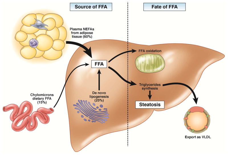
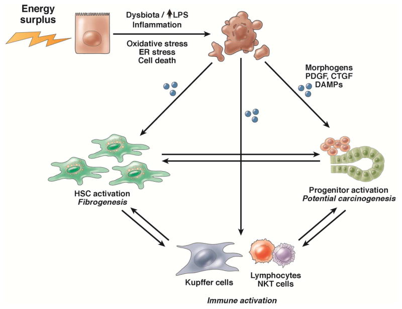
Nonalcoholic steatohepatitis (NASH) is a necro-inflammatory response that ensues when hepatocytes are injured by lipids (lipotoxicity). NASH is a potential outcome of nonalcoholic fatty liver (NAFL), a condition that occurs when lipids accumulate in hepatocytes. NASH may be reversible, but it can also result in cirrhosis and primary liver cancer. We are beginning to learn about the mechanisms of progression of NAFL and NASH. NAFL does not inevitably lead to NASH because NAFL is a heterogeneous condition. This heterogeneity exists because different types of lipids with different cytotoxic potential accumulate in the NAFL, and individuals with NAFL differ in their ability to defend against lipotoxicity. There are no tests that reliably predict which patients with NAFL will develop lipotoxicity. However, NASH encompasses the spectrum of wound-healing responses induced by lipotoxic hepatocytes. Differences in these wound-healing responses among individuals determine whether lipotoxic livers regenerate, leading to stabilization or resolution of NASH, or develop progressive scarring, cirrhosis, and possibly liver cancer. We review concepts that are central to the pathogenesis of NASH.

Keywords: Lipotoxicity; Misrepair; Nonalcoholic Fatty Liver Disease; Wound-Healing Response.


Conflict of interest statement

Conflict of interest: There are no conflicts of interest to state.

Figures

Figure 1
Figure 1. Mechanisms of hepatic steatosis
Hepatic steatosis results from increased influx of lipids to the liver or decreased lipid disposal. The main sources of FA are plasma FFA (arriving mostly from the adipose tissue), de novo lipogenesis, and dietary FA. The liver discards fat by oxidation or by exporting it as VLDL. Alternatively, hepatocytes can shunt excess lipids to the synthesis of triglycerides and storage in lipid droplets. Red boxes highlight rate-limiting enzymes that regulate the main fates of fatty acids in the liver: FAS, fatty acid synthase and ACC, acetyl-CoA carboxylase, which are enzymes in fatty acids synthesis; CPT-1, carnitine palmitoyltransferase 1, enzyme that allows entry of acyl groups into the mitochondria by transferring an acyl group from CoA to carnitine and subsequent transport of acylcarnitine; SCD-1, stearoyl-CoA dessaturase, an enzyme that converts saturated in monounsaturated fatty acids the fatty acids that are preferentially incorporated in triglycerides; DGAT, diglyceride acyltransferase that catalyzes the synthesis of triglycerides from diacylglycerol and acylCoA; MTTP, microsomal triglyceride transfer protein, which controls lipoprotein assembly. Blue boxes indicate transcription factors involved in lipid metabolism: SREBP1C, sterol regulatory element-binding protein-1c; PPARA and PPARG, peroxisome proliferator-activated receptor-α and -γ.
Figure 2
Figure 2. Lipotoxicity and the wound-healing response
Lipid toxicity to hepatocytes causes cell stress and cell death. Dying hepatocytes release signals that induce a wound-healing response, such as damage associated molecular patterns, cytokines, and hedgehog. To promote regeneration or repair, the liver recruits and activates hepatic stellate cells to remove dying and dead cells and sustain the remaining epithelia, and progenitors to replace dead cells. If the wound-healing response cannot be restrained, it promotes inflammation, fibrogenesis, and hepatocarcinogenesis.
Figure 3
Figure 3. The hedgehog signaling pathway
A. The SHH ligand patched 1 constitutively represses SMO, allowing the sequential phosphorylation of Gli by protein kinase A (PKA), glycogen synthase kinase 3 beta (GSK3B), and casein kinase (CK). Phosphorylation of the Gli family zinc finger proteins marks them for ubiquitination and limited proteosomal degradation. N-terminal fragments of Gli proteins translocate to the nucleus where they repress transcription. B. Binding of SHH to patched 1 prevents its inhibition of SMO. This prevents phosphorylation and degradation of Gli proteins. Full-length Gli proteins translocate to the nucleus and promote transcription.

References

    1. Matteoni CA, Younossi ZM, Gramlich T, et al. NAFLD: a spectrum of clinical and pathological severity. Gastroenterology. 1999;116:1413–9. - PubMed
    1. Rafiq N, Bai C, Fang Y, et al. Long-term follow-up of patients with NAFL. Clin Gastroenterol Hepatol. 2009;7:234–8. - PubMed
    1. Musso G, Gambino R, Cassader M, et al. Meta-analysis: natural history of NAFLD and diagnostic accuracy of non-invasive tests for liver disease severity. Ann Med. 2011;43:617–49. - PubMed
    1. Feldstein AE, Canbay A, Angulo P, et al. Hepatocyte apoptosis and fas expression are prominent features of human NASH. Gastroenterology. 2003;125:437–43. - PubMed
    1. Cusi K. Role of obesity and lipotoxicity in the development of NASH: pathophysiology and clinical implications. Gastroenterology. 2012;142:711–725. e6. - PubMed