Case Reports
. 2016 May;4(5):383-9.
doi: 10.1158/2326-6066.CIR-15-0123. Epub 2016 Feb 29.

Autoimmune Bullous Skin Disorders with Immune Checkpoint Inhibitors Targeting PD-1 and PD-L1

Affiliations
Case Reports

Autoimmune Bullous Skin Disorders with Immune Checkpoint Inhibitors Targeting PD-1 and PD-L1

Jarushka Naidoo et al. Cancer Immunol Res. 2016 May.

Abstract

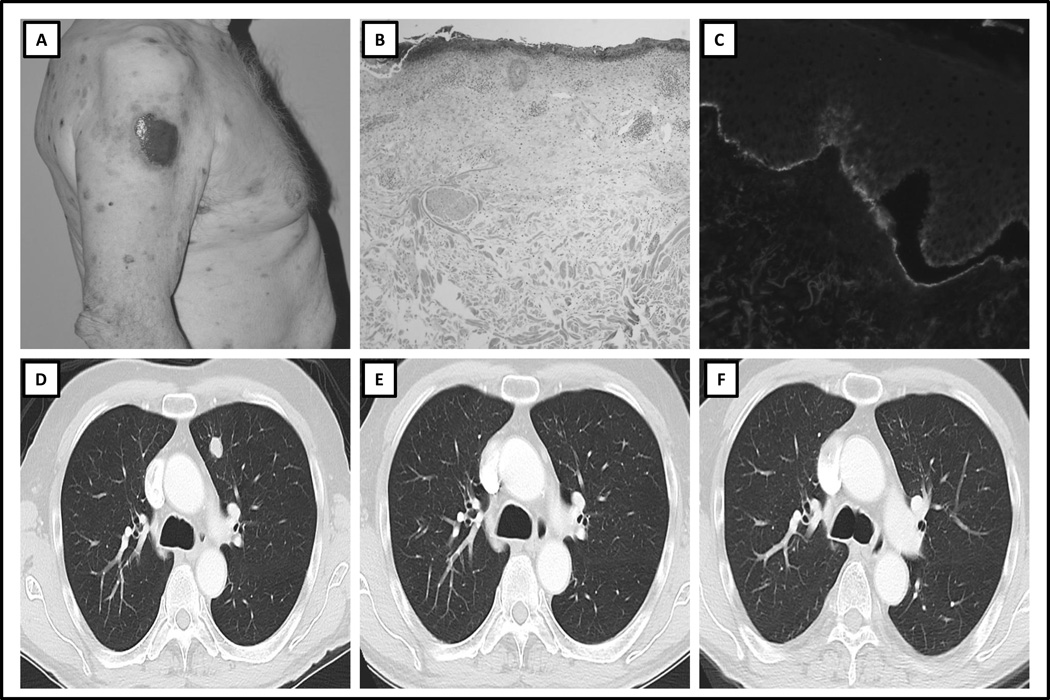
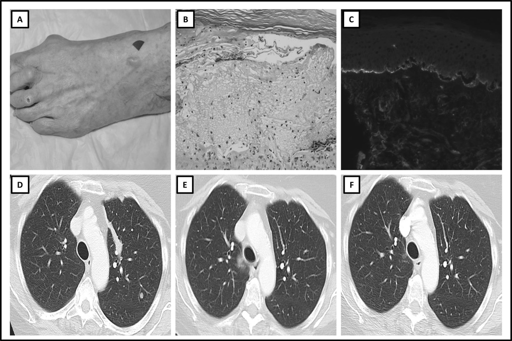
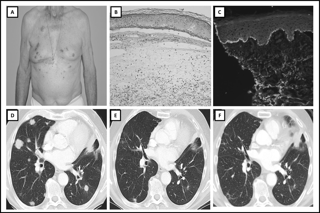
Monoclonal antibodies (mAb) targeting immune checkpoint pathways such as cytotoxic T-lymphocyte-associated protein 4 (CTLA-4) and programmed death 1 (PD-1) may confer durable disease control in several malignancies. In some patients, immune checkpoint mAbs cause cutaneous immune-related adverse events. Although the most commonly reported cutaneous toxicities are mild, a subset may persist despite therapy and can lead to severe or life-threatening toxicity. Autoimmune blistering disorders are not commonly associated with immune checkpoint mAb therapy. We report a case series of patients who developed bullous pemphigoid (BP), an autoimmune process classically attributed to pathologic autoantibody formation and complement deposition. Three patients were identified. Two patients developed BP while receiving the anti-PD-1 mAb nivolumab, and one while receiving the anti-PD-L1 mAb durvalumab. The clinicopathologic features of each patient and rash, and corresponding radiologic findings at the development of the rash and after its treatment, are described. Patients receiving an anti-PD-1/PD-L1 mAb may develop immune-related BP. This may be related to both T-cell- and B-cell-mediated responses. Referral to a dermatologist for accurate diagnosis and management is recommended. Cancer Immunol Res; 4(5); 383-9. ©2016 AACR.


Conflict of interest statement

JN, KS, CAQ, KB, JC, DBP, AW, ASL, EAQ, PKP, MD, SPA : No relevant disclosures

Figures

Figure 1
Figure 1. Clinicopathologic Features and Serial Radiologic Imaging of Case 1
A) Clinical presentation: multiple excoriated blisters on the trunk, B) Hematoxylin and eosin histopathologic slide, demonstrating subepidermal cleft and eosinophilic infiltrate, C) Direct immunofluorescence demonstrating IgG positivity at the dermo-epidermal junction, D) Baseline computed tomography (CT) scan image before treatment with immune checkpoint therapy, demonstrating lung metastasis, E) CT scan image at time of development of bullous pemphigoid, demonstrating partial radiologic response, F) CT scan image 6 months after diagnosis of bullous pemphigoid, demonstrating ongoing partial response.
Figure 2
Figure 2. Clinicopathologic Features and Serial Radiologic Imaging of Case 2
A) Clinical presentation: tense blister on the dorsum of the left foot, B) Hematoxylin and eosin histopathologic slide, demonstrating subepidermal cleft and eosinophilic infiltrate C) Direct immunofluorescence demonstrating subepidermal cleft and C3 deposition at the dermo-epidermal junction, D) Baseline CT scan image prior to treatment with immune checkpoint therapy, demonstrating lung metastasis, E) CT scan image at the time of diagnosis of bullous pemphigoid, demonstrating partial response, F) CT scan image 12 months after discontinuation of treatment, demonstrating no evidence of disease.
Figure 3
Figure 3. Clinicopathologic Features and Serial Radiologic Imaging of Case 3
A) Clinical presentation: multiple papules and blisters on the trunk, B) Hematoxylin and eosin histopathologic slide, demonstrating subepidermal cleft and eosinophilic infiltrate, C) Direct immunofluorescence demonstrating subepidermal cleft and C3 deposition at the dermo-epidermal junction, D) Baseline CT scan image prior to treatment with immune checkpoint therapy, demonstrating lung metastases, E) CT scan image at time of diagnosis of bullous pemphigoid, demonstrating partial response, F) CT scan image 12 months after discontinuation of immune checkpoint therapy, demonstrating ongoing partial response.

References

    1. Hodi FS, O'Day SJ, McDermott DF, Weber RW, Sosman JA, Haanen JB, et al. Improved survival with ipilimumab in patients with metastatic melanoma. The New England journal of medicine. 2010 Aug 19;363(8):711–723. PubMed PMID: 20525992. Pubmed Central PMCID: 3549297. - PMC - PubMed
    1. Robert C, Thomas L, Bondarenko I, O'Day S, M DJ, Garbe C, et al. Ipilimumab plus dacarbazine for previously untreated metastatic melanoma. The New England journal of medicine. 2011 Jun 30;364(26):2517–2526. PubMed PMID: 21639810. Epub 2011/06/07. eng. - PubMed
    1. Topalian SL, Hodi FS, Brahmer JR, Gettinger SN, Smith DC, McDermott DF, et al. Safety, activity, and immune correlates of anti-PD-1 antibody in cancer. The New England journal of medicine. 2012 Jun 28;366(26):2443–2454. PubMed PMID: 22658127. Pubmed Central PMCID: 3544539. - PMC - PubMed
    1. Weber JS, Yang JC, Atkins MB, Disis ML. Toxicities of Immunotherapy for the Practitioner. Journal of clinical oncology : official journal of the American Society of Clinical Oncology. 2015 Apr 27; PubMed PMID: 25918278. Epub 2015/04/29. Eng. - PMC - PubMed
    1. Minkis K, Garden BC, Wu S, Pulitzer MP, Lacouture ME. The risk of rash associated with ipilimumab in patients with cancer: a systematic review of the literature and meta-analysis. Journal of the American Academy of Dermatology. 2013 Sep;69(3):e121–e128. PubMed PMID: 23357570. Epub 2013/01/30. eng. - PubMed

Publication types

MeSH terms