. 2016 Jun:95:96-111.
doi: 10.1016/j.freeradbiomed.2016.02.029. Epub 2016 Feb 27.

Redox regulation of epidermal growth factor receptor signaling during the development of pulmonary hypertension

Affiliations

Redox regulation of epidermal growth factor receptor signaling during the development of pulmonary hypertension

Olga Rafikova et al. Free Radic Biol Med. 2016 Jun.

Abstract

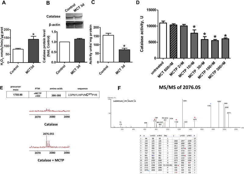
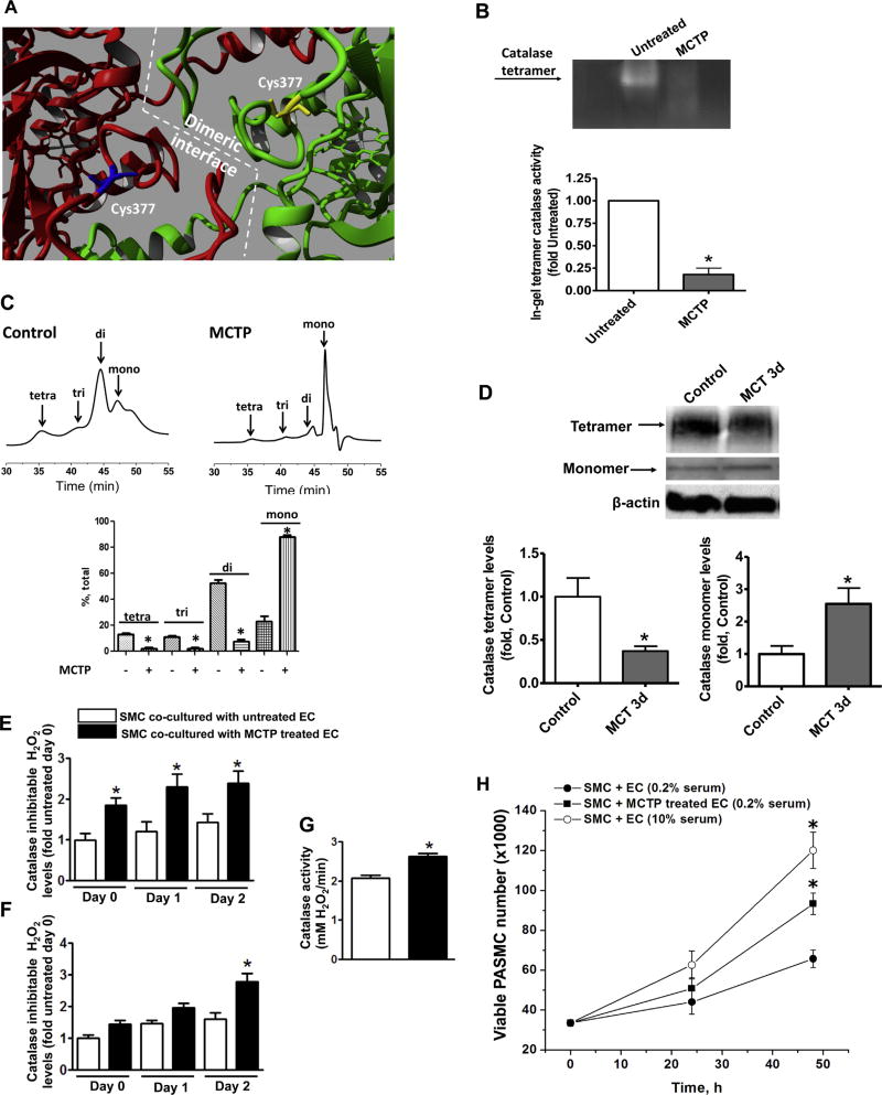
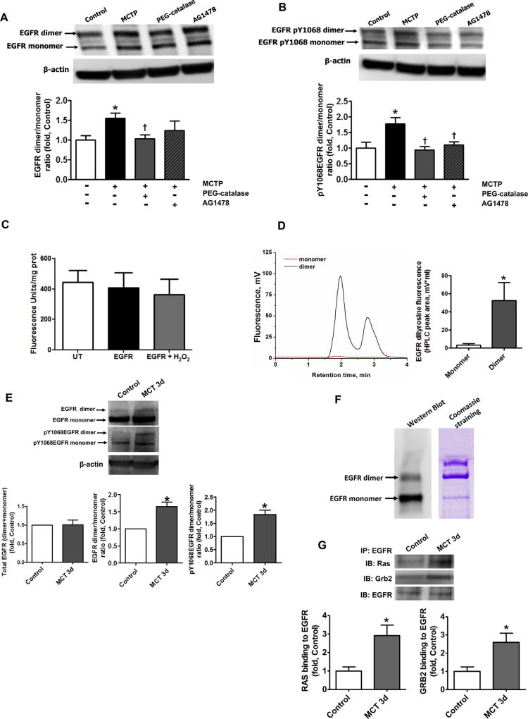
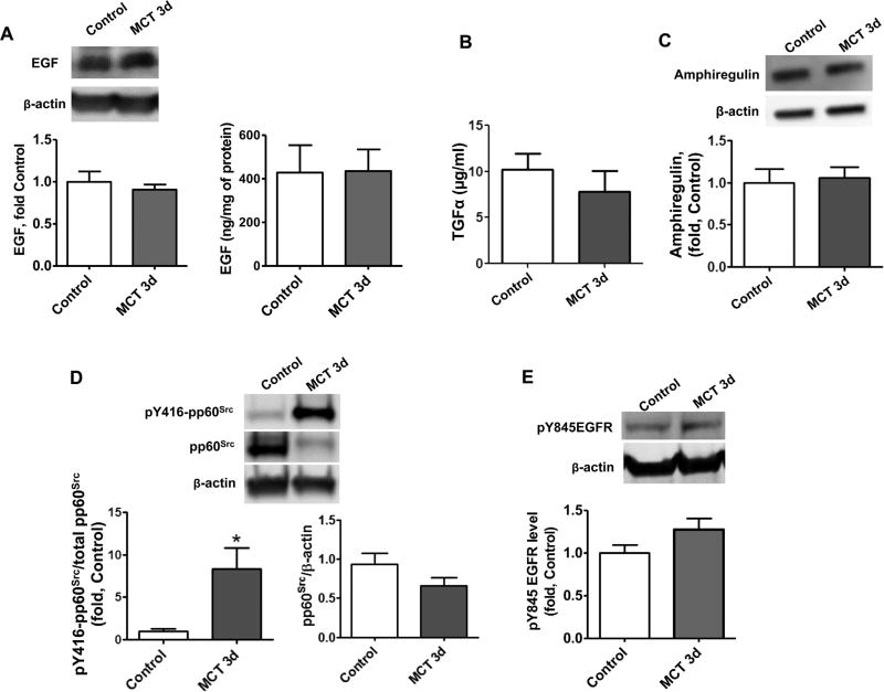
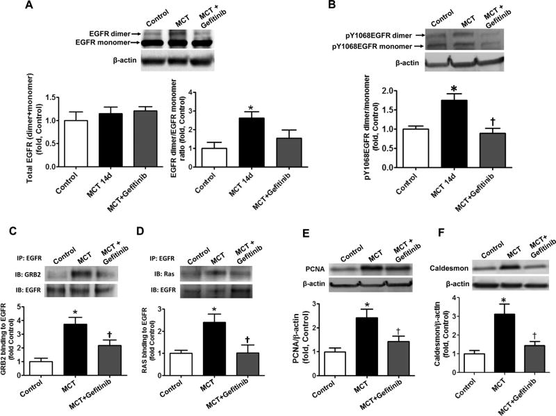
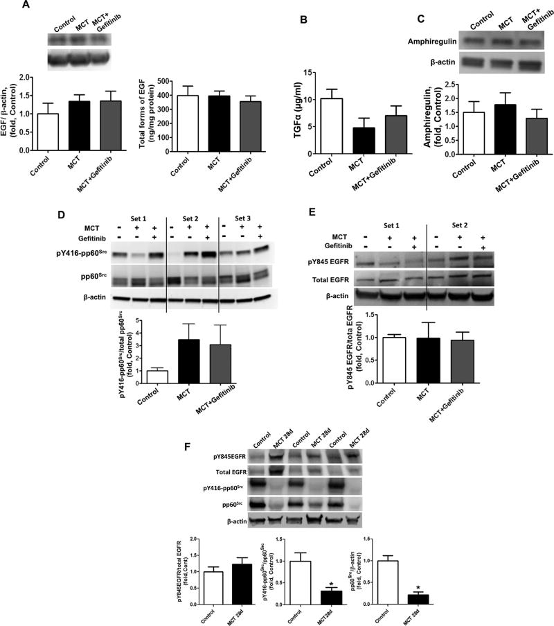
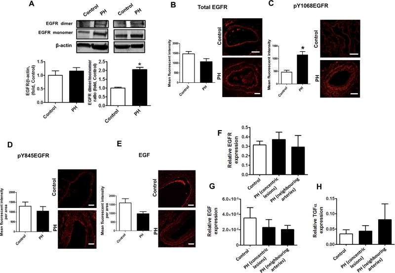
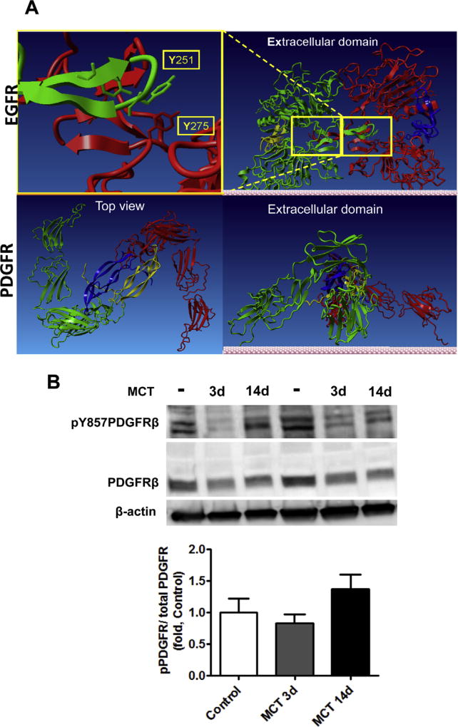
The development of pulmonary hypertension (PH) involves the uncontrolled proliferation of pulmonary smooth muscle cells via increased growth factor receptor signaling. However, the role of epidermal growth factor receptor (EGFR) signaling is controversial, as humans with advanced PH exhibit no changes in EGFR protein levels and purpose of the present study was to determine whether there are post-translational mechanisms that enhance EGFR signaling in PH. The EGFR inhibitor, gefinitib, significantly attenuated EGFR signaling and prevented the development of PH in monocrotaline (MCT)-exposed rats, confirming the contribution of EGFR activation in MCT induced PH. There was an early MCT-mediated increase in hydrogen peroxide, which correlated with the binding of the active metabolite of MCT, monocrotaline pyrrole, to catalase Cys377, disrupting its multimeric structure. This early oxidative stress was responsible for the oxidation of EGFR and the formation of sodium dodecyl sulfate (SDS) stable EGFR dimers through dityrosine cross-linking. These cross-linked dimers exhibited increased EGFR autophosphorylation and signaling. The activation of EGFR signaling did not correlate with pp60(src) dependent Y845 phosphorylation or EGFR ligand expression. Importantly, the analysis of patients with advanced PH revealed the same enhancement of EGFR autophosphorylation and covalent dimer formation in pulmonary arteries, while total EGFR protein levels were unchanged. As in the MCT exposed rat model, the activation of EGFR in human samples was independent of pp60(src) phosphorylation site and ligand expression. This study provides a novel molecular mechanism of oxidative stress stimulated covalent EGFR dimerization via tyrosine dimerization that contributes into development of PH.

Keywords: Catalase; EGFR; Oxidative stress; Proliferation; Pulmonary hypertension.


Figures

Fig. 1
Fig. 1
Monocrotaline pyrrole inhibits catalase activity through adduct formation at cysteine 377. The level of H2O2 produced in snap frozen peripheral lung tissue is increased 3 days after MCT injection (A). Despite no changes in catalase protein as determined by Western blot analysis and normalized to β-actin (B), catalase activity is significantly attenuated in MCT-treated rats. MCTP, but not MCT, attenuates recombinant human catalase (D) and this corresponds to the addition of the pyrole adduct to cysteine 377 located in the peptide sequence 366-LGPNYLHIPVNC*PYR-380 as determined by MS (E). MS/MS spectra (F, upper panel) were analyzed with PEAKS 5.2 software and the resulting ion table is shown (F, lower panel). Due to instability of MCTP modification, the fragmentation was calculated manually. Colored numbers represent predicted y,b ions in MS/MS with MCTP attached to cysteine 377 and colored *-symbols indicate predicted collision products with fragmented MCTP modification. Results are expressed as mean±SEM; n=3–8. *P<0.05 vs. control or untreated group.
Fig. 2
Fig. 2
The monocrotaline pyrrole adduct disrupts the oligomeric structure of catalase. Analysis of the crystal structure of catalase (PBD ID 1QQW) identified C377 as being located at the dimeric interface (A). Semi-native zymography (in gel activity) demonstrates that the activity of recombinant catalase tetramer is significantly attenuated by MCTP (30 µM, 30 min) (B). Analytical gel filtration confirms the disruption of the catalase multimeric structure by MCTP (C). Semi-native gel electrophoresis of peripheral lung tissues of MCT-treated rats and the following western blot analysis identified a significant reduction in the tetrameric form of catalase and accumulation of catalase monomer (D). Pretreatment of PAEC with MCTP (200 µM, 30 min), followed by complete removal of MCTP, causes a significant increase in H2O2 in the media within 2 h and this persists for at least 48 h (E). H2O2 levels increase in the PASMC co-cultured with PAEC pretreated with MCTP after 48 h (F) despite a significant increase in catalase activity (G). The increase in H2O2 in the PASMC correlates with an increase in proliferation (MTS proliferation assay) (H). Results are expressed as mean±SEM; N=3–4. *P<0.05 vs. control or untreated group.
Fig. 3
Fig. 3
Covalent dimerization of EGFR increases EGFR signaling in vitro and in vivo. The levels of EGFR dimer and monomer in PASMC co-cultured with MCTP pretreated PAEC were obtained by performing Western blot analysis of PASMC lysates. The presence of SDS-resistant an EGFR dimer was significantly increased in PASMC co-cultured with PAEC pretreated with MCTP and this was attenuated by pretreatment with PEG-catalase (250 U/ml), but not by the EGFR inhibitor AG1478 (100 nM, A). The ratio of auto-phosphorylated EGFR dimer/auto-phosphorylated EGFR monomer was also significantly increased in PASMC co-cultured with PAEC pretreated with MCTP and attenuated by PEG-catalase or the EGFR kinase inhibitor, AG1478 (B). HEK cells over-expressing EGFR were treated with CuCl2/H2O2 (10 µM/300 µM) to induce oxidative stress. Whole cell lysates were then examined for specific dityrosine fluorescence. Global tyrosine oxidation was unchanged (C) but HPLC analysis of the peptide fragments from dimer and monomer bands of immunoprecipitated and digested EGFR revealed the presence of dityrosine crosslinks only in the EGFR dimer (D). Western blot analysis revealed no change in total EGFR protein levels in peripheral lung tissue lysates prepared from control and MCT-treated rats at 3 days (E, left panel). Loading was normalized to β-actin. However, the EGFR dimer/monomer ratio (E, middle panel) and the levels of auto-phosphorylation, assessed by measuring pY1068EGFR (E, right panel), were both significantly in MCT treated rats. The levels of EGFR dimer assessed by Western blot analysis of EGFR immunoprecipitation (F, left panel) appear to be much less when compared to the band present with Coomassie staining (F, right panel). The paradox may be due to reduced ability of antibodies to recognize the covalently cross-linked EGFR protein. Thus, the amount of EGFR dimer and its contribution into uncontrolled cell growth may be underestimated based on the data obtained by Western blot. The level of EGFR signaling was evaluated by immunopreciptating EGFR and probing for Grb2 and Ras. EGFR/Grb2 and EGFR/Ras interactions were significantly increased in MCT-treated rats (G). Results are expressed as mean±SEM; N=3–7. *P<0.05 vs. control group; †P<0.05 vs. PASMC co-cultured with MCTP treated PAEC.
Fig. 4
Fig. 4
Acute MCT exposure stimulates EGF, TGFα, Amphiregulin- and pp60Src-independent activation of EGFR signaling. The levels of EGF in peripheral lung tissue lysates were measured by Western blot analysis (A, left panel) and ELISA (A, right panel) and no significant changes were observed between control rats and MCT-treated rats at 3d. No changes were observed in the alternate EGFR receptor ligands TGFα (B) or amphiregulin (C). Although pp60Src activity was significantly increased in MCT-treated rats as estimated by Western blot analysis of pY416-pp60Src (D) no significant increase was observed in pp60Src dependent activation of EGFR as estimated by measuring the phosphorylation of Y845EGFR, a pp60Src dependent site of EGFR, by Western blot analysis and normalization to the total level of EGFR in peripheral lung tissue (E). Results are expressed as mean±SEM; N¼6–8. *Po0.05 vs. control group.
Fig. 5
Fig. 5
Stimulation of EGFR signaling is attenuated by selective EGFR kinase inhibition. Total EGFR protein levels were measured by Western blot analysis and normalized with β-actin in peripheral lung lysates prepared from control rats and rats treated for 14 days with MCT in the presence or absence of the EGFR kinase inhibitor, gefitinib (30 mg/kg by oral gavages, daily). No differences in EGFR protein levels were observed between groups (A, left panel). The levels of SDS-resistant EGFR dimer were also evaluated by performing Western blot analysis. The level of SDS-resistant EGFR dimer was significantly increased in lung tissues of MCT treated rats and this increase was not significantly attenuated by gefitinib (A, right panel). However, the increased ratio of autophosphorylated EGFR dimer/autophosphorylated EGFR monomer in lung tissues of MCT treated rats was significantly attenuated by treatment with gefitinib (B). The level of EGFR signaling was evaluated by immunopreciptating EGFR and probing for Grb2 and Ras. EGFR/Grb2 (C) and EGFR/Ras (D) interactions were significantly increased in MCT-treated rats and this was attenuated by gefitinib. The total level of proliferation in peripheral lung tissue was evaluated by measuring PCNA protein levels by Western blot analysis. The levels of PCNA were normalized to β-actin. The levels of PCNA are significantly increased in MCT-treated rats and this is attenuated by gefitinib (E). The proliferation of pulmonary SMC was also evaluated by measuring the levels of the SMC marker, caldesmon by Western blot analysis. The levels of caldesmon were normalized to β-actin. The levels of caldesmon are significantly increased in MCT-treated rats and this is attenuated by gefitinib (F). Results are expressed as mean±SEM; N=3–7. *P<0.05 vs. control group. †P<0.05 vs. MCT alone.
Fig. 6
Fig. 6
EGF, TGFα, Amphiregulin- and pp60Src -independent activation of EGFR signaling after chronic exposure to MCT. The levels of EGF in peripheral lung tissue lysates were measured by Western blot analysis (A, left panel) and ELISA (A, right panel) and no significant changes were observed between control rats or MCT-treated rats in the presence or absence of gefitinib. No changes were observed in the alternate EGFR receptor ligands TGFα (B) or amphiregulin (C). The activity of pp60Src was also analyzed by Western blot analysis. Shown are the Western blot data from three different sets of rats. The levels of p-Y416pp60Src were normalized to the level of pp60Src. The activity of pp60Src was not significantly different between groups (D). No significant differences were observed between groups in pp60Src dependent activation of EGFR, as estimated by measuring the phosphorylation of Y845EGFR, a pp60Src dependent site of EGFR, by Western blot analysis and normalization to the total level of EGFR in peripheral lung tissue (E). Shown are the Western blot data from two different sets of rats (E). No significant differences were observed in phosphorylation of Y845EGFR between control and the 28 days MCT group by Western blot analysis normalized to the total level of EGFR in peripheral lung tissue (F). The levels of p-Y416pp60Src and total pp60Src were significantly reduced between control and the 28 MCT group (F). Results are expressed as mean±SEM; N=4–7. P<0.05 vs. control group. †P<0.05 vs. MCT alone.
Fig. 7
Fig. 7
Ligand independent EGFR activation in pulmonary arteries of patients with advanced pulmonary hypertension. The levels of total EGFR in human peripheral pulmonary tissue were measured by Western blot analysis and normalized on the level of β-actin. There was no significant difference between total EGFR levels in controls and patients with advanced PH (A, left panel). Two representative sets are shown The levels of SDS-resistant EGFR dimer were also evaluated by performing Western blot analysis. Due to differences in expression levels, two different exposure times were used for each blot to measure EGFR monomers and EGFR dimers. Thus, each box represents the same gel but different exposure times. The levels of SDS-resistant EGFR dimer (A, right panel) was significantly increased in lung tissues patients with advanced PH. Immunofluorescence was performed on paraffin tissue sections using antibodies against EGFR (B), pY1068EGFR (C), pY845EGFR (D) and EGF (E) followed by an Alexa Fluor® 546 secondary antibody. Pulmonary artery walls of controls and patients with PH were traced to obtain mean fluorescent signal per area (n=60 pulmonary arteries). Representative images are shown. The fluorescent signal for EGFR (B), pY845EGFR (D) and EGF (E) were not different between controls and patients with advanced PH. However, EGFR auto-phosphorylation (pY1068EGFR) was significantly increased in the pulmonary artery wall of patients with advanced PH (C). Messenger RNA levels were also measured using laser-assisted microdissection followed by qRT-PCR in pulmonary arteries of control and concentric lesions and neighboring arteries in patients with advanced PH. EGFR (F) and EGF (G) and TGFα (H) mRNA levels were not different between controls and patients with advanced PH. Results are expressed as mean±SEM; N=3–17. *P<0.05 vs. controls. Scale bars are equal to 200 µm.
Fig. 8
Fig. 8
PDGF receptor activation. Structural comparison of EGFR and PDGFR dimerization (A). Homology modeling of both EGFR and PDGFR revealed different dimerization characteristics of these receptors. Two EGFR subunits (red and green, upper insert) forms dimeric interface between subunits with stabilization by EGF ligands (yellow and blue). Upper insert indicates two tyrosine residues (Y251 and Y275) from adjacent subunits on the dimeric interface that could involve in dityrosine crosslink. In contrast PDGFR forms a dimer (red and green, lower insert) without direct contact between subunits. Top view (lower insert) indicates that adjacent monomeric subunits of PDGFR (green and red) are separated by PDGF ligands (blue and yellow) and are not capable to form the covalent crosslinks. The activity of PDGFR in pulmonary tissue was analyzed by the level of PDGFR phosphorylation p-Y857 PDGFRβ by Western blot analysis and normalized to the levels of total PDGFRβ. The activity of PDGFRβ was not significantly different between Control and MCT treated animals (B). Results are expressed as mean±SEM; N=6. (For interpretation of the references to color in this figure legend, the reader is referred to the web version of this article.)

References

    1. Bowers R, Cool C, Murphy RC, Tuder RM, Hopken MW, Flores SC, Voelkel NF. Oxidative stress in severe pulmonary hypertension. Am. J. Respir. Crit. Care Med. 2004;169:764–769. - PubMed
    1. Tabima DM, Frizzell S, Gladwin MT. Reactive oxygen and nitrogen species in pulmonary hypertension. Free Radic. Biol. Med. 2012;52:1970–1986. - PMC - PubMed
    1. Perez-Vizcaino F, Cogolludo A, Moreno L. Reactive oxygen species signaling in pulmonary vascular smooth muscle. Respir. Physiol. Neurobiol. 2010;174:212–220. - PubMed
    1. Altenhofer S, Kleikers PW, Radermacher KA, Scheurer P, Rob Hermans JJ, Schiffers P, Ho H, Wingler K, Schmidt HH. The NOX toolbox: validating the role of NADPH oxidases in physiology and disease. Cell Mol. Life Sci. 2012;69:2327–2343. - PMC - PubMed
    1. Higgins P, Dawson J, Lees KR, McArthur K, Quinn TJ, Walters MR. Xanthine oxidase inhibition for the treatment of cardiovascular disease: a systematic review and meta-analysis. Cardiovasc. Ther. 2012;30:217–226. - PubMed

MeSH terms