. 2016 May;1861(5):391-401.
doi: 10.1016/j.bbalip.2016.02.022. Epub 2016 Feb 27.

Peroxidase activation of cytoglobin by anionic phospholipids: Mechanisms and consequences

Affiliations

Peroxidase activation of cytoglobin by anionic phospholipids: Mechanisms and consequences

Jesús Tejero et al. Biochim Biophys Acta. 2016 May.

Abstract

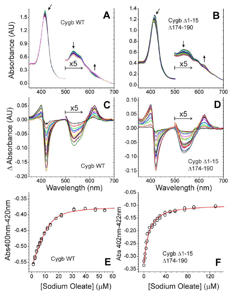
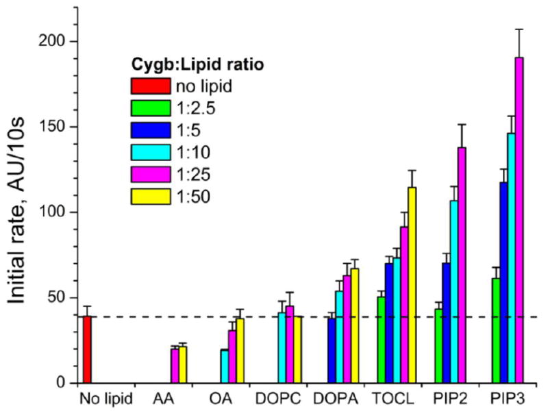
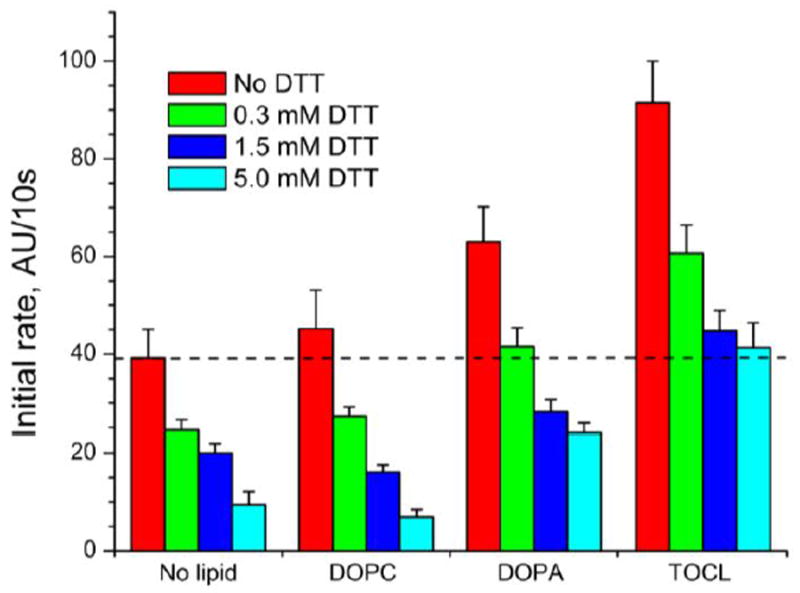
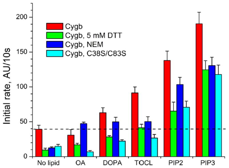
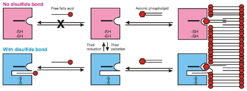
Cytoglobin (Cygb) is a hexa-coordinated hemoprotein with yet to be defined physiological functions. The iron coordination and spin state of the Cygb heme group are sensitive to oxidation of two cysteine residues (Cys38/Cys83) and/or the binding of free fatty acids. However, the roles of redox vs lipid regulators of Cygb's structural rearrangements in the context of the protein peroxidase competence are not known. Searching for physiologically relevant lipid regulators of Cygb, here we report that anionic phospholipids, particularly phosphatidylinositolphosphates, affect structural organization of the protein and modulate its iron state and peroxidase activity both conjointly and/or independently of cysteine oxidation. Thus, different anionic lipids can operate in cysteine-dependent and cysteine-independent ways as inducers of the peroxidase activity. We establish that Cygb's peroxidase activity can be utilized for the catalysis of peroxidation of anionic phospholipids (including phosphatidylinositolphosphates) yielding mono-oxygenated molecular species. Combined with the computational simulations we propose a bipartite lipid binding model that rationalizes the modes of interactions with phospholipids, the effects on structural re-arrangements and the peroxidase activity of the hemoprotein.

Keywords: Cytoglobin; Lipid binding; Peroxidase activity; Phosphatidylinositolphosphates.


Figures

Figure 1
Figure 1. Oleic acid binding to Cygb WT and Cygb Δ1-15Δ174-190
Panels A and B show the spectra after sequential addition of OA to 13 μM Cygb WT (A) or 11 μM Cygb Δ1-15Δ174-190 (B). The arrows indicate the direction of the spectral changes. Panels C and D show the difference spectra along the titration for Cygb WT (C) and Cygb Δ1-15Δ174-190 (D). The 500-700nm region of the spectra is enlarged for clarity. Panels E and F show the fit of the data to a 1:1 binding model (see methods for details). The averaged KD values calculated are 2.3 ± 1.0 μM for Cygb WT (E) and 3.0 ± 1.8 μM for Cygb Δ1-15Δ174-190 (F).
Figure 2
Figure 2. Effect of lipids on Amplex red oxidation by cytoglobin
The peroxidase activity of Cygb was studied in the presence of free FAs (AA, OA) or liposomes containing neutral (DOPC) or anionic (DOPA, TOCL, PIP2, PIP3) phospholipids. The oxidation of the peroxidation substrate Amplex red was monitored following the fluorescence of its oxidation product resorufin. 0.5 μM Cygb was incubated with lipids in Cygb/lipid ratios from 1:2.5 to 1:50.
Figure 3
Figure 3. Effect of cytoglobin thiol reduction on Amplex red oxidation
Cygb was pretreated with different concentrations of DTT to generate increasing amounts of the thiol-reduced protein. Excess DTT was removed before the reaction. The peroxidase activity of Cygb was studied in the absence of lipids or in the presence of DOPC, DOPA or TOCL. The oxidation of the peroxidation substrate Amplex red was monitored following the fluorescence of its oxidation product resorufin. 0.5 μM Cygb was incubated with 12.5 μM of each lipid.
Figure 4
Figure 4. Effect of thiol-modifying agents on Amplex red oxidation by cytoglobin
The peroxidase activity of Cygb with an intramolecular disulfide bond (WT, native), with reduced thiols (Cygb, 5mM DTT), with NEM-blocked thiols (Cygb, NEM), or a mutant Cygb without cysteine groups (Cygb, C38S/C83S) was studied using Amplex red as substrate. The reaction was monitored in the absence of lipids or in the presence of OA or anionic phospholids (DOPA, TOCL, PIP2, PIP3). 0.5 μM Cygb was incubated with 12.5 μM of each lipid.
Figure 5
Figure 5. EPR assessments of the cytoglobin peroxidase activity in the presence of TOCL
The peroxidase activity of Cygb with an intramolecular disulfide bond (Panel A) or after DTT-treatment (Panel B) was monitored by the formation of etoposide phenoxyl radicals. The inset shows the typical EPR signal of etoposide phenoxyl radicals. The encircled part of the spectrum was recorded repeatedly to obtain the time course of etoposide radical formation. Panel B, the EPR signal of thiol-reduced Cygb in the absence (i) or presence (ii) of TOCL is shown. Panel C, effect of lipids on etoposide radical generation by native or thiol-reduced Cygb measured 3 min after the addition of H2O2. 4 μM Cygb was incubated with different concentrations of lipids in 20 mM HEPES, 100 μM DTPA, pH 7.4 for 5 minutes. Measurements of the time course of etoposide radical formation were started 1 minute after the addition of H2O2 to the incubation mixture.
Figure 6
Figure 6. LC-MS/MS analysis and quantitation of phospholipid oxidation by Cygb/H2O2 system
Panel A, the bar graph shows the amount of phospholipid oxidized by the Cygb/H2O2 system. In the presented values, control experiments (without Cygb) were subtracted (mean ± SD from three experiments). Panel B, confirmation of SAPA (top spectrum) and the oxidized SAPA (bottom spectrum) by MS2 fragmentation. The bold labelled m/z indicates FA fragments (283.264-stearoyl, 303.233-arachidonoyl). The underlined m/z indicates an oxidized arachidonoyl fragment (319.227) and a fragment representing a lost water molecule from the oxidized arachidonoyl fragment (301.127). Panel C, confirmation of SAPIP3 (top spectrum) and oxidized SAPIP3 (bottom spectrum) by MS2 fragmentation. Labels are as indicated for panel B.
Figure 7
Figure 7. Formation of disulfide bond between Cys38 and Cys83 induces the bending of the E helix and an increase of the distance between E and F helices
The figure shows the overlay of the hexa-coordinated crystal structure of the C38S/C83S mutant of human cytoglobin (purple, PDB ID: 1UT0) with a model of the penta-coordinated, ferric wild-type structure (cyan). The yellow dashed line indicates the distance between the alpha carbons of Ala88 (E helix) and Val105 (F helix). Molecular dynamics simulations indicate that the formation of the disulfide bond causes an increase in this interhelical distance (8.04 ± 0.64 Å vs 7.2 Å, respectively), allowing for the binding of hydrophobic chains in the hydrophobic core of Cygb.
Figure 8
Figure 8. Binding models for cytoglobin and OA, DOPA and PIP3
Panels A and B, predicted binding model for OA. The aliphatic chain extends into the hydrophobic core of the protein; the carboxylate group interacts with the Thr91 side chain. Panel A shows the view from the heme plane. Panel B shows a top view. Panels C and D, predicted binding model for DOPA. Panel C, view from the heme plane; Panel D, top view. The model shows Thr91 and Arg84 stabilizing the phosphate group of DOPA; one oleic chain extends into the hydrophobic core of the protein (yellow) whereas the other hydrophobic chain is either interacting with the solvent or occupying a secondary binding site in the distal heme pocket (pink). Panels E and F, predicted binding model for PIP3. Panel E, view from the heme plane; Panel F, top view. The phosphate groups of PIP3 are shown interacting with Lys111 and Lys116. Arg84 and Thr91 interact with the ester groups from the FA chains. The phosphate group missing in PIP2 is the phosphate group interacting with Lys 116. As observed in the DOPA models, one oleic chain extends into the hydrophobic core of the protein (yellow) whereas the other hydrophobic chain is either interacting with the solvent or occupying a secondary binding site in the distal heme pocket (pink). The lipids, heme group and selected Cygb residues are shown as ball and sticks. Lipid carbon atoms are shown in yellow/pink, heme and Cygb side chain carbon atoms are shown in cyan.
Figure 9
Figure 9. Bipartite binding model for cytoglobin-lipid binding
The binding of aliphatic chains to Cygb is regulated by the formation of the disulfide bridge between Cys38 and Cys83. The state of the disulfide bond is regulated by the cell environment and can shift between the free thiol conformations (pink) and the disulfide bond state (blue). Free FAs may bind to Cygb only when the disulfide bond is formed (bottom left). Anionic phospholipids may bind to the cytoglobin surface independently of the formation of the disulfide bond (right side). The formation of the disulfide bond allows for the additional binding of one phospholipid FA chain, increasing the affinity for the lipid.

References

    1. Reeder BJ. The redox activity of hemoglobins: from physiologic functions to pathologic mechanisms. Antioxid Redox Signal. 2010;13:1087–123. - PubMed
    1. Kagan VE, Bayir HA, Belikova NA, Kapralov O, Tyurina YY, Tyurin VA, Jiang J, Stoyanovsky DA, Wipf P, Kochanek PM, Greenberger JS, Pitt B, Shvedova AA, Borisenko G. Cytochrome c/cardiolipin relations in mitochondria: a kiss of death. Free Radic Biol Med. 2009;46:1439–53. - PMC - PubMed
    1. Pesce A, De Sanctis D, Nardini M, Dewilde S, Moens L, Hankeln T, Burmester T, Ascenzi P, Bolognesi M. Reversible hexa- to penta-coordination of the heme Fe atom modulates ligand binding properties of neuroglobin and cytoglobin. IUBMB Life. 2004;56:657–64. - PubMed
    1. Genova ML, Lenaz G. Functional role of mitochondrial respiratory supercomplexes. Biochim Biophys Acta. 2014;1837:427–43. - PubMed
    1. Kagan VE, Tyurin VA, Jiang J, Tyurina YY, Ritov VB, Amoscato AA, Osipov AN, Belikova NA, Kapralov AA, Kini V, Vlasova II, Zhao Q, Zou M, Di P, Svistunenko DA, Kurnikov IV, Borisenko GG. Cytochrome c acts as a cardiolipin oxygenase required for release of proapoptotic factors. Nat Chem Biol. 2005;1:223–32. - PubMed

Publication types

MeSH terms

LinkOut - more resources