. 2016 Mar 1;6(1):1-14.
doi: 10.1002/9780470942390.mo150182.

Analysis of Mitochondrial Respiratory Chain Supercomplexes Using Blue Native Polyacrylamide Gel Electrophoresis (BN-PAGE)

Affiliations

Analysis of Mitochondrial Respiratory Chain Supercomplexes Using Blue Native Polyacrylamide Gel Electrophoresis (BN-PAGE)

Pooja Jha et al. Curr Protoc Mouse Biol. .

Abstract

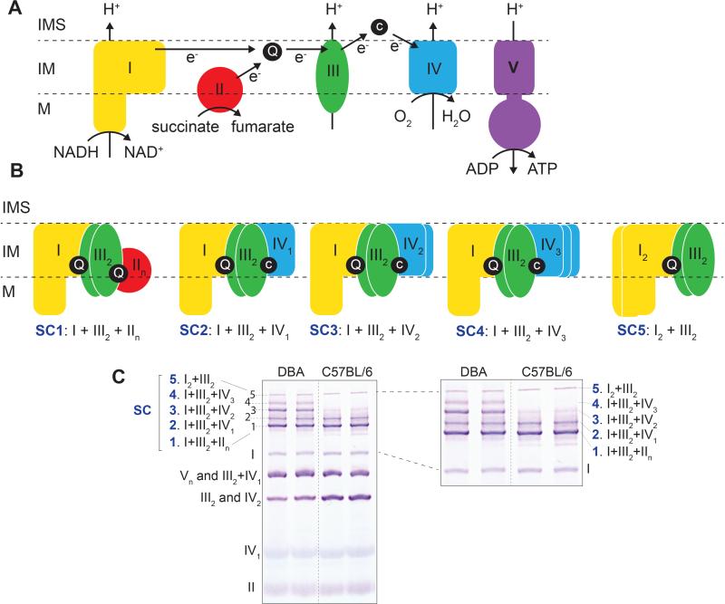
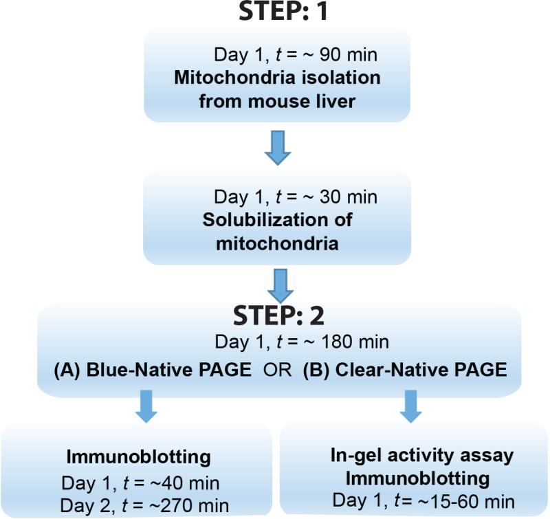
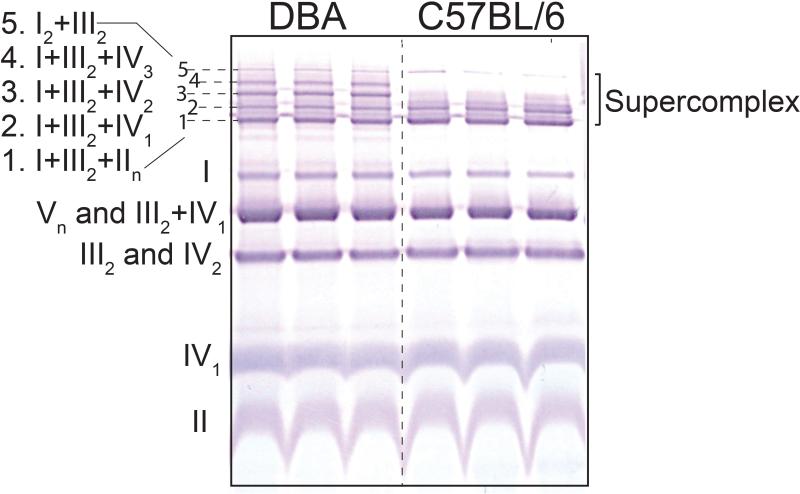
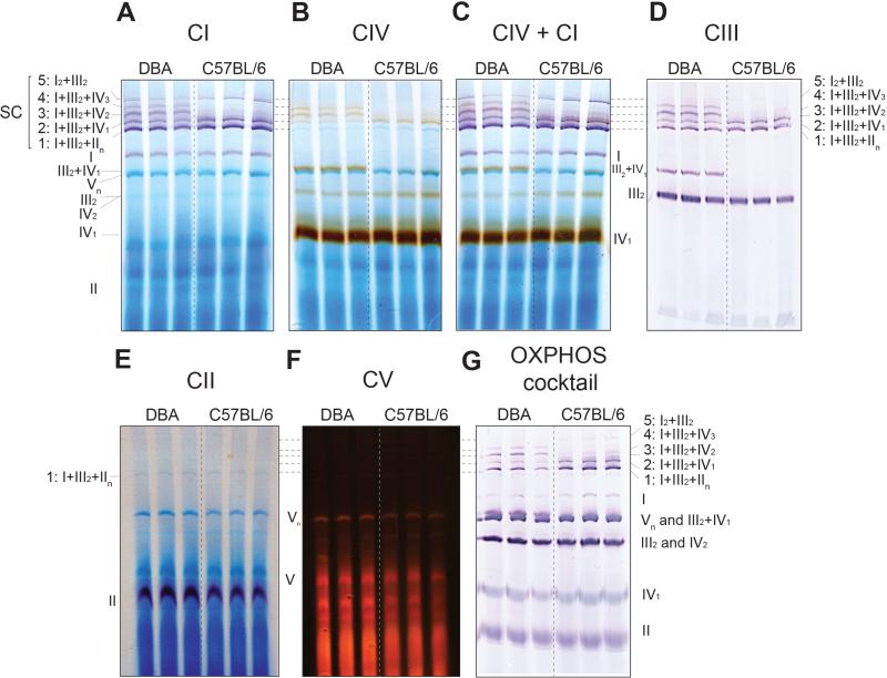
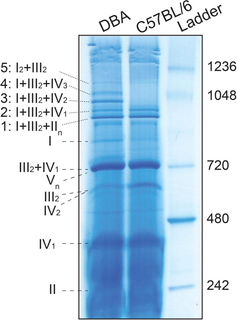
Mitochondria are cellular organelles that harvest energy in the form of ATP through a process termed oxidative phosphorylation (OXPHOS), which occurs via the protein complexes of the electron transport chain (ETC). In recent years it has become unequivocally clear that mitochondrial complexes of the ETC are not static entities in the inner mitochondrial membrane. These complexes are dynamic and in mammals they aggregate in different stoichiometric combinations to form supercomplexes (SCs) or respirasomes. It has been proposed that the net respiration is more efficient via SCs than via isolated complexes. However, it still needs to be determined whether the activity of a particular SC is associated with a disease etiology. Here we describe a simplified method to visualize and assess in-gel activity of SCs and the individual complexes with good resolution using blue native polyacrylamide gel electrophoresis (BN-PAGE).

Keywords: Cox7a2l; SCAFI; in-gel activity; mitochondria; oxidative phosphorylation; supercomplex.


Figures

Figure 1
Figure 1. Schematic representation of the oxidative phosphorylation (OXPHOS) system showing its individual complexes, supercomplexes and its nomenclature
(A) Mitochondrial electron transport chain (ETC) and ATP synthesis. In TCA cycle, NADH and succinate are electron donors, oxidized by NADH dehydrogenase (CI) and succinate dehydrogenase (CII), respectively. Ubiquinone (Q) is one of the electron acceptors which transfers the electrons to cytochrome c (c) by cytochrome c reductase (CIII). The electrons then pass to their terminal acceptor molecular oxygen (O2) to release water by cytochrome c oxidase (CIV). This electron transfer is coupled with the transfer of protons (H+) across mitochondrial inner membrane, except complex II. The electrochemical proton gradient generated drives the generation of chemical energy in the form of adenosine triphosphate (ATP) through ATP synthase (CV). Note that in this figure, for simplicity, all components are shown as monomers, however in reality their stoichiometry is more complex e.g. complex III exists as dimers (as shown in the figure). Dimers and multiple aggregation of CIV are also present (not shown). (B and C) Representative stoichiometry and nomenclature of mitochondrial supercomplexes shown as a cartoon (B), and in a BN-PAGE (C). I, II, III, IV, V: complexes I to V; SC, supercomplex; M, matrix; IM, inner membrane; IMS, inner membrane space.
Figure 2
Figure 2. Schematic representation of the workflow
STEP 1 involves isolation and solubalization of mitochondria. In STEP 2, one can choose to either do BN-PAGE followed by immunoblotting (STEP 2(A)) or CN-PAGE (minor modification of BN-PAGE) followed by in-gel activity of the supercomplexes and complexes, with or without additional immunoblotting of a separate gel, run simultaneously (STEP 2(B)).
Figure 3
Figure 3. BN-PAGE showing mitochondrial complexes and supercomplexes
Liver mitochondria were isolated form DBA and C57BL6/ mice. 50 μg of mitochondrial protein (8g/g digitonin to protein ratio) was used for BN-PAGE for a run time of 30 min at 150 V and 1 h at 250 V followed by immunoblotting with an OXPHOS antiboty cocktail. The five SC stoichiometric combinations are indicated as numbers 1 to 5. Note, in C57BL/6 mice only 3 SC bands are observed (1, 2 and 5) in contrast to the 5 bands present in DBA mice.
Figure 4
Figure 4. BN-PAGE, followed by in gel activity assay and immunoblotting to define the SC bands in DBA and C57BL/6 (representing the 2 typical patterns of SCs in rodents)
Liver mitochondria were isolated and 50 μg of mitochondrial protein (4g/g digitonin to protein ratio) was used for BN-PAGE for a run time of 30 min at 150 V and 150 min at 250 V. In-gel activity assays and western blot was performed thereafter. (A) CI activity is shown in violet (20 min incubation). (B) CIV in brown (40 min incubation). (C) CIV+CI in brown and violet, respectively (40 min incubation in CIV substrate followed by 20 min in CI substrate). (D) CIII is shown by immunoblotting against the UQCRC2 subunit of CIII. (E) CII activity is shown in violet (40 min incubation). (F) CV activity is shown in red (16 h incubation). (G) Western blot developed with an OXPHOS antibody cocktail. Note, SCs 3, 4 and III2+IV1 are absent in C57BL/6 mice.
Figure 5
Figure 5. BN-PAGE, followed by Coomassie staining and destaining
Liver mitochondria were isolated and 150 μg of mitochondrial protein (4g/g digitonin to protein ratio) was used for BN-PAGE for a run time of 30min at 150V and 150min at 250V. The gel was stained and then destained using the colloidal blue staining kit.

References

    1. Acin-Perez R, Fernandez-Silva P, Peleato ML, Perez-Martos A, Enriquez JA. Respiratory active mitochondrial supercomplexes. Molecular cell. 2008;32:529–539. - PubMed
    1. Adams S, Pacharinsak C. Mouse anesthesia and analgesia. Current protocols in mouse biology. 2015;5:51–63. - PubMed
    1. Barrientos A, Ugalde C. I function, therefore I am: overcoming skepticism about mitochondrial supercomplexes. Cell Metab. 2013;18:147–149. - PMC - PubMed
    1. Budde SM, van den Heuvel LP, Janssen AJ, Smeets RJ, Buskens CA, DeMeirleir L, Van Coster R, Baethmann M, Voit T, Trijbels JM, Smeitink JA. Combined enzymatic complex I and III deficiency associated with mutations in the nuclear encoded NDUFS4 gene. Biochemical and biophysical research communications. 2000;275:63–68. - PubMed
    1. Chaban Y, Boekema EJ, Dudkina NV. Structures of mitochondrial oxidative phosphorylation supercomplexes and mechanisms for their stabilisation. Biochimica et biophysica acta. 2014;1837:418–426. - PubMed

Publication types

MeSH terms

Substances

LinkOut - more resources