Review
. 2016 May;14(5):918-30.
doi: 10.1111/jth.13302. Epub 2016 Apr 9.

Platelet signaling: a complex interplay between inhibitory and activatory networks

Affiliations
Review

Platelet signaling: a complex interplay between inhibitory and activatory networks

A P Bye et al. J Thromb Haemost. 2016 May.

Abstract

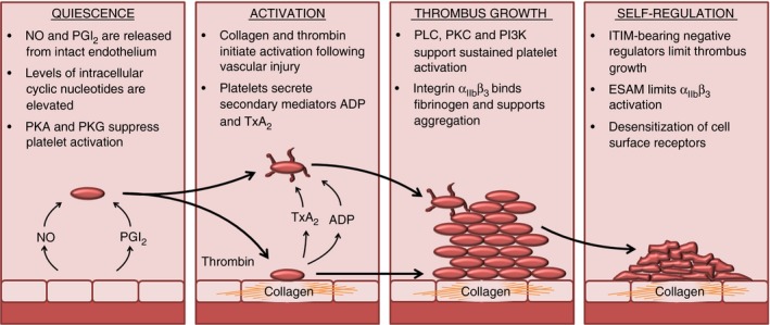
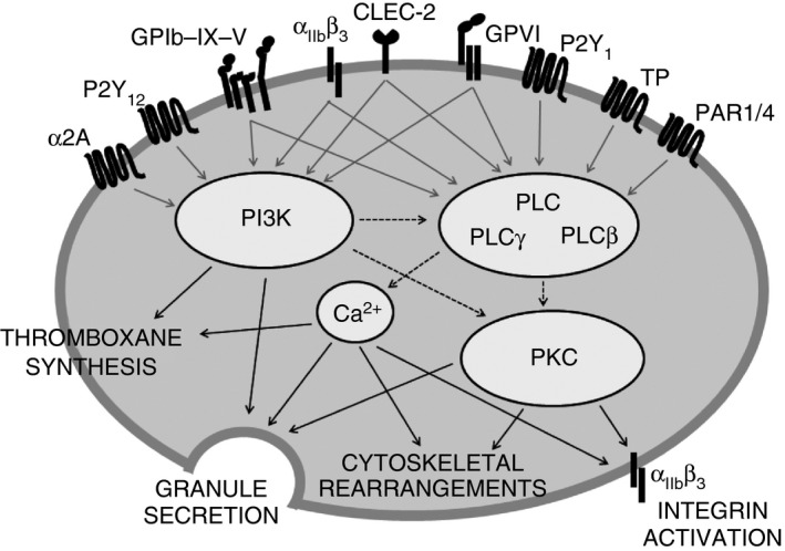
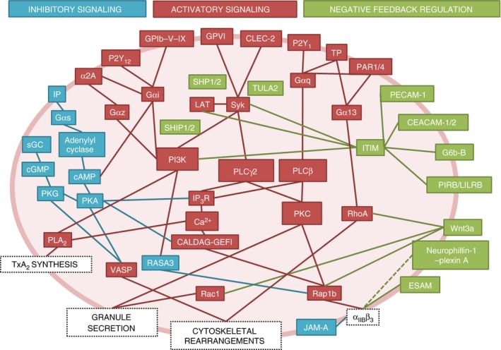
The role of platelets in hemostasis and thrombosis is dependent on a complex balance of activatory and inhibitory signaling pathways. Inhibitory signals released from the healthy vasculature suppress platelet activation in the absence of platelet receptor agonists. Activatory signals present at a site of injury initiate platelet activation and thrombus formation; subsequently, endogenous negative signaling regulators dampen activatory signals to control thrombus growth. Understanding the complex interplay between activatory and inhibitory signaling networks is an emerging challenge in the study of platelet biology, and necessitates a systematic approach to utilize experimental data effectively. In this review, we will explore the key points of platelet regulation and signaling that maintain platelets in a resting state, mediate activation to elicit thrombus formation, or provide negative feedback. Platelet signaling will be described in terms of key signaling molecules that are common to the pathways activated by platelet agonists and can be described as regulatory nodes for both positive and negative regulators.

Keywords: blood platelets; hemostasis; platelet activation; review; thrombosis.


Figures

Figure 1
Figure 1
Stages of platelet activation and thrombus formation. Platelets in the circulation are kept in a quiescent state by nitric oxide (NO) and prostacyclin (prostaglandin I2; PGI 2), which are released by the vascular endothelium. In platelets, NO and PGI 2 increase the levels of cGMP and cAMP, and suppress platelet activity by the activation of protein kinase A (PKA) and protein kinase G (PKG). Following vessel injury, components of the subendothelial matrix are exposed, including collagen, which provides an adhesive surface for platelets to attach to and initiate signaling events and platelet activation. Local production of thrombin and secretion of secondary mediators also contribute to the initiation of platelet activation. Key components of platelet signaling pathways are activated, including phospholipase C (PLC), protein kinase C (PKC), and phosphatidylinositide‐3‐kinase (PI3K), supporting sustained platelet activation and thrombus formation through the initiation of cytoskeletal rearrangements, granule secretion, and activation of integrin αII bβ3. So as to limit thrombus growth and prevent the formation of occlusive thrombi, platelets contain self‐regulating negative feedback mechanisms that counteract positive signaling. These negative regulators include immunoreceptor tyrosine‐based inhibition motif (ITIM)‐containing receptors, endothelial cell‐selective adhesion molecule (ESAM), which negatively regulates integrin αII bβ3 activity, phosphatases that counteract phosphorylation‐dependent positive signaling, and receptor desensitization, which reduces the platelets’ response to secondary mediator signaling. TxA2, thromboxane A2.
Figure 2
Figure 2
Phospholipase C (PLC), protein kinase C (PKC) and phosphatidylinositide‐3‐kinase (PI3K) are key mediators of platelet activation. All main activating platelet agonist receptors identified to date activate at least one of the following key activatory signaling mediators: PLC, PKC, or PI3K. These signaling nodes underlie several key processes required for platelet activation, including secretion of secondary mediators and activation of integrin αII bβ3, facilitating fibrinogen binding and platelet aggregation, and also cytoskeletal rearrangements, which enable platelet shape change and spreading. CLEC‐2, C‐type lectin receptor 2; GPVI, glycoprotein VI (collagen receptor); GPIb–IX–V, glycoprotein Ib–IX–V (von Willebrand factor receptor); PAR, protease‐activated receptor; P2Y1/P2Y12, ADP receptors; TP, thromboxane A2 receptor; αII bβ3, receptor for fibrinogen; α2A, adrenergic receptor.
Figure 3
Figure 3
Complexity of platelet signaling networks. Platelet signaling models frequently describe signaling pathways activated by individual agonists; however, platelet signaling in vivo is highly complex, and involves simultaneous activation by multiple agonists and negative regulators, which form a complex signaling network. Several key signaling molecules, i.e. phospholipase C (PLC), protein kinase C (PKC), and phosphatidylinositide‐3‐kinase (PI3K), are common between the different pathways and form key nodes of platelet regulation. Blue boxes and lines represent mediators of inhibitory signaling that act to suppress platelet function in the absence of platelet activators in the healthy endothelium. Red boxes and lines represent mediators of activatory signaling following platelet activation by platelet agonists. Green boxes and lines represent mediators of negative feedback and inhibitory signaling that act to limit platelet activation following stimulation by platelet agonists. CALDAG‐GEFI, Ca2+‐dependent Rap1 guanine nucleotide exchange factor; CEACAM‐1/2, carcinoembryonic antigen cell adhesion molecule‐1/2; CLEC‐2, C‐type lectin receptor 2; ESAM, endothelial cell‐selective adhesion molecule; GP, glycoprotein; IP, prostaglandin receptor; IP 3 R, inositol trisphosphate receptor; ITIM, immunoreceptor tyrosine‐based inhibition motif; JAM‐A, junctional adhesion molecule A; LAT, linker of activated T cells; PAR, protease‐activated receptor; PECAM‐1, platelet endothelial cell adhesion molecule‐1; PIRB, paired immunoglobulin‐like receptor B; PKA, protein kinase A; PKG, protein kinase G; PLA 2, phospholipase A2; sGC, soluble guanyl cyclase; TULA2, T‐cell ubiquitin ligand‐2; TxA2, thromboxane A2; VASP, vasodilator‐stimulated phosphoprotein; α2A, adrenergic receptor.

References

    1. Aarts PA, van den Broek SA, Prins GW, Kuiken GD, Sixma JJ, Heethaar RM. Blood platelets are concentrated near the wall and red blood cells, in the center in flowing blood. Arteriosclerosis 1988; 8: 819–24. - PubMed
    1. Smolenski A. Novel roles of cAMP/cGMP‐dependent signaling in platelets. J Thromb Haemost 2012; 10: 167–76. - PubMed
    1. Mellion BT, Ignarro LJ, Ohlstein EH, Pontecorvo EG, Hyman AL, Kadowitz PJ. Evidence for the inhibitory role of guanosine 3′,5′‐monophosphate in ADP‐induced human platelet aggregation in the presence of nitric oxide and related vasodilators. Blood 1981; 57: 946–55. - PubMed
    1. Dutta‐Roy AK, Sinha AK. Purification and properties of prostaglandin E1/prostacyclin receptor of human blood platelets. J Biol Chem 1987; 262: 12685–91. - PubMed
    1. Gorman RR, Bunting S, Miller OV. Modulation of human platelet adenylate cyclase by prostacyclin (PGX). Prostaglandins 1977; 13: 377–88. - PubMed

Publication types

MeSH terms