Review
. 2016 Jun;15(6):1773-90.
doi: 10.1074/mcp.O115.057638. Epub 2016 Feb 29.

Maturing Glycoproteomics Technologies Provide Unique Structural Insights into the N-glycoproteome and Its Regulation in Health and Disease

Affiliations
Review

Maturing Glycoproteomics Technologies Provide Unique Structural Insights into the N-glycoproteome and Its Regulation in Health and Disease

Morten Thaysen-Andersen et al. Mol Cell Proteomics. 2016 Jun.

Abstract

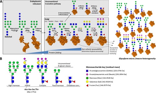
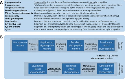
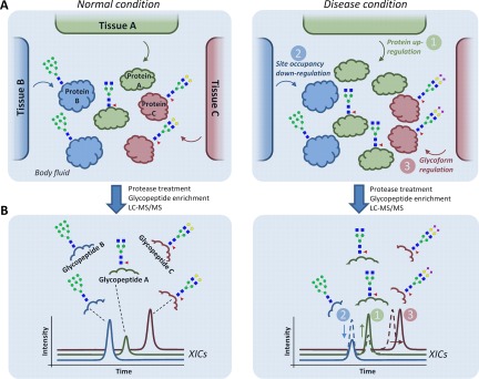
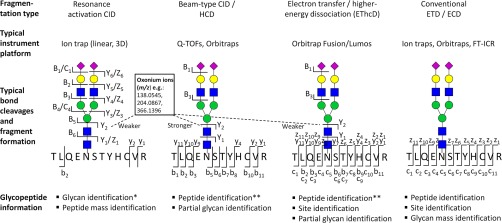
The glycoproteome remains severely understudied because of significant analytical challenges associated with glycoproteomics, the system-wide analysis of intact glycopeptides. This review introduces important structural aspects of protein N-glycosylation and summarizes the latest technological developments and applications in LC-MS/MS-based qualitative and quantitative N-glycoproteomics. These maturing technologies provide unique structural insights into the N-glycoproteome and its synthesis and regulation by complementing existing methods in glycoscience. Modern glycoproteomics is now sufficiently mature to initiate efforts to capture the molecular complexity displayed by the N-glycoproteome, opening exciting opportunities to increase our understanding of the functional roles of protein N-glycosylation in human health and disease.


Figures

Fig. 1.
Fig. 1.
Overview of the biosynthesis and structural classes of mammalian protein N-glycosylation. A, Schematic summary of the biosynthetic machinery of N-glycoproteins. The enzymatic processing, which is initiated while the glycoproteins are still being translated, translocated, and folded, may terminate at any point in the enzymatic sequence depending partially on the Asn solvent accessibility of the maturely folded glycoprotein (3). This generates site-, cell-, and even subcellular-specific glycoform heterogeneity forming one of the functionally most important features of the glycoproteome (5), and also creates substantial analytical challenges. TGN: trans-Golgi network. B, Mammalian N-glycoproteins are typically divided into three main N-glycan classes: high mannose, hybrid, and complex type. Unusual paucimannosidic and chitobiose core type N-glycans arising from unconventional truncation pathways (dashed box) have been reported in specific cell types and physiological conditions (21). Monosaccharide residues are depicted according to the establish nomenclature (180) with residual monoisotopic masses provided.
Fig. 2.
Fig. 2.
A, Definitions and explanations of commonly used nomenclature in glycoproteomics. B, Generic workflow illustrating important components of a glycoproteomics experiment, which crudely can be divided into segments related to glycopeptide sample preparation (top box) and detection (bottom box). Typical examples of the individual components are provided. *Additional sample handling including glycoprotein derivatization and glycopeptide labeling for quantitation purposes may be introduced at this step.
Fig. 3.
Fig. 3.
Three fundamental levels of molecular dysregulation of multiple tissues contributing to an altered secreted N-glycoproteome during disease. A, Hypothetical example illustrating three sources of dysregulation: 1) protein level (green), 2) site occupancy (blue), or 3) glycosylation micro-heterogeneity (red) from three separate tissues (Tissue A-C) contributing to a joint secreted N-glycoproteome (Protein A-C) in a body fluid derived from disease (right) and 'normal' healthy (left) condition. B, After proteolysis and enrichment, the altered abundance of the resulting glycopeptides can be detected using LC-MS/MS-based label-free quantitative glycoproteomics as shown by color-coded traces representing extracted ion chromatograms (XICs). However, establishing which of the three mechanisms causes the glycopeptide alterations for the detected glycoproteins may be challenging solely with glycopeptide analysis, especially in glycoproteomes arising from multiple tissues. Parallel quantitative proteomics and “deglycoproteomics” (detection of formerly occupied N-sites) of the same samples can assist in this task.
Fig. 4.
Fig. 4.
The main dissociation methods in glycoproteomics, typically used in conjunction i.e. resonance activation (ion trap type) CID, beam-type (Q-TOF type) CID or HCD, hybrid-type EThcD, and the conventional ETD (or ECD) fragmentation. The resulting bond cleavages and fragment ion formations when applied to N-glycopeptides are indicated including the presence of diagnostic oxonium ions (insert) (116, 117). *N-Glycan identification is usually restricted to specification of the monosaccharide composition and the partial or complete topology. **The b/y-ions in the beam-type CID (HCD) and EThcD dissociation schemes usually lose the conjugated glycan and therefore do not provide information about the glycosylation site.
Fig. 5.
Fig. 5.
Nonchronological summary of the N-glycoproteome coverage as evaluated by the number of reported unique intact N-glycopeptides of glycoproteomics studies published over the past decade. Dark blue bars represent recent glycoproteomics studies (2014–present) covered in this review (see also Table II) and light blue bars represent older studies (2005–2014) covered elsewhere (59).

References

    1. Varki A., and Sharon N. (2009) Historical Background and Overview. In: Varki A., Cummings R. D., Esko J. D., Freeze H. H., Stanley P., Bertozzi C. R., Hart G. W., and Etzler M. E., eds. Essentials of Glycobiology, 2nd Ed., Cold Spring Harbor (NY) - PubMed
    1. Shrimal S., Cherepanova N. A., and Gilmore R. (2015) Cotranslational and posttranslocational N-glycosylation of proteins in the endoplasmic reticulum. Semin. Cell Dev. Biol. 41, 71–78 - PMC - PubMed
    1. Thaysen-Andersen M., and Packer N. H. (2012) Site-specific glycoproteomics confirms that protein structure dictates formation of N-glycan type, core fucosylation and branching. Glycobiology 22, 1440–1452 - PubMed
    1. Rudd P. M., and Dwek R. A. (1997) Glycosylation: heterogeneity and the 3D structure of proteins. Crit. Rev. Biochem. Mol. Biol. 32, 1–100 - PubMed
    1. Lee L. Y., Lin C. H., Fanayan S., Packer N. H., and Thaysen-Andersen M. (2014) Differential site accessibility mechanistically explains subcellular-specific N-glycosylation determinants. Front. Immunol. 5, 404. - PMC - PubMed

Publication types