. 2016 Mar 15;113(11):3078-83.
doi: 10.1073/pnas.1512603113. Epub 2016 Feb 29.

Ablation of sensory neurons in a genetic model of pancreatic ductal adenocarcinoma slows initiation and progression of cancer

Affiliations

Ablation of sensory neurons in a genetic model of pancreatic ductal adenocarcinoma slows initiation and progression of cancer

Jami L Saloman et al. Proc Natl Acad Sci U S A. .

Abstract

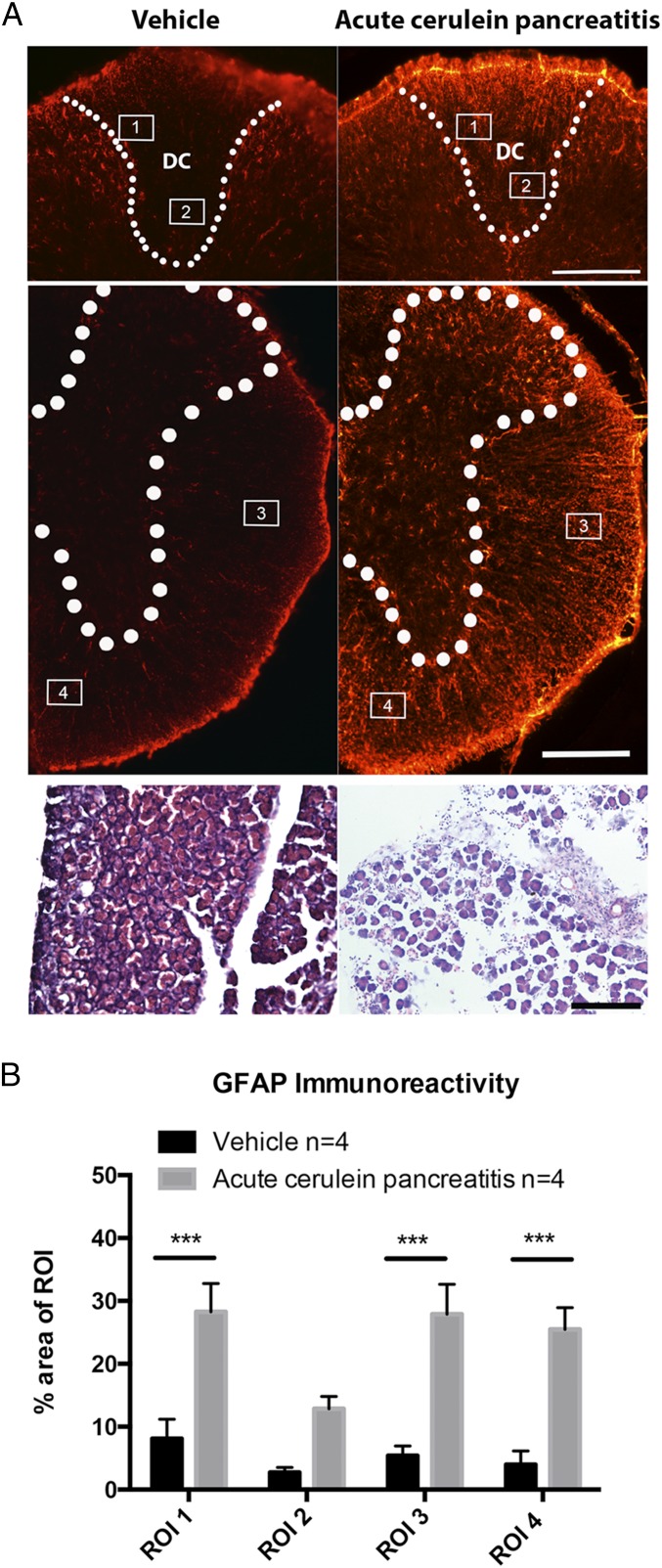
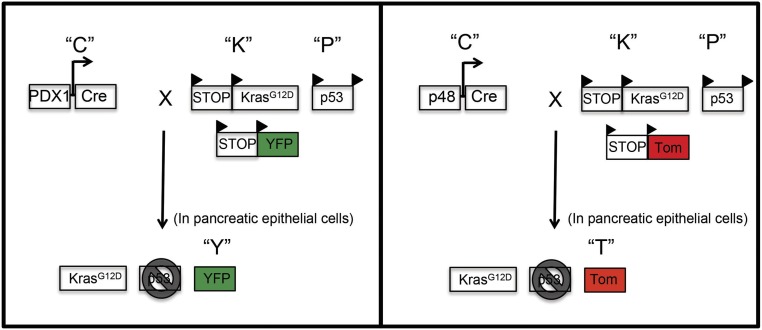
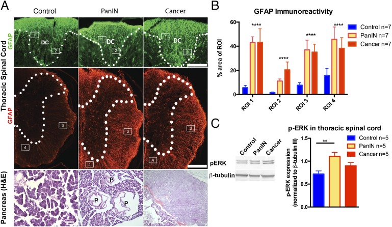
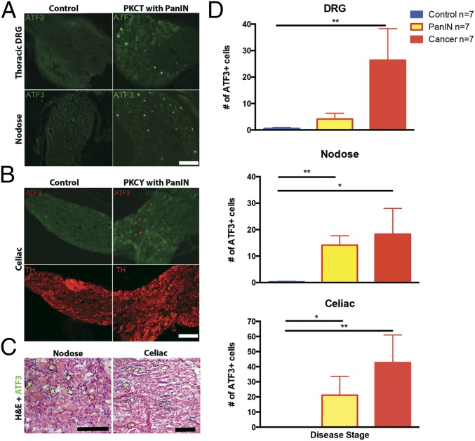
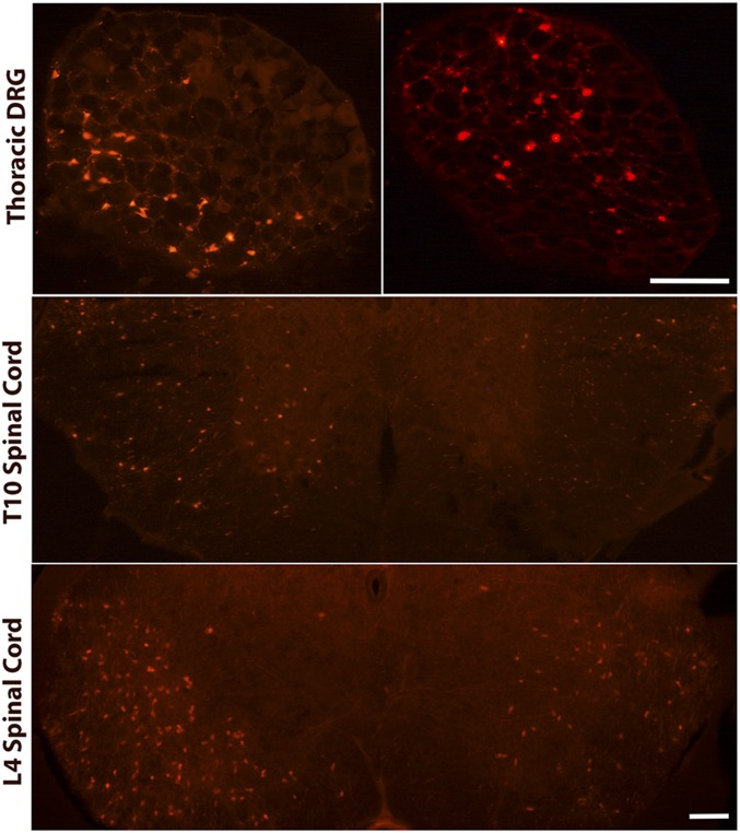
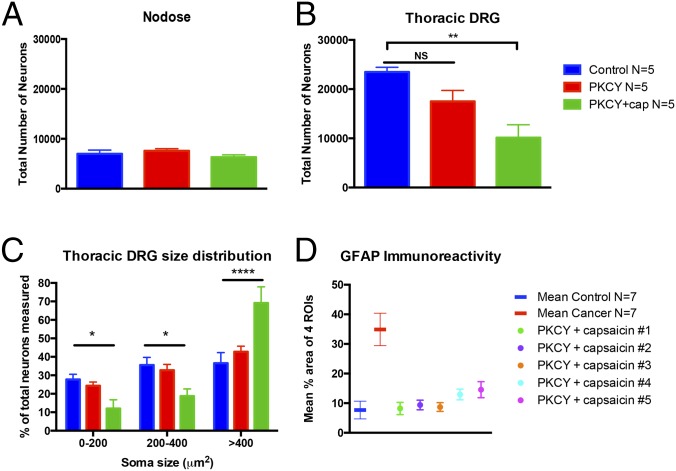
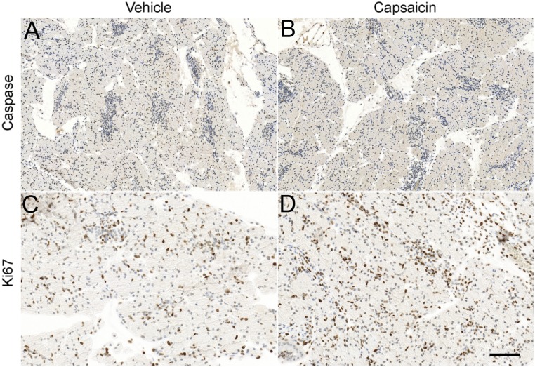
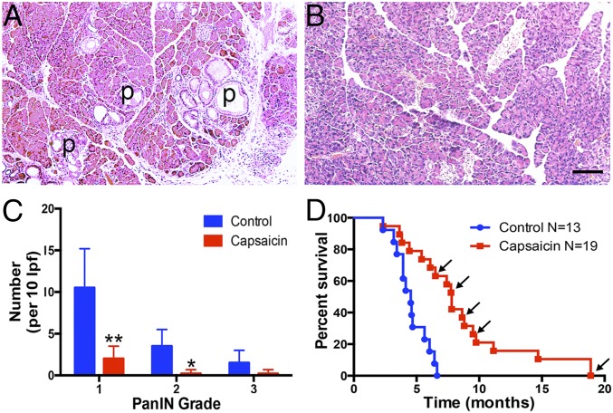
Pancreatic ductal adenocarcinoma (PDAC) is characterized by an exuberant inflammatory desmoplastic response. The PDAC microenvironment is complex, containing both pro- and antitumorigenic elements, and remains to be fully characterized. Here, we show that sensory neurons, an under-studied cohort of the pancreas tumor stroma, play a significant role in the initiation and progression of the early stages of PDAC. Using a well-established autochthonous model of PDAC (PKC), we show that inflammation and neuronal damage in the peripheral and central nervous system (CNS) occurs as early as the pancreatic intraepithelial neoplasia (PanIN) 2 stage. Also at the PanIN2 stage, pancreas acinar-derived cells frequently invade along sensory neurons into the spinal cord and migrate caudally to the lower thoracic and upper lumbar regions. Sensory neuron ablation by neonatal capsaicin injection prevented perineural invasion (PNI), astrocyte activation, and neuronal damage, suggesting that sensory neurons convey inflammatory signals from Kras-induced pancreatic neoplasia to the CNS. Neuron ablation in PKC mice also significantly delayed PanIN formation and ultimately prolonged survival compared with vehicle-treated controls (median survival, 7.8 vs. 4.5 mo; P = 0.001). These data establish a reciprocal signaling loop between the pancreas and nervous system, including the CNS, that supports inflammation associated with oncogenic Kras-induced neoplasia. Thus, pancreatic sensory neurons comprise an important stromal cell population that supports the initiation and progression of PDAC and may represent a potential target for prevention in high-risk populations.

Keywords: PanIN; inflammation; pancreatic ductal adenocarcinoma; sensory neuron; tumorigenesis.


Conflict of interest statement

The authors declare no conflict of interest.

Figures

Fig. 1.
Fig. 1.
Cerulein-induced acute pancreatitis causes spinal inflammation. (A) The thoracic spinal cord has increased GFAP immunoreactivity in cerulein-treated mice. H&E staining (Lower) shows cerulein-induced disruption in pancreatic histology. (B) Quantification of GFAP immunoreactivity across ROIs encompassing the dorsal columns, anterolateral, and medioventral white matter tracts of vehicle and cerulein-treated groups. ***P < 0.001, n = 4 per group. DC, dorsal columns. (Scale bar, 200 µm.)
Fig. S1.
Fig. S1.
Generation of PDAC transgenic mice (PKCT and PKCY). PDAC mice express a conditional activated (mutant) Kras allele targeted to the endogenous Kras locus under Lox-Stop-Lox control (KrasG12D) and a conditional TP53 allele with LoxP sites in intron 1 and intron 10 of the Tp53 gene (p53). PDAC mice were crossed with PDX1-Cre (PKC) or Ptf1a-p48-Cre (p48-Cre) mice. PKC mice expressing PDX1-Cre were crossed with a ROSA-YFP mouse to make PKCY mice (Left). PKC mice expressing p48-Cre were crossed with a ROSA Tdtomato reporter strain to make PKCT mice (Right) (1, 29).
Fig. 2.
Fig. 2.
Spinal inflammation is detected in early phases of PDAC. (A) Disease-stage–specific changes occur in GFAP staining in the thoracic spinal cord and pancreatic histology in PKCT and PKCY mice. (B) Astrocyte reactivity increases in regions of the spinal cord at the PanIN and cancer stage. (C) p-ERK is up-regulated in thoracic spinal cord of PKCT mice at the PanIN stage. ****P < 0.0001, n = 5–7 per group. DC, dorsal columns; P, PanIN lesion. (Scale bar, 200 µm.)
Fig. 3.
Fig. 3.
Pancreatic disease increases ATF3 in sensory afferents. (A) Nuclear localization of ATF3 immunoreactivity in DRG (Top) and nodose ganglia (Bottom) of PKCT mice at the PanIN stage. (B) ATF3 is also elevated in celiac ganglia of PKCY mice. (C) H&E staining shows ATF3-IR in neurons of nodose and celiac ganglia. (D) Summary of increases in neuronal ATF3 expression in sensory and autonomic ganglia during PanIN and cancer stages. *P < 0.05, **P < 0.01, n = 7 per group. (Scale bar, 100 µm.)
Fig. 4.
Fig. 4.
Pancreatic cells invade the DRG and spinal cord before PDAC development. Individual Tdtomato positive cells are present in thoracic DRG and thoracic and lumbar regions of the spinal cord. (Scale bar, 200 µm for DRG and 500 µm for spinal cord sections.)
Fig. 5.
Fig. 5.
Neonatal capsaicin ablates DRG neurons. (A) The total number of nodose ganglion neurons is unchanged in adult mice treated with capsaicin at P2. (B) In contrast, capsaicin causes a significant loss of thoracic DRG neurons. (C) A rightward shift in somal diameters of the remaining neurons indicates selective reduction of small diameter afferents. (D) Neonatal capsaicin prevents spinal inflammation. The mean percent area of the four ROIs (designated in Fig. 2A) covered by GFAP immunoreactivity is plotted as a mean (n = 7 per group) and compared with the mean of the four ROIs for individual animals treated with neonatal capsaicin. PKCY mice treated with capsaicin exhibit GFAP immunoreactivity levels similar to control. *P < 0.05, **P < 0.01, ****P < 0.0001.
Fig. S2.
Fig. S2.
Six P2 pups were euthanized 2 h post injection of capsaicin or vehicle. Vehicle-treated pancreata were processed for cleaved caspase 3 (A) or Ki67 (C) immunodetection as previously described (79, 80). Slides were counterstained with hematoxylin. Neonatal capsaicin treatment had no effect on cleaved caspase 3 (B) or Ki67 (D) expression. (Scale bar, 200 μm for A and B and 100 µm for C and D.)
Fig. 6.
Fig. 6.
Neonatal capsaicin slows development of PanIN lesions and prolongs survival of PKCY mice. Pancreata of 10-wk-old PKC mice were treated with vehicle (A) or capsaicin (B). Vehicle-treated mice exhibit numerous PanIN lesions (P), whereas pancreata from capsaicin-treated mice do not. (C) There were significantly fewer total PanIN lesions and virtually no lesions above grade 1 in capsaicin-treated mice; *P < 0.05, **P < 0.001. (D) Capsaicin-treated mice survive longer than vehicle-treated PKCY mice. Arrows indicate mice that were euthanized for histological analysis while otherwise healthy (no tumors upon ultrasound).

Comment in

References

    1. Rhim AD, et al. EMT and dissemination precede pancreatic tumor formation. Cell. 2012;148(1-2):349–361. - PMC - PubMed
    1. Rhim AD, et al. Detection of circulating pancreas epithelial cells in patients with pancreatic cystic lesions. Gastroenterology. 2014;146(3):647–651. - PMC - PubMed
    1. Carobi C. Capsaicin-sensitive vagal afferent neurons innervating the rat pancreas. Neurosci Lett. 1987;77(1):5–9. - PubMed
    1. Fasanella KE, Christianson JA, Chanthaphavong RS, Davis BM. Distribution and neurochemical identification of pancreatic afferents in the mouse. J Comp Neurol. 2008;509(1):42–52. - PMC - PubMed
    1. Lindsay TH, et al. Pancreatic cancer pain and its correlation with changes in tumor vasculature, macrophage infiltration, neuronal innervation, body weight and disease progression. Pain. 2005;119(1-3):233–246. - PubMed

Publication types

MeSH terms