

Structural basis for DNA cleavage by the potent antiproliferative agent (-)-lomaiviticin A
- PMID: 26929332
- PMCID: PMC4801295
- DOI: 10.1073/pnas.1519846113
Structural basis for DNA cleavage by the potent antiproliferative agent (-)-lomaiviticin A
Abstract
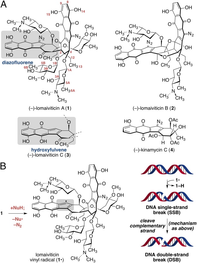
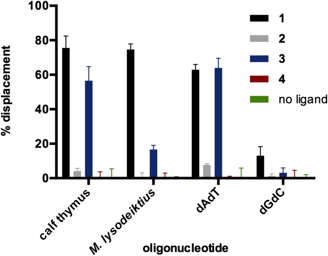
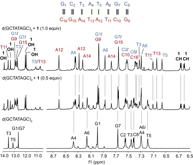
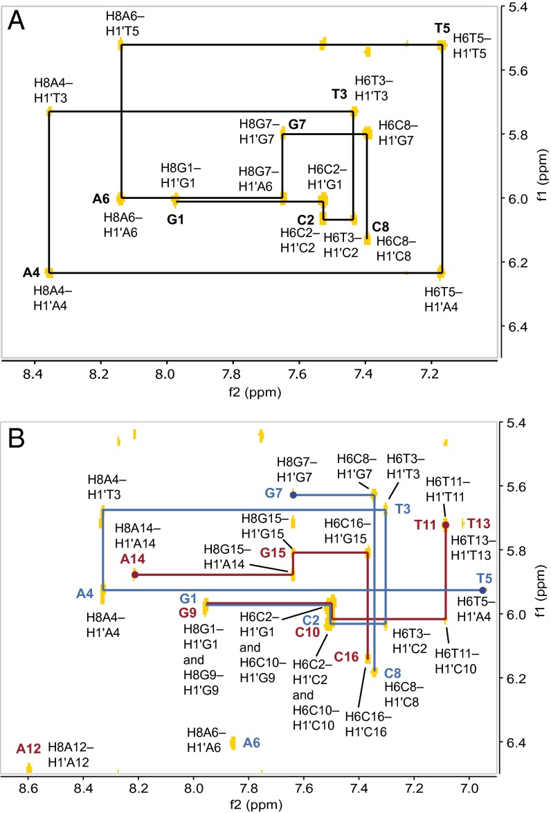
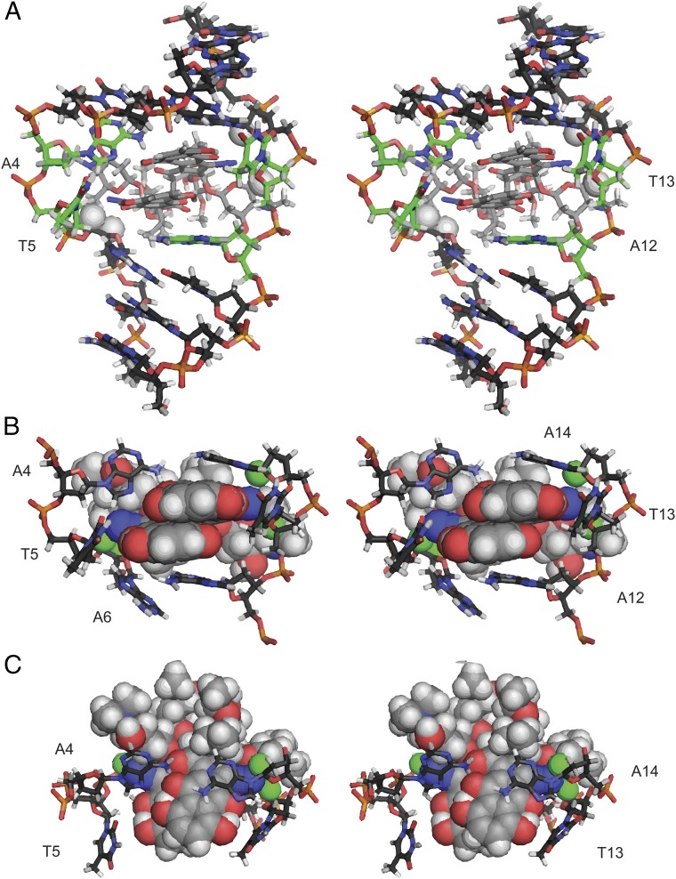
(-)-Lomaiviticin A (1) is a complex antiproliferative metabolite that inhibits the growth of many cultured cancer cell lines at low nanomolar-picomolar concentrations. (-)-Lomaiviticin A (1) possesses a C2-symmetric structure that contains two unusual diazotetrahydrobenzo[b]fluorene (diazofluorene) functional groups. Nucleophilic activation of each diazofluorene within 1 produces vinyl radical intermediates that affect hydrogen atom abstraction from DNA, leading to the formation of DNA double-strand breaks (DSBs). Certain DNA DSB repair-deficient cell lines are sensitized toward 1, and 1 is under evaluation in preclinical models of these tumor types. However, the mode of binding of 1 to DNA had not been determined. Here we elucidate the structure of a 1:1 complex between 1 and the duplex d(GCTATAGC)2 by NMR spectroscopy and computational modeling. Unexpectedly, we show that both diazofluorene residues of 1 penetrate the duplex. This binding disrupts base pairing leading to ejection of the central AT bases, while placing the proreactive centers of 1 in close proximity to each strand. DNA binding may also enhance the reactivity of 1 toward nucleophilic activation through steric compression and conformational restriction (an example of shape-dependent catalysis). This study provides a structural basis for the DNA cleavage activity of 1, will guide the design of synthetic DNA-activated DNA cleavage agents, and underscores the utility of natural products to reveal novel modes of small molecule-DNA association.
Keywords: DNA; NMR; double-strand break; natural product.
Conflict of interest statement
The authors declare no conflict of interest.
Figures
References
-
- He H, et al. Lomaiviticins A and B, potent antitumor antibiotics from Micromonospora lomaivitiensis. J Am Chem Soc. 2001;123(22):5362–5363. - PubMed
-
- Woo CM, Beizer NE, Janso JE, Herzon SB. Isolation of lomaiviticins C-E, transformation of lomaiviticin C to lomaiviticin A, complete structure elucidation of lomaiviticin A, and structure-activity analyses. J Am Chem Soc. 2012;134(37):15285–15288. - PubMed
-
- Itŏ S, Matsuya T, Ōmura S, Otani M, Nakagawa A. A new antibiotic, kinamycin. J Antibiot (Tokyo) 1970;23(6):315–317. - PubMed
-
- Hata T, Ōmura S, Iwai Y, Nakagawa A, Otani M. A new antibiotic, kinamycin: Fermentation, isolation, purification and properties. J Antibiot (Tokyo) 1971;24(6):353–359. - PubMed
Publication types
MeSH terms
Substances
Associated data
- Actions
Grants and funding
LinkOut - more resources
Full Text Sources
Other Literature Sources
Miscellaneous
