. 2016 Mar 15;113(11):2868-73.
doi: 10.1073/pnas.1520244113. Epub 2016 Feb 29.

Efficient delivery of genome-editing proteins using bioreducible lipid nanoparticles

Affiliations

Efficient delivery of genome-editing proteins using bioreducible lipid nanoparticles

Ming Wang et al. Proc Natl Acad Sci U S A. .

Abstract

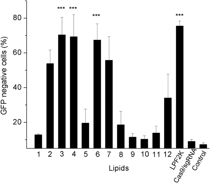
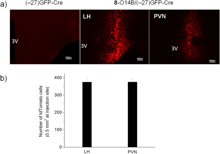
A central challenge to the development of protein-based therapeutics is the inefficiency of delivery of protein cargo across the mammalian cell membrane, including escape from endosomes. Here we report that combining bioreducible lipid nanoparticles with negatively supercharged Cre recombinase or anionic Cas9:single-guide (sg)RNA complexes drives the electrostatic assembly of nanoparticles that mediate potent protein delivery and genome editing. These bioreducible lipids efficiently deliver protein cargo into cells, facilitate the escape of protein from endosomes in response to the reductive intracellular environment, and direct protein to its intracellular target sites. The delivery of supercharged Cre protein and Cas9:sgRNA complexed with bioreducible lipids into cultured human cells enables gene recombination and genome editing with efficiencies greater than 70%. In addition, we demonstrate that these lipids are effective for functional protein delivery into mouse brain for gene recombination in vivo. Therefore, the integration of this bioreducible lipid platform with protein engineering has the potential to advance the therapeutic relevance of protein-based genome editing.

Keywords: CRISPR/Cas9; Cre recombinase; genome editing; lipid nanoparticle; protein delivery.


Conflict of interest statement

The authors declare no conflict of interest.

Figures

Fig. 1.
Fig. 1.
Design of bioreducible lipid-like materials and negatively supercharged protein for effective protein delivery and genome editing.
Fig. 2.
Fig. 2.
Synthesis of bioreducible lipid-like materials. (A) Synthesis route and lipid nomenclature. (B) Chemical structures of amines used as head groups for lipid synthesis.
Fig. 3.
Fig. 3.
Cellular uptake (A) and DsRed expression profile (B) of HeLa-DsRed cells treated with (–27)GFP-Cre alone or different lipid complexes. For the cellular uptake study, cells were treated with complexes of 25 nM protein and 2 µg/mL lipid for 6 h, and DsRed expression was quantified 24 h after delivery. **P < 0.01, ***P < 0.001 compared with treatment with protein alone (no lipid).
Fig. S1.
Fig. S1.
TEM image of (–27)GFP-Cre/8-O14B nanoparticles; 25 nM protein and 2 µg/mL lipid were mixed in 25 mM phosphate buffer (pH = 7.4) for TEM imaging. (Scale bar, 100 nm.)
Fig. S2.
Fig. S2.
Lipid and protein binding study. (A) CD. (B) Fluorescence spectra of (–27)GFP-Cre in the presence and absence of 8-O14B nanoparticles; 0.5 µM (–27)GFP-Cre protein was mixed with 24 µg 8-O14B nanoparticles in 25 mM phosphate buffer (pH = 7.4) for CD and GFP fluorescence (excitation: 480 nm) spectra measurement.
Fig. S3.
Fig. S3.
Cellular uptake of (–27)GFP-Cre/8-O14B nanoparticle was suppressed by endocytosis inhibitors. HeLa-DsRed cells were pretreated with sucrose (450 mM), M-β-CD (2 mM), dynasore (80 µM), or nystatin (100 nM) for 1 h before exposed to 25 nM (–27)GFP-Cre nanoparticles. ***P < 0.001 compared with normalized control.
Fig. 4.
Fig. 4.
CLSM images of HeLa-DsRed cells treated with (–27)GFP-Cre alone (12.5 nM protein) or complexed with 1 µg/mL lipid 8-O14B for 6 h. Endosome/lysosome was stained using LysoTracker Red. (Scale bar, 20 µm.)
Fig. 5.
Fig. 5.
(A) Protein dose-dependent DsRed expression of HeLa-DsRed cells treated with (–27)GFP-Cre in the absence and presence of lipid 8-O14B (2 µg/mL). (B) Tail length of bioreducible lipid determines Cre recombination efficiency. HeLa-DsRed cells were treated with 25 nM (–27)GFP-Cre complexed with 2 µg/mL lipid derived from amine 8 featuring different hydrophobic tail length. ***P < 0.001 compared with 8-O14B.
Fig. S4.
Fig. S4.
Cytotoxicity assay of (–27)GFP-Cre/8-O14B nanoparticles. HeLa-DsRed cells were treated with the nanocomplex of 2 µg/mL lipid and increased concentration of protein for 24 h, followed by cell viability measurement using MTT assay.
Fig. S5.
Fig. S5.
(A) Encapsulation efficiency and (B) intracellular delivery of (–27)GFP-Cre ino HeLa-DsRed cells using lipid with different hydrocarbon tails. See SI Materials and Methods for experimental details. ***P < 0.001 compared with 8-O14B–mediated delivery.
Fig. S6.
Fig. S6.
(A) Encapsulation efficiency and (B) intracellular delivery of Cre recombinase fused with different supernegative GFP variants ino HeLa-DsRed cells using 8-O14B nanoparticles. See SI Materials and Methods for experimental details. **P < 0.01, ***P < 0.001 compared with the encapsulation of (–27)GFP-Cre.
Fig. 6.
Fig. 6.
The charge density of negatively supercharged protein determines Cre-mediated recombination efficiency. HeLa-DsRed cells were treated with the complex of 8-O14B (2 µg/mL) and 25 nM Cre protein fused to GFP with the charge as indicated. ***P < 0.001 compared with the delivery of (–27) GFP-Cre.
Fig. 7.
Fig. 7.
Delivery of Cas9:sgRNA complex into cultured human cells for genome editing. GFP stably expressing HEK cells were treated with 25 nM Cas9:sgRNA complex targeting the GFP locus, with and without lipid (6 µg/mL). The GFP KO efficiency was quantified after 3 d. ***P < 0.001 compared with Cas9/sgRNA alone treated cells.
Fig. S7.
Fig. S7.
TEM image of 3-O14B/Cas9:sgRNA nanoparticle; 50 pmol Cas9:sgRNA complex was mixed with 8 µg 3-O14B in 25 mM phosphate buffer (pH = 7.4) for TEM imaging. (Scale bar, 200 nm.)
Fig. 8.
Fig. 8.
In vivo delivery of Cre recombinase to mouse brain. Rosa26tdTomato mouse was microinjected with 0.1 µL 50 µM (–27)GFP-Cre alone or the same amount of protein complexed with lipid 8-O14B. After 6 d, the tdTomato expression indicative of Cre-mediated recombination in dorsomedial hypothalamic nucleus (DM; X = +0.20, Y = −1.6, Z = −5.0), mediodorsal thalamic nucleus (MD; X = +0.25, Y = −1.4, Z = −2.2), and bed nucleus of the stria terminalis (BNST; X = +0.9, Y = +0.4, Z = −4.0) was visualized using fluorescent microscopy. (Scale bar, 100 µm.)
Fig. S8.
Fig. S8.
In vivo delivery of Cre recombinase to mouse brain. Rosa26tdTomato mouse was microinjected with 0.1 µL 50 µM (–27)GFP-Cre alone or the sample amount of protein complexed with lipid 8-O14B. After 6 d, the tdTomato expression indicative of Cre-mediated recombination in dentate gyrus (DG; X = +0.25, Y = −1.5, Z = −2.2), cortex (X = +0.25, Y = −1.4, Z = −1.0), and ventral lateral septal nucleus (LSV; X = +0.8, Y = +0.4, Z = −3.5) was visualized using fluorescent microscopy. (Scale bar, 100 µm.)
Fig. S9.
Fig. S9.
In vivo delivery of Cre recombinase to hypothalamus of mouse brain. Rosa26tdTomato mouse was microinjected with 0.1 µL 50 µM (–27)GFP-Cre alone or the same amount of protein complexed with lipid 8-O14B. After 6 d, the tdTomato expression indicative of Cre-mediated recombination in hypothalamus was visualized using fluorescence microscopy (A) or counted in 0.5 mm2 at the injection sites of each brain slice (B). (Scale bar, 200 µm.)
Fig. S10.
Fig. S10.
Accumulation of (–27)GFP-Cre and tdTomato expression in DM (A), MD (B), and BNST (C) following (–27)GFP-Cre/8-O14B nanoparticle injections. (Scale bar, 50 µm in A and B; 100 µm in C.)

References

    1. Leader B, Baca QJ, Golan DE. Protein therapeutics: A summary and pharmacological classification. Nat Rev Drug Discov. 2008;7(1):21–39. - PubMed
    1. Doudna JA, Charpentier E. Genome editing. The new frontier of genome engineering with CRISPR-Cas9. Science. 2014;346(6213):1258096. - PubMed
    1. Hsu PD, Lander ES, Zhang F. Development and applications of CRISPR-Cas9 for genome engineering. Cell. 2014;157(6):1262–1278. - PMC - PubMed
    1. Zuris JA, et al. Cationic lipid-mediated delivery of proteins enables efficient protein-based genome editing in vitro and in vivo. Nat Biotechnol. 2015;33(1):73–80. - PMC - PubMed
    1. Davis KM, Pattanayak V, Thompson DB, Zuris JA, Liu DR. Small molecule-triggered Cas9 protein with improved genome-editing specificity. Nat Chem Biol. 2015;11(5):316–318. - PMC - PubMed

Publication types

MeSH terms

LinkOut - more resources