. 2016 Apr 22;291(17):9025-41.
doi: 10.1074/jbc.M116.715474. Epub 2016 Feb 29.

Structural and Functional Analysis of a Novel Interaction Motif within UFM1-activating Enzyme 5 (UBA5) Required for Binding to Ubiquitin-like Proteins and Ufmylation

Affiliations

Structural and Functional Analysis of a Novel Interaction Motif within UFM1-activating Enzyme 5 (UBA5) Required for Binding to Ubiquitin-like Proteins and Ufmylation

Sabrina Habisov et al. J Biol Chem. .

Abstract

The covalent conjugation of ubiquitin-fold modifier 1 (UFM1) to proteins generates a signal that regulates transcription, response to cell stress, and differentiation. Ufmylation is initiated by ubiquitin-like modifier activating enzyme 5 (UBA5), which activates and transfers UFM1 to ubiquitin-fold modifier-conjugating enzyme 1 (UFC1). The details of the interaction between UFM1 and UBA5 required for UFM1 activation and its downstream transfer are however unclear. In this study, we described and characterized a combined linear LC3-interacting region/UFM1-interacting motif (LIR/UFIM) within the C terminus of UBA5. This single motif ensures that UBA5 binds both UFM1 and light chain 3/γ-aminobutyric acid receptor-associated proteins (LC3/GABARAP), two ubiquitin (Ub)-like proteins. We demonstrated that LIR/UFIM is required for the full biological activity of UBA5 and for the effective transfer of UFM1 onto UFC1 and a downstream protein substrate both in vitro and in cells. Taken together, our study provides important structural and functional insights into the interaction between UBA5 and Ub-like modifiers, improving the understanding of the biology of the ufmylation pathway.

Keywords: LC3/GABARAP; LIR; UBA5; UFIM; UFM1; isothermal titration calorimetry (ITC); nuclear magnetic resonance (NMR); protein motif; signal transduction; x-ray crystallography.


Figures

FIGURE 1.
FIGURE 1.
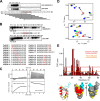
UBA5 interacts with UFM1 and LC3/GABARAP proteins via an evolutionary conserved combined LIR/UFIM motif. A, multiple sequence alignment of UBA5 C termini from different species (ClustalW2 algorithm). The degrees of homology (identity plus similarity) for domains are given as percentages (%) for each region. Asterisk (*), fully conserved residue; colon (:), strongly similar conservation; period (.), weakly similar conservation. The phylogenetic tree (upper right plot) was built for the indicated phylogenetic groups according to the “greatest likelihood” algorithm. UBA5 secondary structure elements were predicted by JPRED algorithms. B, alignment of LIR/UFIM sequences from human UBA5 with published LIRs from indicated human proteins. Multiple sequence alignment was performed using the ClustalW2 algorithm. C, identification of a GABARAP-interacting LIR motif in UBA5. Arrays of 20-mer peptides covering full-length UBA5 were synthesized and prepared on cellulose membranes. Each peptide was shifted three amino acids relative to the previous peptide. The arrays were probed with GST-GABARAP, and binding was detected with anti-GST antibodies. Sequences of the GABARAP-interacting peptides are shown in black, and non-interacting peptides are in gray. D, a schematic map of UBA5. Schematic domain organization and deletion/mutant constructs used in this study are shown. E, analysis of UBA5 interaction with UBLs by a GST pulldown assay. Lysates of HEK293 cells expressing GFP or the indicated GFP-UBA5 constructs were precipitated by immobilized GST or GST-UBLs (LC3/GABARAPs and UFM1). Co-precipitated proteins were detected using an anti-GFP antibody. Ponceau S staining of immunoblot membranes shows loading of GST fusion proteins. Representative results from three independent experiments are shown. WB, Western blotting. F, GST pulldown assay demonstrates interaction of wild-type UBA5 with GABARAPL2, UFM1, and UFC1. LIR/UFIM is required for binding of UBA5 to UFM1 and GABARAPL2, and the C terminus contains a UFC1-binding domain (UFC1 BD). G, co-immunoprecipitation of GFP-UBA5 and endogenous UFM1 (endogenous GABARAPL2 fails to be detected in the co-immunoprecipitated samples). The indicated forms of human HA-tagged UBA5 were transiently expressed in HEK293 cell, and immunoprecipitated (IP) by an anti-HA antibody. Co-precipitated UBLs were detected using the indicated antibodies. Representative results from three independent experiments are shown.
FIGURE 2.
FIGURE 2.
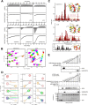
LIR/UFIM of UBA5 specifically interacts with UFM1 and LC3/GABARAP proteins. A, demonstration of dual specificity of LIR/UFIM for UFM1 and GABARAPL2. Arrays of 20-mer peptides covering the C terminus of UBA5 were synthesized and prepared on PVDF membranes. Each peptide was shifted three amino acids relative to the previous peptide. The arrays were probed with the indicated GST fusion protein, and binding was detected with anti-GST antibodies. Sequences of UBL-interacting peptides are shown in black, and non-interacting peptides are in gray. B, confirmation of the core LIR/UFIM sequence by alanine scanning. PVDF membranes with spotted 20-mer peptides (with indicated alanine substitutions marked in red in the peptide sequences) were incubated with the indicated GST-UBLs and probed with an anti-GST antibody to reveal bound proteins. C, ITC analysis of the interaction between UBA5 LIR/UFIM and UFM1. A peptide spanning the UBA5 LIR/UFIM sequence (left panel) or p62/SQSTM1 LIR as a negative control (right panel, without correction of the dilution heat) was titrated to UFM1. The upper graphs represent the raw data; the integrated heat of each injection is displayed in the lower graphs for each titration. Thermodynamic parameters are summarized in Table 5. D, NMR data for interaction between UBA5 LIR/UFIM and UFM1. An overlay of representative areas of the 1H-15N HSQC spectra of 15N-UFM1 to which the nonlabeled UBA5 LIR/UFIM peptide was added stepwise. The rainbow color code indicates increasing molar ratios upon titration from free UFM1 (red) to saturation (molar ratio 1:2 (blue)). E, one-dimensional and three-dimensional mapping of CSP induced in UFM1 NMR spectra upon UBA5 LIR/UFIM binding. CSPs calculated for all assigned resonances are shown as bars, and the dashed lines represent S.D. (1× S.D., yellow; 2× S.D., red). HN resonances for residues in the loop L1 connecting β1 and β2 (**), as well as V23 in the bound state (*) could not be assigned (gray bars showing intensity of the neighbor signals). CSPs were mapped on the UFM1 structure (PDB ID: 1WXS) presented schematically on the left plot and as a surface representation in two projections on the right plot. Residues that are not affected or are slightly (CSP < 1× S.D.), intermediately (1× S.D. < CSP < 2× S.D.), or strongly (CSP > 2× S.D.) affected by the binding are colored in gray, yellow, and red, respectively. The mostly non-assigned loop L1 is shown in cyan.
FIGURE 3.
FIGURE 3.
LIR/UFIM of UBA5 specifically interacts with UFM1 and LC3/GABARAP proteins (continued from Fig. 2 legend). A, ITC analysis of the interactions between LIR/UFIM and LC3/GABARAPs. A peptide spanning the LIR/UFIM sequence was titrated to indicated LC3/GABARAP proteins. The upper graphs represent the raw data; the integrated heat of each injection is displayed in the lower graphs for each titration. Thermodynamic parameters are summarized in Table 5. B, NMR data for interaction between UBA5 LIR/UFIM and GABARAPL2 versus UBA5 LIR/UFIM and LC3B. An overlay of representative areas of the 15N-1H HSQC spectra of 15N-GABARAPL2 (left plot) and 15N-LC3B (right plot) to which the nonlabeled UBA5 LIR/UFIM was added stepwise. The rainbow color code indicates increasing peptide molar ratios upon titration from free GABARAPL2 and LC3B (red) to saturation (magenta, 1:2 in the case of GABARAPL2 and 1:4 for LC3B). C, three-dimensional modeling of the interaction between UBA5 LIR/UFIM and GABARAPL2 versus UBA5 LIR/UFIM and LC3B. CSPs upon interaction with the UBA5 LIR/UIFM were calculated for the GABARAPL2 (upper plot) and LC3B (lower plot) residues and compared with the well characterized LC3B-optineurin (OPTN)-LIR interaction (shown in the boxed insert). The dashed lines represent S.D. over all resonances, except the three most perturbed ones. CSP mapping of the respective structures (GABARAPL2 (PDB ID: 1EO6) and LC3B (PDB ID: 3VTU)) is shown. Residues that are not or only slightly affected (CSP < 1× S.D.) or intermediately (1× S.D. < CSP < 2× S.D.) or strongly (CSP > 2× S.D.) affected by the UBA5 LIR/UIFM peptide binding are colored in gray, yellow, and red, respectively. D, demonstration of UFM1 and GABARAPL2 competition for LIR/UFIM by an NMR competition assay. 15N-1H HSQC spectra of labeled 15N-UFM1 were recorded for the unbound form (red) to which the nonlabeled LIR/UFIM peptide from human UBA5 was added to saturation (yellow, 1:2 molar ratio). To this complex, nonlabeled GABARAPL2 was added to a molar ratio of 1:2:2 (green) and 1:2:4 (blue). Competition binding is indicated by the return of all UFM1 resonances to the unbound form due to binding of the LIR/UIFM to GABARAPL2. Well resolved HN resonance of binding-relevant Phe-35 is shown here as a representative example. The solid lines represent the visible Phe-35 HN resonance at each stage of the experiment, and the broken lines show the positions of Phe-35 HN resonance at previous stages. Additionally, the well resolved HN resonance of binding-relevant residues Phe-40 and Lys-34 are shown. The solid lines represent visible Phe-40 and Lys-34 HN resonances at each stage of the experiment, and the broken lines show the positions of Phe-40 and Lys-34 HN resonances at previous stages. E, demonstration of UFM1 and GABARAPL2 competition for LIR/UFIM by a GST pulldown assay. Immobilized GST-UBA5 with pre-bound UFM1 was incubated with increasing concentrations of GABARAPL2 (0.01 nm–100 μm). Samples were analyzed by immunoblotting using antibodies to the indicated proteins. Ponceau S staining shows the relative loading of GST fusion proteins. Representative results from three independent experiments are shown.
FIGURE 4.
FIGURE 4.
Structure of UBA5 LIR/UFIM in complex with UFM1. A, general structural characteristics of the UBA5 LIR/UFIM·UFM1 complex (PDB ID: 5HKH). The asymmetric unit comprises two UFM1 molecules (green and cyan; schematic representation) and one UBA5 LIR/UFIM peptide (orange; backbone is shown schematically and side chains of Trp-341 and Ile-343 as sticks). Although the first UFM1 molecule (green) represents the most relevant interactions to LIR/UFIM, the second one stabilizes the complex and could be considered a crystallographic artifact (however, we cannot completely exclude such a dual UFM1 coordination near UBA5 LIR/UFIM). The LIR/UFIM of UBA5 binds to a hydrophobic pocket on the first UFM1 molecule (similar to HP2 for LC3/GABARAP proteins) mainly over the Ile-343 side chain. B, specific structural characteristics of the UBA5 LIR/UFIM·UFM1 complex (PDB ID: 5HKH). The left plot shows a schematic representation of the UFM1 first molecule (green) with the key residues shown as sticks (labeled in green). The UBA5 LIR/UFIM (ball-stick presentation; carbon, oxygen, and nitrogen atoms shown in orange, red, and blue, respectively) binds to a hydrophobic pocket similar to HP2 for LC3/GABARAP proteins. The right plot shows a two-dimensional LigPlot diagram with the intermolecular interface between the first UFM1 molecule and the UBA5 LIR/UFIM in a complex. Hydrophobic interactions are represented by red semicircles and hydrogen bonds by green dashed lines. C, comparison of the UFM1·UBA5-LIR/UFIM complex with those of Ub·UIM, SUMO·SBM, and LC3·LIR. Surface and schematic diagrams of UBLs are in the same orientation, with β2 and α1/α3 at the front. Interacting peptides (orange) are shown for the UBA5 LIR/UFIM·UFM1 complex (PDB ID: 5HKH; first UFM1 molecule is colored green), the human Ub in complex with the VPS27 UIM (PDB ID: 2KDI), the human SUMO-1 in complex with the SUMO-binding motif (SBM) from PIASX (PDB ID: 2ASQ), and human LC3B in complex with the p62 LIR peptide (PDB ID: 2K6Q; all light blue). D, schematic representation of the UBA5 LIR/UFIM orientation in complex with UFM1. Typical for the UBL, β-strand β2 and α-helix α1 (α3 in LC3/GABARAP proteins) are shown in the presence of UBA5 LIR/UFIM peptide (backbones are shown as sticks; reverse rainbow color code: blue for the N terminus and red for the C terminus). The arrows indicate the direction of the peptide (from N to C terminus). Examples of parallel and antiparallel orientations to β2 peptide in known SUMO and LC3/GABARAP proteins are listed. E, superimposition of the solved crystal structure of UFM1 (green) over those of Ub (left plot, blue), SUMO1 (middle plot), and LC3B (right plot). The structures are represented schematically, root-mean-square deviation values for positions of the Cα atoms in each pair are given in Å, and the PDB ID code for each structure is indicated.
FIGURE 5.
FIGURE 5.
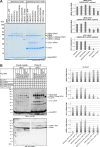
Formation of UFM1 conjugates depends on intact LIR/UFIM within UBA5. A, demonstration of functional significance of LIR/UFIM for UBA5 function by in vitro UFM1-UBA5 and UFM1-UFC1 thioester formation assay. Purified UFM1ΔC2 (with exposed C-terminal glycine) and UBA5, or the indicated UBA5 mutants, were incubated with UFC1 (lanes 1–7) or without UFC1 (lanes 8–14) at 25 °C for 5 min. As a negative control, the reactions were performed in the absence of ATP (lanes 1 and 8). The samples were resolved on a nonreducing NuPAGE followed by Coomassie Brilliant Blue staining. Relative ratios of UFM1-UBA5/UBA5 and UFM1-UFC1/UFC1 were determined by the intensity of the bands. Data shown are representative of three independent experiments. Error bars represent ± S.E.; p values were determined by unpaired t test (*, p < 0.05; **, p < 0.01; ***, p < 0.001). B, demonstration of the functional significance of LIR/UFIM for UBA5 function by the cellular ufmylation assay. HEK293 cells, in which UBA5 was deleted by CRISPR/Cas9 technology, were transfected with the indicated plasmids, and immunoprecipitation experiments (IP) with anti-FLAG M2-agarose were performed. FLAG-IP (right half of the gel) showed a remarkable reduction of the UFC1-UFM1 intermediate and the UFM1-UFBP1 and X-UFM1 conjugates (where “X” represents an uncharacterized substrate protein) in the samples transfected with UBA5 LIR/UFIM mutants or C-terminal deletion construct (1–330 aa). Relative ratios of UBA5-UFM1/UFM1, X-UFM1/UFM1, UFC1-UFM1/UFM1, and UFBP1-UFM1/UFBP1 (derived by FLAG-IP) were determined by the intensity of the bands. Data shown are representative of three independent experiments. Error bars represent ± S.E.; p values were determined by unpaired t test (*, p < 0.05; **, p < 0.01; ***, p < 0.001). WB, Western blotting.
FIGURE 6.
FIGURE 6.
Model for functional interactions between UBA5, UFC1, and UBLs. The LIR/UFIM motif in the C terminus of UBA5 (E1 enzyme) attracts UFM1. Because of this interaction, UFM1 may occupy the activation site near the ATP-binding site in the catalytic (adenylation) domain with a higher efficiency. LIR/UFIM may further be required for the binding of activated UFM1 for its transfer onto UFC1 (E2 enzyme), which is recruited to UBA5 via its UFC1 binding domain (UFC1 BD). UFL1 (E3 enzyme) promotes the transfer of the UFM1 molecule to the substrate protein (target). LC3/GABARAPs might compete UFM1 out of the LIR/UFIM motif. Their function in the ufmylation pathway is presently unknown.

References

    1. Komatsu M., Chiba T., Tatsumi K., Iemura S., Tanida I., Okazaki N., Ueno T., Kominami E., Natsume T., and Tanaka K. (2004) A novel protein-conjugating system for Ufm1, a ubiquitin-fold modifier. EMBO J. 23, 1977–1986 - PMC - PubMed
    1. Kang S. H., Kim G. R., Seong M., Baek S. H., Seol J. H., Bang O. S., Ovaa H., Tatsumi K., Komatsu M., Tanaka K., and Chung C. H. (2007) Two novel ubiquitin-fold modifier 1 (Ufm1)-specific proteases, UfSP1 and UfSP2. J. Biol. Chem. 282, 5256–5262 - PubMed
    1. Yoo H. M., Kang S. H., Kim J. Y., Lee J. E., Seong M. W., Lee S. W., Ka S. H., Sou Y. S., Komatsu M., Tanaka K., Lee S. T., Noh D. Y., Baek S. H., Jeon Y. J., and Chung C. H. (2014) Modification of ASC1 by UFM1 is crucial for ERα transactivation and breast cancer development. Mol. Cell 56, 261–274 - PubMed
    1. Tatsumi K., Sou Y. S., Tada N., Nakamura E., Iemura S., Natsume T., Kang S. H., Chung C. H., Kasahara M., Kominami E., Yamamoto M., Tanaka K., and Komatsu M. (2010) A novel type of E3 ligase for the Ufm1 conjugation system. J. Biol. Chem. 285, 5417–5427 - PMC - PubMed
    1. Zhang M., Zhu X., Zhang Y., Cai Y., Chen J., Sivaprakasam S., Gurav A., Pi W., Makala L., Wu J., Pace B., Tuan-Lo D., Ganapathy V., Singh N., and Li H. (2015) RCAD/Ufl1, a Ufm1 E3 ligase, is essential for hematopoietic stem cell function and murine hematopoiesis. Cell Death Differ. 22, 1922–1934 - PMC - PubMed

Publication types

MeSH terms