Review
. 2016 Feb 9:9:1-11.
doi: 10.2147/JIR.S82320. eCollection 2016.

The macrophages in rheumatic diseases

Affiliations
Review

The macrophages in rheumatic diseases

Antonella Laria et al. J Inflamm Res. .

Abstract

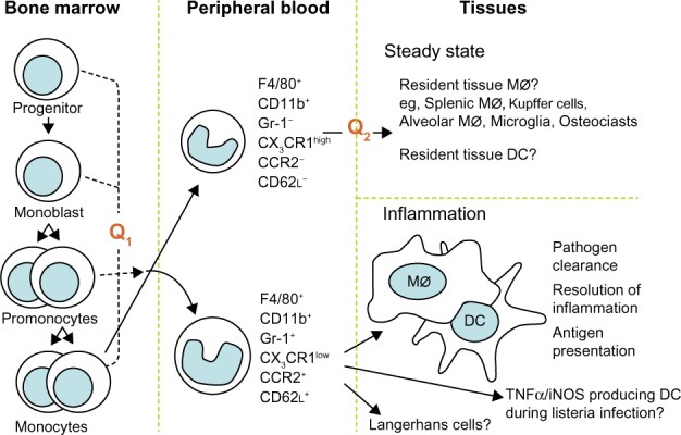
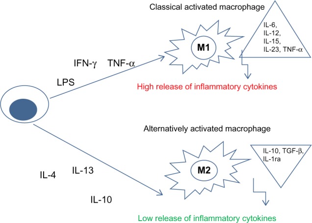
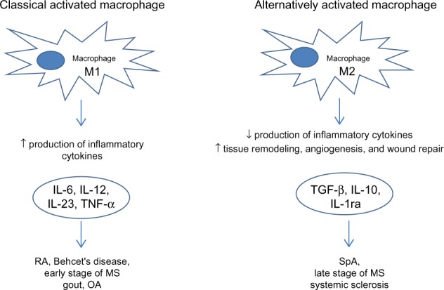
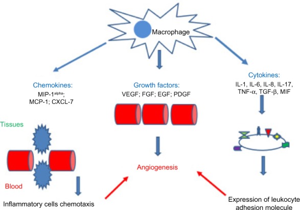
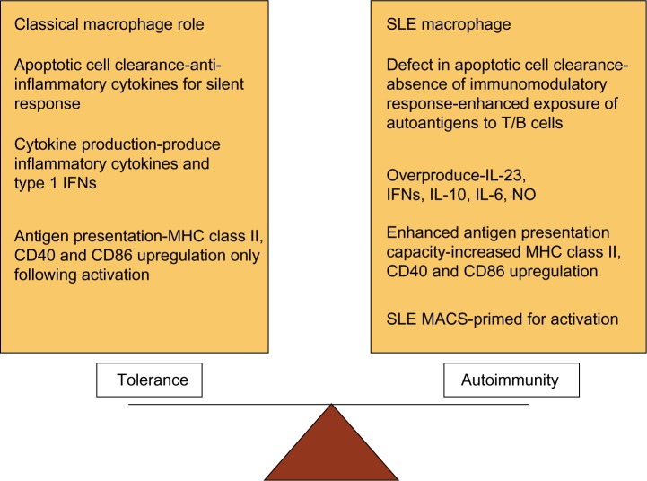
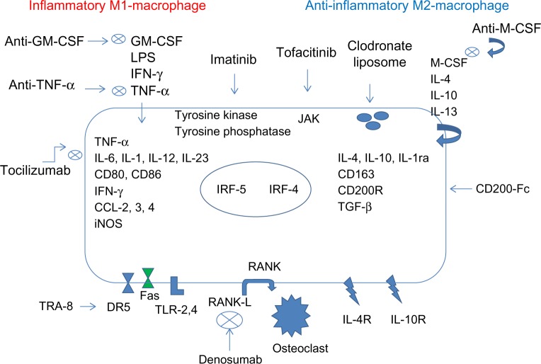
Macrophages belong to the innate immune system giving us protection against pathogens. However it is known that they are also involved in rheumatic diseases. Activated macrophages have two different phenotypes related to different stimuli: M1 (classically activated) and M2 (alternatively activated). M1 macrophages release high levels of pro-inflammatory cytokines, reactive nitrogen and oxygen intermediates killing microorganisms and tumor cells; while M2 macrophages are involved in resolution of inflammation through phagocytosis of apoptotic neutrophils, reduced production of pro-inflammatory cytokines, and increased synthesis of mediators important in tissue remodeling, angiogenesis, and wound repair. The role of macrophages in the different rheumatic diseases is different according to their M1/M2 macrophages phenotype.

Keywords: macrophage; rheumatic diseases.


Figures

Figure 1
Figure 1
Schematic representation of the development of the monocyte subsets. Abbreviation: DC, dendritic cells.
Figure 2
Figure 2
Schematic representation of macrophage polarization. Abbreviations: LPS, lipopolysaccharide; IFN-γ, interferon-gamma; TNF-α, tumor necrosis factor α; IL, interleukin; IL-1ra, interleukin 1 receptor antagonist; TGF-β, transformer growth factor β.
Figure 3
Figure 3
Wright stain images of a patient with rheumatoid arthritis. Notes: (A) Activated macrophage in synovial fluid of rheumatoid arthritis. wright stain, synovial fluid smear from a patient with rheumatoid arthritis 1,000×; (B) and (C) macrophages phagocytizing a small lymphocyte and neutrophil granulocytes. wright stain 1,000× (rheumatoid arthritis).
Figure 4
Figure 4
Macrophages in rheumatic diseases. Abbreviations: RA, rheumatoid arthritis; OA, osteoarthritis; MS, multiple sclerosis; SpA, spondyloarthritis; TNF-α, tumor necrosis factor α; IL, interleukin; IL-1ra, interleukin 1 receptor antagonist; TGF-β, transformer growth factor β.
Figure 5
Figure 5
Chemokines, growth factors, and cytokines involved in the angiogenic activity of macrophages. Abbreviations: MIP-1alpha, macrophage inflammatory protein-1 alpha; MCP-1, monocyte chemotactic protein 1; CXCL-7, CXC chemokine ligand 7; VEGF, vascular endothelial growth factor; FGF, fibroblast growth factor; EGF, epidermal growth factor; PDGF, platelet derived growth factor; TNF-α, tumor necrosis factor α; IL, interleukin; TGF-β, transformer growth factor β; MIF, macrophage migration inhibitory factor.
Figure 6
Figure 6
Dysregulation of macrophage function in systemic lupus erythematosus (SLE). Abbreviation: MHC, major histocompatibility complex; INFs, Interferons; MHC, major histocompatibility complex; CD40, cluster of differentiation 40; CD86, cluster of differentiation 86; IL, interleukin; NO, nitrous oxide; SLE MACS, SLE macrophages.
Figure 7
Figure 7
Schematic representation of the role of the inflammasome in processing Pro-IL-1β. Abbreviations: MSU, monosodium urate monohydrate; LRR, leucine-rich repeat domain; PAMP, pathogen associated molecular pattern; DAMP, damage-associated molecular pattern; RNA, acid ribonucleic; ATP, adenosine triphosphate; NALP-3, PYD domains-containing protein 3; ASC, caspase activation and recruitment domain; NATCH, nucleotide-binding domain; CARD, caspase-recruitment domain; Pro-IL1β, pro-interleukin 1β.
Figure 8
Figure 8
Macrophage polarization in RA synovium and related therapeutic targets. Abbreviation: RA, rheumatoid arthritis; GM-CSF, Granulocyte-macrophage colony-stimulating factor; LPS, Lipopolysaccharide; TNF-α, Tumor necrosi factor; INF-γ, Interferon-γ; CD, cluster of differentation; iNOS, Inducible nitric oxide synthase; CCL-2,3,4, chemokine (C-C motif) ligand 2,3,4; IL, Interleukin; JAK, Janus kinase; IRF, interferon regulatory factor; M-CSF, macrophage colony-stimulating factor; CD200-Fc, cluster of differentation 200-fragment crystallizable; TGF-β, Transformer growth factor β; RANK, Receptor Activator of NF-KB; RANK-L, Receptor Activator of NF-KB ligand; TRA-8, monomeric monoclonal antibody targeting DR5; DR5, death receptor 5; FAS, a death receptor; TLR-2,4, Toll-like receptor 2,4.

References

    1. Volkman A, Gowans JL. The Origin of Macrophages from Bone Marrow in the Rat. Br J Exp Pathol. 1965;46:62–70. - PMC - PubMed
    1. Burke B; Sumner S, Maitland N, Lewis CE. Macrophages in gene therapy: cellular delivery vehicles and in vivo targets. J Leukoc Biol. 2002 Sep;72(3):417–428. - PubMed
    1. Passlick B, Flieger D, Ziegler-Heitbrock HW. Identification and characterization of a novel monocyte subpopulation in human peripheral blood. Blood. 1989;74(7):2527–2534. - PubMed
    1. Weber C, Belge KU, von Hundelshausen P, et al. Differential chemokine receptor expression and function in human monocyte subpopulations. J Leukoc Biol. 2000;67:699–704. - PubMed
    1. Randolph GJ, Sanchez-Schmitz G, Liebman RM, Schakel K. The CD16(+) (FcgammaRIII(+)) subset of human monocytes preferentially becomes migratory dendritic cells in a model tissue setting. J Exp Med. 2002;196:517–527. - PMC - PubMed

LinkOut - more resources