. 2016 Mar 1;11(3):e0150360.
doi: 10.1371/journal.pone.0150360. eCollection 2016.

Distinct Contributions of Astrocytes and Pericytes to Neuroinflammation Identified in a 3D Human Blood-Brain Barrier on a Chip

Affiliations

Distinct Contributions of Astrocytes and Pericytes to Neuroinflammation Identified in a 3D Human Blood-Brain Barrier on a Chip

Anna Herland et al. PLoS One. .

Abstract

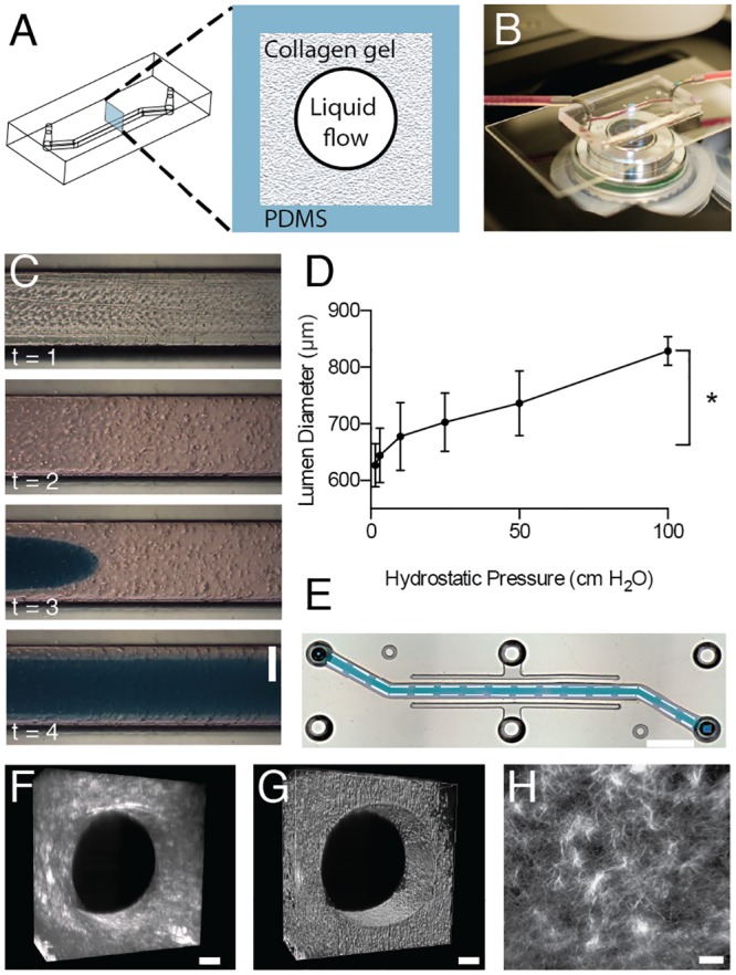
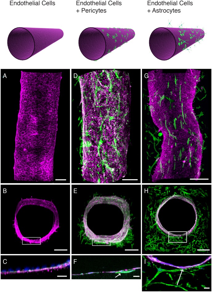
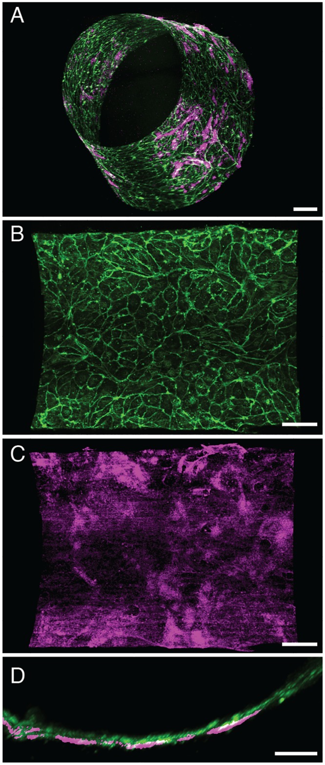
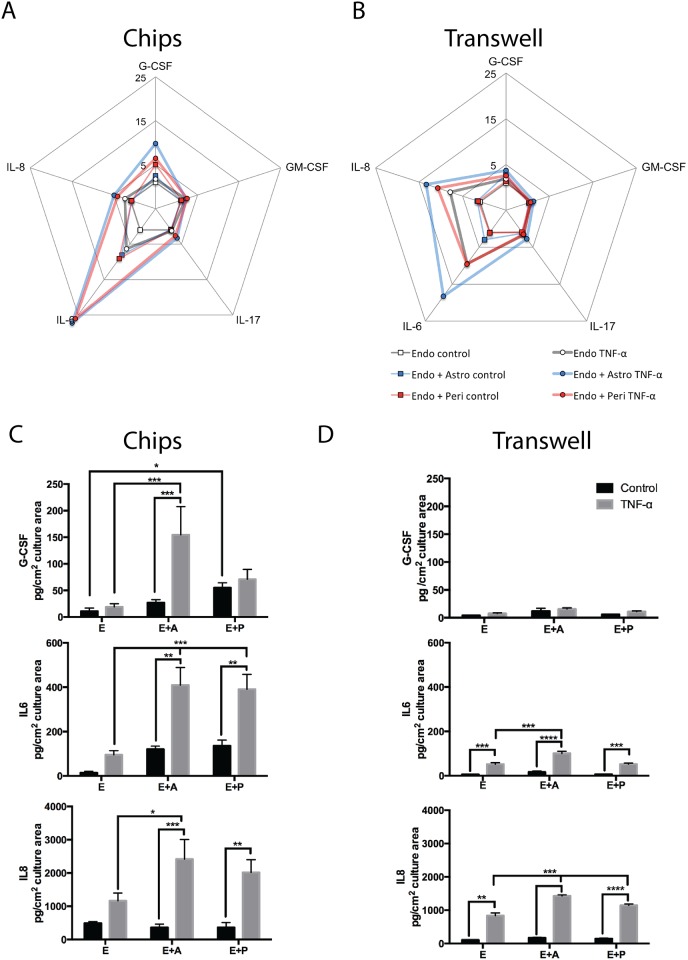
Neurovascular inflammation is a major contributor to many neurological disorders, but modeling these processes in vitro has proven to be difficult. Here, we microengineered a three-dimensional (3D) model of the human blood-brain barrier (BBB) within a microfluidic chip by creating a cylindrical collagen gel containing a central hollow lumen inside a microchannel, culturing primary human brain microvascular endothelial cells on the gel's inner surface, and flowing medium through the lumen. Studies were carried out with the engineered microvessel containing endothelium in the presence or absence of either primary human brain pericytes beneath the endothelium or primary human brain astrocytes within the surrounding collagen gel to explore the ability of this simplified model to identify distinct contributions of these supporting cells to the neuroinflammatory response. This human 3D BBB-on-a-chip exhibited barrier permeability similar to that observed in other in vitro BBB models created with non-human cells, and when stimulated with the inflammatory trigger, tumor necrosis factor-alpha (TNF-α), different secretion profiles for granulocyte colony-stimulating factor (G-CSF) and interleukin-6 (IL-6) were observed depending on the presence of astrocytes or pericytes. Importantly, the levels of these responses detected in the 3D BBB chip were significantly greater than when the same cells were co-cultured in static Transwell plates. Thus, as G-CSF and IL-6 have been reported to play important roles in neuroprotection and neuroactivation in vivo, this 3D BBB chip potentially offers a new method to study human neurovascular function and inflammation in vitro, and to identify physiological contributions of individual cell types.


Conflict of interest statement

Competing Interests: DEI is a founder and holds equity in Emulate, Inc., and chairs its scientific advisory board. AvdM is a consultant for Emulate, Inc. and receives compensation for these services. This does not alter the authors' adherence to PLOS ONE policies on sharing data and materials.

Figures

Fig 1
Fig 1. The pressure-driven viscous fingering method used to generate the cylindrical collagen gel in the 3D BBB chip.
A) Schematic diagram of the PDMS structure used to generate the 3D BBB chip (left) and an illustration of a cross-section through the chip showing the PDMS channel containing the collagen gel made with viscous fingering and a central lumen (right). B) Photograph of the 3D BBB chip on the stage of an inverted microscope. C) Time-lapse images of the fingering method showing the microchannel before (t = 1) and after infusion of a neutralized collagen gel containing dispersed human astrocytes (t = 2), which was then followed by injection of a low viscosity liquid (dyed blue here) driven by hydrostatic pressure to initiate “finger” formation in the center of the gel (t = 3), and eventually a continuous hollow cylindrical lumen throughout the length of the device (t = 4). The time course from t = 1 to 4 is user dependent but normally less than 30 sec (bar, 500 μm). D) Graph showing the correlation between the hydrostatic pressures used to drive the fingering process and the resulting lumen diameter (* p<0.05 Student’s t-test, n = 3). E) Low magnification micrograph of an entire device containing a lumen filled with blue fluid, formed as described in C (dashed lines, delineate the edges of the channel; black dotted rectangle indicates where images shown in F and G were recorded (bar, 3 mm). F) Second harmonic generation image of the collagen distribution in the 3D BBB chip, and an intensity generated voxel illustration of the lumen based on this information (G) (bar, 100 μm). H) High magnification of the second harmonic generation image showing of collagen microstructure in the cylindrical gel within the 3D BBB chip (bar, 50 μm).
Fig 2
Fig 2. Co-culture of human brain microvascular endothelial cells, pericytes and astrocytes in the 3D BBB chip.
Schematic illustrations of the cells populating the 3D vessel structures for the three experimental set-ups are shown at the top, and fluorescence confocal micrographs of the engineered brain microvessel viewed from the top (A, D, G) or shown in cross-section at either low (B, E, H) or high (C, F, I) magnification (rectangles in lower magnifications images indicate respective areas shown at higher magnification below). The fluorescence micrographs show the cell distributions in 3D BBB chips containing brain microvascular endothelium alone (A-C), endothelium with prior plating of brain pericytes on the surface of the gel in the central lumen (D-F) or endothelium with brain astrocytes embedded in the surrounding gel (G-I). High-magnification cross-sections are projections of confocal stacks (bars, 200 μm in A,B,D,E,G,H and 30 μm in C, F, I). Green indicates F-actin staining, blue represents Hoechst-stained nuclei, and magenta corresponds to VE-Cadherin staining, except for G where morphology and intensity masks were used to discriminate astrocytes (green) from endothelial cells (magenta); original image can be seen in S2 Movie. Arrows indicate contact points between endothelium and pericytes (F) or astrocytes (I).
Fig 3
Fig 3. Production of an abluminal basement membrane by brain endothelial cells in the 3D BBB chip.
A) Perspective view of a 3D reconstruction of a confocal fluorescence micrograph showing a monolayer of brain microvascular endothelial cells lining the lumen of a engineered vessel in the 3D BBB chip (green, F-actin staining; magenta, collagen IV staining). Higher magnification views of staining for F-actin (B) and collage IV (C), and a cross-sectional view (D) showing the accumulation of a linear pattern of basement membrane collagen IV (magenta) staining beneath the F-actin (green) containing endothelial cells (bars, 100 μm in A; 80 μm in B, C; 40 μm in D).
Fig 4
Fig 4. Establishment of a low permeability barrier by the engineered brain microvascular endothelium in the 3D BBB chip.
A) Fluorescence micrograph of a chip containing a cylindrical collagen gel viewed from above with (left) or without (right) a lining endothelial monolayer after five days of culture (left; Cell-lined lumen) compared to a chip with an empty collagen lumen (right). The images were recorded at 0 (top) and 500 (bottom) sec after injection of fluorescently-labeled 3 kDa dextran to analyze the dynamics of dextran diffusion and visualize endothelial barrier function in the 3D BBB chip. Note that the presence of the endothelium significantly restricts dye diffusion compared to gels without cells (left versus right). B) Apparent permeabilities of the endothelium cultured in the 3D BBB chip calculated from the diffusion of 3 kDa dextran with an endothelial monolayer (Endo; n = 6), an endothelial monolayer surrounded by astrocytes (Endo+Astro; n = 3) and an endothelial monolayer surrounded by pericytes (Endo+Peri; n = 3). Error bars indicate S.E.M.; * p<0.05, Student’s t-test.
Fig 5
Fig 5. Comparison of cytokine release profiles after inflammatory stimulation with TNF-α in the microfluidic 3D BBB chip versus static Transwell cultures.
A-B) Diagrammatic representation of the profile of cytokine release for 5 inflammatory cytokines (G-CSF, GM-CSF, IL-6, IL-8, IL-17) in the 3D BBB chips (A) versus Transwells (B). All data were normalized to the levels of cytokines released by endothelial cells cultured alone; concentric scales indicate fold increase. C-D) Release of G-CSF, IL-6 and IL-8 in the 3D BBB chips (C) and Transwells (D) under basal conditions and when stimulated with TNF-α, normalized for culture area (* p<0.05, ** p<0.01, *** p<0.001, **** p<0.0001 Multiple-comparison ANOVA with Bonferroni's comparisons test; n = 4–7 for 3D BBB chips and n = 3 for Transwells).

References

    1. Abbott NJ, Patabendige AA, Dolman DE, Yusof SR, Begley DJ. Structure and function of the blood–brain barrier. Neurobiology of disease. 2010;37(1):13–25. 10.1016/j.nbd.2009.07.030 - DOI - PubMed
    1. Minagar A, Alexander JS. Blood-brain barrier disruption in multiple sclerosis. Multiple sclerosis. 2003;9(6):540–9. - PubMed
    1. Bell RD, Zlokovic BV. Neurovascular mechanisms and blood–brain barrier disorder in Alzheimer’s disease. Acta neuropathologica. 2009;118(1):103–13. 10.1007/s00401-009-0522-3 - DOI - PMC - PubMed
    1. Wolburg H, Wolburg-Buchholz K, Kraus J, Rascher-Eggstein G, Liebner S, Hamm S, et al. Localization of claudin-3 in tight junctions of the blood-brain barrier is selectively lost during experimental autoimmune encephalomyelitis and human glioblastoma multiforme. Acta neuropathologica. 2003;105(6):586–92. - PubMed
    1. Hawkins BT, Davis TP. The blood-brain barrier/neurovascular unit in health and disease. Pharmacological reviews. 2005;57(2):173–85. - PubMed

Publication types

MeSH terms