

doi: 10.1164/rccm.201507-1508LE.
Intrapulmonary Bronchopulmonary Anastomoses and Plexiform Lesions in Idiopathic Pulmonary Arterial Hypertension
Affiliations
- PMID: 26930433
- PMCID: PMC4824924
- DOI: 10.1164/rccm.201507-1508LE
Item in Clipboard
Intrapulmonary Bronchopulmonary Anastomoses and Plexiform Lesions in Idiopathic Pulmonary Arterial Hypertension
Am J Respir Crit Care Med.
.
No abstract available
Figures
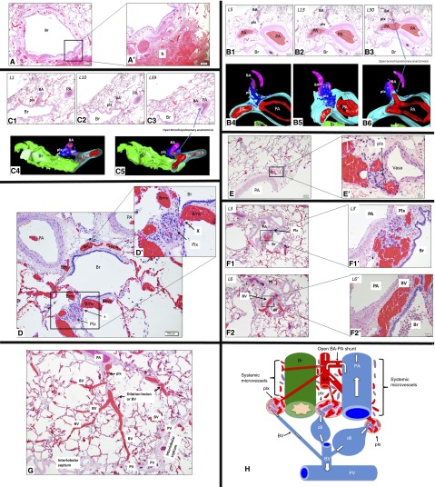
(A) The bronchial vasculature is overloaded because of shunted blood via an open intrapulmonary bronchopulmonary anastomotic pathway (IBA). Bronchial vessels are markedly congested around a bronchiole (Br) (magnification ×4). (A’) High magnification shows a hemorrhage (h). (B) A plexiform lesion (plx) is connected to the bronchial circulation and is in close proximity to an open bronchial artery (BA)–pulmonary artery (PA) anastomotic connection. Hematoxylin and eosin (H&E) sections (B1–B3) show that the plx bridges the BA and the wall of a large PA (L5–13). Deeper sections (L30) and three-dimensional images (B4–B6) show that the same BA connects to a large PA and forms an open BA–PA anastomosis. Three-dimensional images show that the plx is connected to the BA and to the vasa vasorum (vasa) of the PA (magnification ×2). (C) A plx is connected to the bronchial circulation and is in close proximity to an open BA–PA anastomotic connection as shown by H&E images (C1–C3). The plx bridges the BA and the microvessels within the wall of a larger (Br) (L1–10). Deeper section (L39) and three-dimensional images (C4 and C5) show that the same BA connects to a larger PA via an open BA–PA anastomosis. Three-dimensional images show that the plx bridges the BA and the microvessels of the airway (Br) (magnification ×2). (D) A plx is connected (X) with dilated and congested bronchial microcirculation (Bmv). (D') High magnification shows open connections between bronchial microvessels and the plx (magnification ×10). (E) A plx develops on the vasa of a large PA (magnification ×4). (E’) High magnification confirms that the plx is intimately associated with the vasa. (F) A plx is located between a large PA and Br where the bronchial microvessels are normally located (F1/L3) (magnification ×4). (F1’) Higher magnification confirms that the plx is located where the systemic microvessels naturally reside. Deeper sections (F2/L6) show that the same plx in F1 becomes a dilated bronchial vein (BV), as confirmed by a high-magnification image (F2’). (G) A plx is located at the site of vasa of a large PA that connects to a dilation lesion or dilated and congested BV that is eventually drained by a pulmonary vein (PV) that normally resides within the interlobular septum. (H) Prominent IBAs are present in distal lungs with idiopathic pulmonary arterial hypertension. The BA has an open connection with PA and blood shunts from the PA to the BA (white arrows depict blood postulated flow directions). Systemic microvessels surrounding the PA (vasa), and Brs (peribronchial microvessels) are supplied by the BA and drained by the BV, respectively. Because of the markedly increased shear stress due to shunted blood exerted on bronchial microvessels, a plx develops at the sites where systemic microvessels originally reside. Dilatation lesions (dil) are distal to the plx and represent blood overload and a dilated BV that is drained by the PV.
Comment in
-
New Thoughts about the Origin of Plexiform Lesions.Am J Respir Crit Care Med. 2016 Mar 1;193(5):484-5. doi: 10.1164/rccm.201510-1959ED. Am J Respir Crit Care Med. 2016. PMID: 26930432 No abstract available.
References
-
- Cool CD, Stewart JS, Werahera P, Miller GJ, Williams RL, Voelkel NF, Tuder RM. Three-dimensional reconstruction of pulmonary arteries in plexiform pulmonary hypertension using cell-specific markers. Evidence for a dynamic and heterogeneous process of pulmonary endothelial cell growth. Am J Pathol. 1999;155:411–419. - PMC - PubMed
-
- Robertson B. Postnatal formation and obliteration of arterial anastomoses in the human lung: a microangiographic and histologic study. Pediatrics. 1969;43:971–979. - PubMed
Publication types
MeSH terms
Grants and funding
LinkOut - more resources
Full Text Sources
Other Literature Sources
