Review
. 2016 Apr:39:93-100.
doi: 10.1016/j.ceb.2016.02.013. Epub 2016 Feb 27.

Adhesion protein networks reveal functions proximal and distal to cell-matrix contacts

Affiliations
Review

Adhesion protein networks reveal functions proximal and distal to cell-matrix contacts

Adam Byron et al. Curr Opin Cell Biol. 2016 Apr.

Abstract

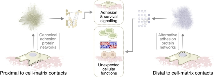
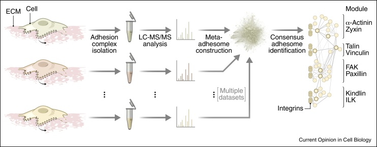
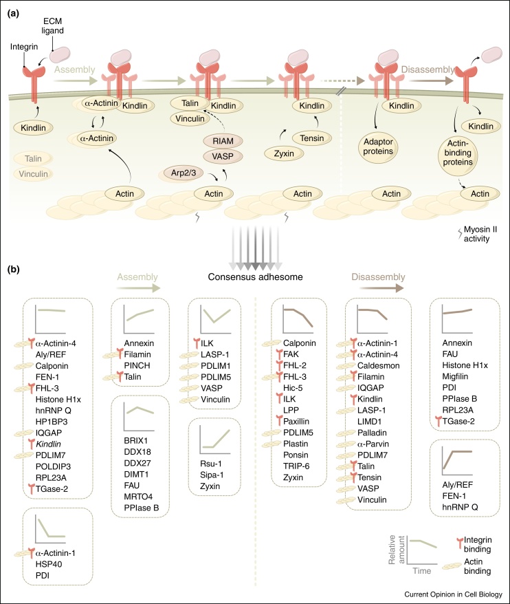
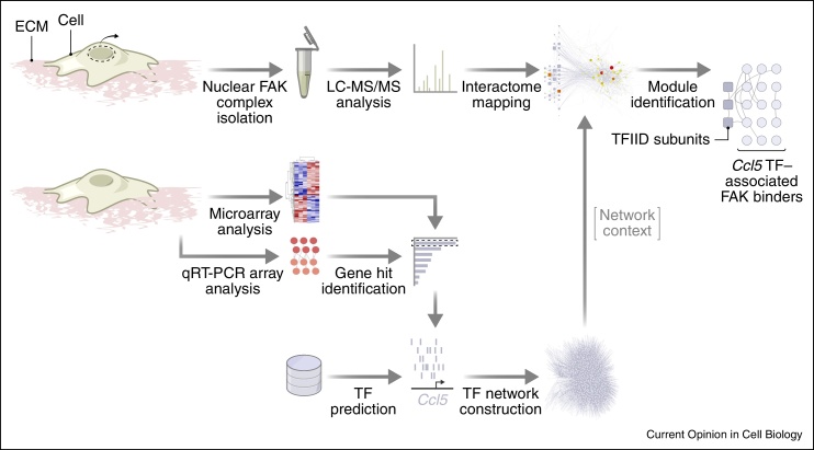
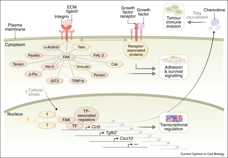
Cell adhesion to the extracellular matrix is generally mediated by integrin receptors, which bind to intracellular adhesion proteins that form multi-molecular scaffolding and signalling complexes. The networks of proteins, and their interactions, are dynamic, mechanosensitive and extremely complex. Recent efforts to characterise adhesions using a variety of technologies, including imaging, proteomics and bioinformatics, have provided new insights into their composition, organisation and how they are regulated, and have also begun to reveal unexpected roles for so-called adhesion proteins in other cellular compartments (for example, the nucleus or centrosomes) in diseases such as cancer. We believe this is opening a new chapter on understanding the wider functions of adhesion proteins, both proximal and distal to cell-matrix contacts.


Figures

None
Graphical abstract
Figure 1
Figure 1
Definition of a consensus adhesome. Adhesion complexes induced by the integrin ligand fibronectin were stabilised and purified (curly arrows) and their proteomes were characterised by quantitative mass spectrometry (LC–MS/MS) in multiple studies using different cell types. Integration of these datasets generated a meta-adhesome, from which a core consensus adhesome was established [15••]. Network nodes (circles) represent interacting proteins; thick node borders indicate proteins that define the axes of emergent consensus adhesome modules (labelled, right).
Figure 2
Figure 2
Temporal dynamics of adhesion complex composition. (a) Hierarchical recruitment of adhesion proteins to integrins (assembly, left), as determined by fluorescence microscopy studies. Some proteins may exist as pre-formed complexes in the cytoplasm (such as talin and vinculin, grey font). α-Actinin aggregates are transiently incorporated into developing adhesions and link integrins to the actin cytoskeleton. In membrane protrusions, RIAM binds Ena/VASP (brown nodes) and talin to link integrins to actin. Recruitment of talin to β1 integrin tails and maturation of adhesions requires myosin II activity, as indicated. Loss of adhesion proteins during disassembly (right), as suggested by proteomic experiments, also appears to occur hierarchically. (b) Assembly (left) and disassembly (right) dynamics of consensus adhesome proteins. Line profiles for each cluster show trends of protein abundance over time, as quantified by mass spectrometry [15••]. Integrin-binding and actin-binding proteins are indicated. Kindlin in the assembly dataset is kindlin-3 (italics), whereas kindlin-2 is the family member in the consensus adhesome.
Figure 3
Figure 3
Integrative analysis of non-canonical FAK function. FAK complexes were isolated from purified SCC cell nuclei (curly arrow) and their interactomes were characterised by quantitative mass spectrometry (LC–MS/MS). The discovered protein interaction network was contextualised by mapping onto a network neighbourhood of transcription factors (TFs). Selected TFs were predicted to bind promoters of genes regulated by FAK (e.g. Ccl5), as identified by microarray and quantitative reverse transcription polymerase chain reaction (qRT-PCR) array analyses. This identified predicted TFs of FAK-regulated genes (e.g. transcription factor II D (TFIID) subunits) and their upstream regulators that interact with nuclear FAK [57••].
Figure 4
Figure 4
Role of FAK in the interplay between the microenvironment, adhesion receptor complexes and the nucleus. The network of FAK interaction partners from the consensus adhesome, and their predicted interactions (light grey lines; FAK interactions, dark grey lines), is shown associated with integrins at the plasma membrane. Synergistic integrin and growth factor receptor signalling regulates cell adhesion, migration and proliferation. Several FAK-interacting partners have been reported in the nucleus (brown nodes). Upon cellular stress, FAK localises to the nucleus, where it associates with chromatin and binds transcription factors and their regulators to control gene expression, including that of the chemokine Ccl5, leading to regulatory T cell (Treg) recruitment and immune evasion [57••].

References

    1. Byron A., Humphries J.D., Humphries M.J. Defining the extracellular matrix using proteomics. Int J Exp Pathol. 2013;94:75–92. - PMC - PubMed
    1. Hynes R.O. The extracellular matrix: not just pretty fibrils. Science. 2009;326:1216–1219. - PMC - PubMed
    1. Charras G., Sahai E. Physical influences of the extracellular environment on cell migration. Nat Rev Mol Cell Biol. 2014;15:813–824. - PubMed
    1. Engler A.J., Humbert P.O., Wehrle-Haller B., Weaver V.M. Multiscale modeling of form and function. Science. 2009;324:208–212. - PMC - PubMed
    1. Humphries J.D., Byron A., Humphries M.J. Integrin ligands at a glance. J Cell Sci. 2006;119:3901–3903. - PMC - PubMed

Publication types

LinkOut - more resources