Review
. 2016 Mar 1;8(3):a018754.
doi: 10.1101/cshperspect.a018754.

A Structural Perspective on Readout of Epigenetic Histone and DNA Methylation Marks

Affiliations
Review

A Structural Perspective on Readout of Epigenetic Histone and DNA Methylation Marks

Dinshaw J Patel. Cold Spring Harb Perspect Biol. .

Abstract

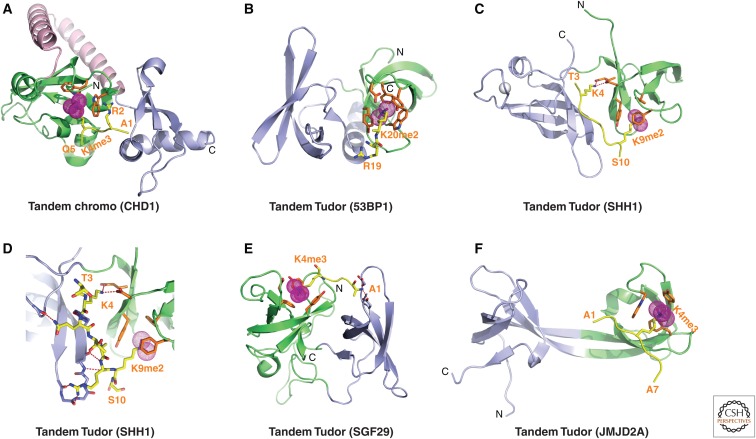
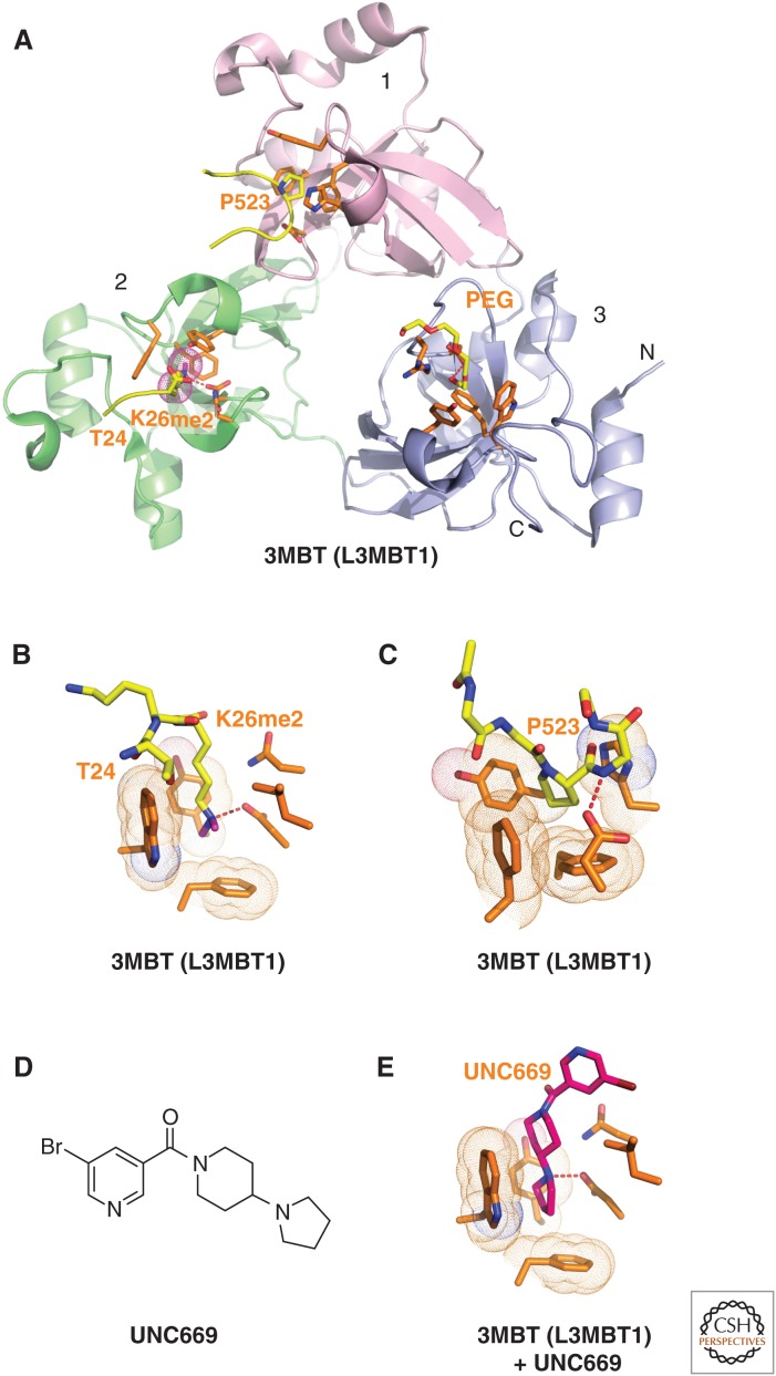
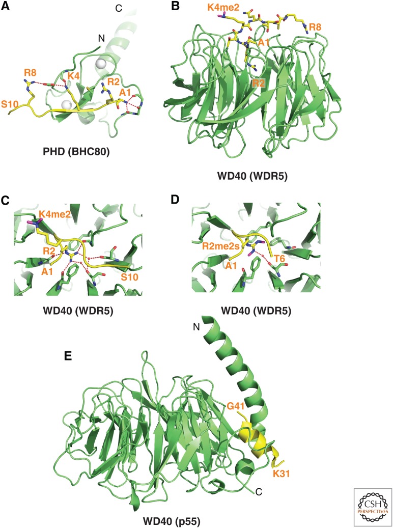
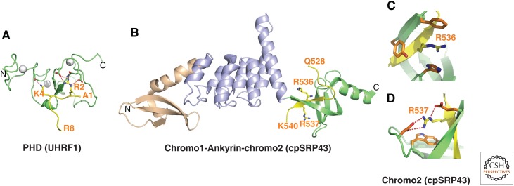
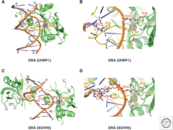
This article outlines the protein modules that target methylated lysine histone marks and 5mC DNA marks, and the molecular principles underlying recognition. The article focuses on the structural basis underlying readout of isolated marks by single reader molecules, as well as multivalent readout of multiple marks by linked reader cassettes at the histone tail and nucleosome level. Additional topics addressed include the role of histone mimics, cross talk between histone marks, technological developments at the genome-wide level, advances using chemical biology approaches, the linkage between histone and DNA methylation, the role for regulatory lncRNAs, and the promise of chromatin-based therapeutic modalities.


Figures

Figure 1.
Figure 1.
Sequence of H3 tail and distribution of PTMs. (A) Sequence of H3 tail and positioning of methyllysine (Kme) and methylarginine (Rme) marks. (B) Positioning of adjacent marks within the H3 tail at R2-T3-K4, A7-R8-K9-S10, and A25-R26-K27-S28 segments. Kme marks are partitioned between those that are activating and those that are repressive.
Figure 2.
Figure 2.
Structure of BPTF PHD finger bound to H3K4me3-containing peptide. (A) 2.0-Å crystal structure of the complex of the BPTF PHD finger bound to H3(1-15)K4me3 peptide (PDB: 2F6J). The PHD finger (as part of a PHD finger-bromo cassette) in a ribbon representation is in green, with two stabilizing bound Zn ions in silver balls. The bound peptide from A1 to T6 is shown in yellow with the trimethyl group of Kme3 shown by dotted balls in magenta. The residues forming the aromatic-lined cage are colored in orange. (B) Details showing the antiparallel alignment of the β-strands of the bound H3K4me3-containing peptide and PHD finger, resulting in formation of an antiparallel β-pleated sheet on complex formation. Note that the positively charged amino terminus is anchored in its own pocket. (C) Positioning of the K4me3 group within the aromatic-lined cage in the complex. (D) Positioning of R2 and K4me3 side chains in adjacent open surface pockets (surface groove mode), separated by the indole ring of an invariant Trp in the complex. The PHD finger and peptide are shown in surface- and space-filling representations, respectively. (E) Positioning of the K4me2 group into an engineered pocket, containing a Glu residue replacing the Tyr residue in C (PDB: 2RIJ).
Figure 3.
Figure 3.
Structures of BAH domains of mammalian ORC1 and plant ZMET2 bound to methylated lysine histone peptides. (A) 1.95-Å crystal structure of the complex of mouse ORC1 BAH domain bound to H4(14-25)K20me2 peptide (PDB: 4DOW). The bound K20me2-containing H4 peptide can be traced from G14 to R23. (B) Enlargement of (A) showing details of the alignment of the K20me2-containing H4 peptide from G14 to R23 positioned on the mouse ORC1 BAH domain in the complex. The dimethylammonium group of H4K20 inserts into an aromatic-lined pocket in the BAH domain. (C) 2.7-Å crystal structure of the complex of maize ZMET2 BAH domain bound to H3(1-32)K9me2 peptide (PDB: 4FT4). The chromodomain, methyltransferase, and BAH domains are colored in pink, blue, and green, respectively. The bound K9me2-containing H3 peptide in yellow can be traced from Q5 to T11. (D) Enlargement showing details of the alignment of the K9me2-containing H3 peptide from Q5 to T11 positioned on the maize ZMET2 BAH domain in the complex. The dimethylammonium group of H3K9me2 inserts into an aromatic-lined pocket in the BAH domain.
Figure 4.
Figure 4.
Structures of single Royal Family modules bound to methylated lysine histone peptides. (A) 2.4-Å crystal structure of the complex containing an HP1 chromodomain bound to H3(1-15)K9me3 peptide (PDB: 1KNE). The bound K9me3-containing H3 peptide can be traced from Q5 to S10. The HP1 residues in orange illustrate the aromatic cage that captures K9me3. (B) 2.35-Å crystal structure of the complex between the male-specific lethal (MSL)3 chromodomain bound to a H4(9-31)K20me1 peptide in the presence of duplex DNA (in surface representation) (PDB: 3OA6). The bound K20me1-containing H4 peptide can be traced from H18 to L22. (C) 1.85-Å crystal structure of the complex containing the PHF1 (a Polycomb-like protein) Tudor domain bound to H3(31-40)K36me3 peptide (PDB: 4HCZ). The bound K36me3-containing H3 peptide can be traced from S31 to R40. (D) 1.5-Å crystal structure of the complex of Brf1 PWWP domain bound to H3(22-42)K36me3 peptide complex (PDB: 2X4W). The bound K36me3-containing H3 peptide can be traced from S28 to R40.
Figure 5.
Figure 5.
Structures of tandem Royal Family modules bound to methylated lysine histone peptides. (A) 2.4-Å crystal structure of the complex of the human CDH1 tandem chromodomains bound to H3(1-19)K4me3 peptide (PDB: 2B2W). Chromodomains 1 and 2 are colored in green and blue, respectively, with the connecting helix-turn-helix linker in pink. The bound K4me3-containing H3 peptide can be traced from A1 to Q5. (B) 1.7-Å crystal structure of the complex of 53BP1 tandem Tudor domains bound to H4(15-24)K20me2 peptide (PDB: 2IG0). Tudor domains 1 and 2 are colored in green and blue, respectively. The bound K20me2-containing H4 peptide can be traced for the R19-K20me2 step. (C) 2.7-Å crystal structure of the complex of tandem Tudor domains of A. thalania SHH1 protein bound to a H3(1-15)K9me2 peptide (PDB: 4IUT). A bound zinc ion is shown in a silver ball. Tudor domains 1 and 2 are colored in green and blue, respectively. The bound K9me2-containing H3 peptide can be traced from T3 to S10. (D) The enlargement shows details of the alignment of the K9me2-containing H3 peptide from T3 to S10 positioned on the A. thaliana SHH1 domain in the complex with intermolecular interactions formed with both Tudor domains. (E) 1.26-Å crystal structure of the complex of tandem Tudor domains of SGF29 bound to H3(1-11)K4me3 peptide (PDB: 3MEA). Tudor domains 1 and 2 are colored in blue and green, respectively. The bound K4me3-containing peptide can be traced from A1 to K4me3. (F) 2.1-Å crystal structure of the complex of tandem Tudor domains of JMJD2A bound to H3(1-10)K4me3 peptide (PDB: 2GFA). Individual Tudor domains are colored in green and blue, respectively. The bound K4me3-containing peptide can be traced from A1 to A7.
Figure 6.
Figure 6.
Structures of L3MBTL1 bound to methylated lysine histone peptides and an inhibitor. (A) 1.66-Å crystal structure of the complex-containing L3MBTL1 bound to H1(22-26)K26me2 peptide (PDB: 2RHI). A carboxy-terminal peptide from an adjacent L3MBTL1 in the crystal lattice inserts Pro523 into the aromatic-lined pocket of MBT domain 1 (in pink). The dimethylammonium group of bound K26me2 inserts into the aromatic-lined pocket of MBT domain 2 (in green) with the K26me2-containing H1 peptide traced from T24 to K26me2. A polyethylene glycol (PEG) molecule inserts into the aromatic-lined pocket of MBT domain 3 (in blue). (B) Details of how the H1K26me2 dimethylammonium group inserts into the aromatic-lined pocket of MBT domain 2. (C) Structural detail showing how the proline from an adjacent L3MBTL1 in the crystal lattice inserts into the aromatic-lined pocket of MBT domain 1. This pocket is shallower than the one shown in B. (D) Chemical formula of UNC669. (E) Details of how UNC669 inserts into the aromatic-lined pocket of MBT domain 2, based on the 2.55-Å crystal structure of L3MBTL1 bound to UNC669 (PDB: 3P8H).
Figure 7.
Figure 7.
Structures of expanded and paired modules bound to methylated lysine histone marks. (A) 1.6-Å crystal structure of the complex containing the ADD domain of ATRX bound to the H3(1-15)K9me3 peptide (PDB: 3QLA). The ADD GATA-1 and PHD fingers are colored in blue and green, respectively. Bound Zn ions are shown as silver balls. The H3 peptide containing K9me3 is traced from A1 to S10. (B) Enlargement of A showing intermolecular contacts between the K9me3-containing H3 peptide, traced from A1 to S10, complexed with the ADD domain. The me3 are shown as magenta spheres. (C) Ribbon and stick representation of K9me3 positioned to interact with the GATA-1 and PHD finger domains in the complex. (D) Surface and space-filling representation of surface complementarity between K9me3 and the walls of the pocket lined by the GATA-1 and PHD finger domains in the complex. (E) 1.7-Å crystal structure of the ternary complex of the Pygo PHD finger (in green) bound to H3(1-7)K4me2 peptide (in yellow) in the presence of the HD1 domain of BCL9 (in pink) (PDB: 2VPE). (F) 2.99-Å crystal structure of the complex consisting of the G9a ankyrin repeats (green) bound to an H3(1-15)K9me2 peptide traced from A7 to G13 in yellow (PDB: 3B95). The K9me2 aromatic-lined binding pocket is positioned between the fourth and fifth ankyrin repeats of G9a.
Figure 8.
Figure 8.
Structures of expanded and paired modules bound to methylated arginine histone marks. (A) NMR solution structure of the complex containing the SMN Tudor domain (green ribbon representation) bound to a symmetrical Rme2s-containing peptide (yellow) (PDB: 4A4E). The methyl groups are illustrated with magenta spheres and the aromatic-lined cage in orange. (B) 2.8-Å crystal structure of the complex containing the SND1 extended Tudor module bound to the amino-terminal PIWI peptide, traced from R10 to R17, with the R14me2s modification (PDB: 3NTI). The core fold of the Tudor domain is shown in green, whereas the extensions are shown in blue. (C) Enlargement of B showing the positioning of R14me2s in the aromatic-lined cage of the SND1 Tudor domain in the complex.
Figure 9.
Figure 9.
Structures of reader modules bound to lysines and arginines. (A) 1.43-Å crystal structure of the complex of the PHD finger of BHC80 bound to H3(1-10) peptide (in yellow) (PDB: 2PUY). The bound H3 peptide can be traced from A1 to S10. Zinc ions are shown as silver balls. (B) 1.5-Å crystal structure of the complex of the WD40 motif of WDR5 bound to H3(1-9)K4me2 peptide. The bound K9me2-containing peptide can be traced from A1 to R8 (PDB: 2H6N). (C) Insertion of R2 into the central channel of the WD40 motif in the H3(1-9)K9me2-WDR5 complex. (D) Insertion of R2me2s into the central channel of the WD40 motif in the H3(1-15)R2me2sK9me2-WDR5 complex solved at 1.9 Å (PDB: 4A7J). (E) 3.2-Å crystal structure of the complex of the WD40 motif of p55 bound to H4(15-41) peptide (PDB: 3C9C). The bound H4 peptide can be traced from K31 to G41.
Figure 10.
Figure 10.
Structures of PHD finger and chromodomains bound to unmodified arginines. (A) 1.8-Å crystal structure of the complex of the PHD finger of UHRF1 bound to H3(1-9) peptide (PDB: 3SOU). The bound H3 peptide can be traced from A1 to R8. Zinc ions are shown by silver balls. (B) 3.18-Å crystal structure of the complex of the chromodomains and Ankyrin repeats of A. thaliana chloroplast signal recognition particle (cpSRP)43 bound to an RRKR (Arg-Arg-Lys-Arg)-containing peptide (yellow) (PDB: 3UI2). The side chains of R536 and R537 of the bound RRKR-containing peptide are positioned in adjacent pockets at the interface between the fourth Ankyrin repeat (purple) and the second chromodomain (green) in the complex. (C) Positioning of Arg536 of the RRKR-containing peptide within an aromatic-lined cage in the complex. (D) Positioning of Arg537 of the RRKR-containing peptide in a pocket lined by a Trp and two acidic side chains in the complex.
Figure 11.
Figure 11.
Structures of PHD-bromo cassettes involved in multivalent readout. (A) 2.0-Å crystal structure of the PHD-bromo cassette of BPTF bound to H3(1-15)K4me3 peptide (PDB: 2F6Z). A separate 1.8-Å crystal structure of the BPTF bromodomain bound to H4(12-21)K16ac peptide was also solved (PDB: 3QZS) and that information was superpositioned on the structure shown in this panel. The bound H3(1-15)K4me3-containing peptide can be traced from A1 to T6, whereas the bound H4(12-21)K16ac-containing peptide can be traced from K14 to V21 in the complexes. (B) 1.9-Å crystal structure of the MLL1 PHD-bromo cassette bound to H3(1-9)K4me3 peptide (PDB: 3LQJ). The bound H3(1-9)K4me3-containing peptide can be traced from A1 to T6 in the complex. (C) 2.7-Å crystal structure of the TRIM33 PHD-bromo cassette bound to H3(1-22)K9me3K18ac peptide (PDB: 3U5O). The bound H3(1-22)K9me3K18ac peptide can be traced from A1 to L20 in the complex. (D) The crystal structures of the TRIM24 PHD-bromo cassette bound to H3(1-10) peptide (2.0 Å) (PDB: 3O37) and bound to H3(13-32)K23ac peptide (1.9 Å) (PDB: 3O37). The structures were superpositioned to generate the composite structure shown in this panel. The bound H3(13-32)K23ac peptide can only be traced from T22 to T32 in the complex.
Figure 12.
Figure 12.
Structures of linked binding modules involved in multivalent readout at the peptide and nucleosomal levels. (A) 2.9-Å crystal structure of the UHRF1 tandem Tudor-PHD finger cassette bound to H3(1-13)K9me3 peptide (PDB: 3ASK). The tandem Tudor domains are shown in cyan and purple, whereas the PHD finger is shown in green. Zinc ions are shown as silver balls. The bound H3(1-13)K9me3-containing peptide can be traced from A1 to S10. (B) Enlargement of A, showing details of the intermolecular contacts between H3(1-13)K9me3-containing peptide (A1 to S10) and the tandem Tudor-PHD finger cassette of UHRF1. (C) 1.47-Å crystal structure of the complex of the tandem PHD finger cassette of MOZ bound to H3(1-18)K14ac peptide (PDB: 3V43). There is a bound acetate (in space-filling representation) from buffer bound in a pocket in the amino-terminal PHD finger (in blue). The bound H3(1-18)K14ac-containing peptide can be traced from A1 to A7, in which it is bound to the carboxy-terminal PHD finger (in green). (D) glutathione S-transferase (GST) pull-down of modified nucleosomes with semisynthetic histones produced by expressed protein ligation. Nucleosomes containing dual marks involving H4K12ac, H4K16ac, or H4K20ac in combination with H3K4me3 are pulled down with resin bound GST-BPTF PHD-bromo cassette and detected by autoradiography after native gel electrophoresis. (D, Reprinted from Ruthenburg et al. 2011.)
Figure 13.
Figure 13.
Structures of BPTF PHD-bromo cassette with cis and trans linker prolines. (A) 1.72-Å crystal structure of the MLL1 PHD-bromo cassette with a cis linker proline (circled in red) in the free state (PDB: 3LQH). The PHD finger and bromodomain are colored in green and blue, respectively. (B) Model of the MLL1 PHD-bromo cassette with a trans linker proline (circled in red) with this alignment stabilized by a bound RRM domain of CyP33 (magenta). (C) NMR solution structure of the complex containing the MLL1 PHD3 fragment (1603–1619, in green) and RRM domain of CyP33 (2-82, in magenta) (PDB: 2KU7).
Figure 14.
Figure 14.
Structures of methylcytosine-binding proteins bound to fully methylated 5mCpG DNA. (A) NMR solution structure of MBD1 protein bound to fully methylated 5mCpG-containing DNA duplex (PDB: 1IG4). Two loops L1 and L2 are colored in yellow. The methyl groups of 5mC’s are marked by magenta dotted circles. (B) Schematic of intermolecular contacts centered about the 5mCpG/5mCpG site involving loops L1 and L2, and the amino-terminal α-helix adjacent to L2. Methyl group of 5mC is represented by the magenta circle. Hydrophobic interactions between the 5mC residues and side chains of MBD1 are indicated by magenta colored arrows. (C) 2.5-Å crystal structure of MeCP2 protein bound to fully methylated 5mCpG-containing DNA duplex (PDB: 3C2I). The methyl groups of 5mCs are marked by magenta dotted circles. (D) Intermolecular contacts between hydrophilic amino acids of MeCP2 (green stick representation) and the 5mC groups (magenta dotted circles) in the major groove of the duplex, including C—H••O hydrogen bonds to tightly bound water molecules.
Figure 15.
Figure 15.
Structures of methylcytosine-binding zinc-finger proteins bound to fully methylated 5mCpG DNA. (A) 2.8-Å crystal structure of three zinc fingers of Kaiso protein bound to a pair of fully methylated 5mCpG-containing DNA duplex (PDB: 4F6N). The first, second, and third zinc fingers are colored in green, blue, and pink, respectively. Note that although the majority of the intermolecular contacts are with the major groove, involving zing fingers 1 (green) and 2 (blue), there are also contacts with the minor groove, involving zinc finger 3 and the carboxy-terminal extension of Kaiso. Zinc ions are shown as silver balls. The methyl groups of 5mC are marked by magenta dotted circles. (B) Intermolecular contacts between amino acids of the first zinc finger (in green) of Kaiso and 5mC groups in the major groove of the duplex. (C) 0.99-Å crystal structure of two zinc fingers of Zfp57 protein bound to a fully methylated 5mCpG-containing DNA duplex (PDB: 4GZN). Zinc ions are shown as silver balls. The methyl groups of 5mC are marked by magenta dotted circles. (D) One of the 5mC groups in the Zfp57-DNA complex is involved in hydrophobic interactions through positioning between an Arg side chain and a neighboring guanine. (E) The second 5mC in the Zfp57-DNA complex interacts with a layer of ordered water molecules (red circles).
Figure 16.
Figure 16.
Structures of SRA domain-containing proteins bound to hemimethylated 5mCpG DNA. (A) 1.6-Å crystal structure of SRA domain of UHRF1 bound to a hemimethylated 5mCpG-containing DNA duplex (PDB: 2ZKD). The stoichiometry of the complex is one SRA domain per DNA duplex. The methyl groups of 5mC are marked by magenta dotted circles. (B) Alignment of the flipped-out 5mC (ring is shaded for clarity) in a pocket within the SRA domain of UHRF1. (C) 2.37-Å crystal structure of SRA domain of plant SUVH5 bound to a hemimethylated 5mCpG-containing DNA duplex (PDB: 3Q0D). The stoichiometry of the complex is two SRA domains per DNA duplex. 5mC’s methyl groups are marked by magenta dotted circles. (D) Alignment of the flipped-out 5mC (ring is shaded for clarity) in a pocket within the SRA domain of plant SUVH5.

References

    1. Ali M, Yan K, Lalonde ME, Degerny C, Rothbart SB, Strahl BD, Cote J, Yang XJ, Kutateladze TG. 2012. Tandem PHD fingers of MORF/MOZ acetyltransferases display selectivity for acetylated histone H3 and are required for the association with chromatin. J Mol Biol 424: 328–338. - PMC - PubMed
    1. Allfrey VG, Faulkner R, Mirsky AE. 1964. Acetylation and methylation of histones and their possible role in the regulation of RNA synthesis. Proc Natl Acad Sci 51: 786–794. - PMC - PubMed
    1. Allis CD, Muir TW. 2011. Spreading chromatin into chemical biology. Chembiochem 12: 264–279. - PubMed
    1. Allis CD, Jeuwein T, Reinberg D. 2014. Overview and concepts. Cold Spring Harb Perspect Biol 10.1101/cshperspect.a018739. - DOI
    1. Amir RE, Van den Veyver IB, Wan M, Tran CQ, Francke U, Zoghbi HY. 1999. Rett syndrome is caused by mutations in X-linked MECP2, encoding methyl-CpG-binding protein 2. Nat Genet 23: 185–188. - PubMed

MeSH terms

LinkOut - more resources