. 2016 Mar 2:6:22451.
doi: 10.1038/srep22451.

A new approach to chemotherapy: drug-induced differentiation kills African trypanosomes

Affiliations

A new approach to chemotherapy: drug-induced differentiation kills African trypanosomes

Tanja Wenzler et al. Sci Rep. .

Abstract

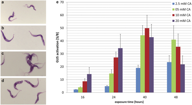
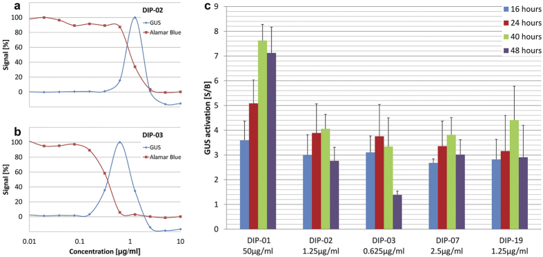
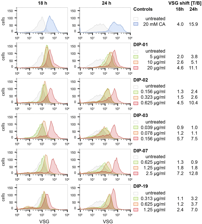
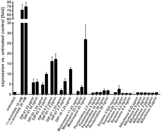
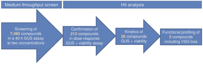
Human African trypanosomiasis (sleeping sickness) is a neglected tropical disease caused by Trypanosoma brucei spp. The parasites are transmitted by tsetse flies and adapt to their different hosts and environments by undergoing a series of developmental changes. During differentiation, the trypanosome alters its protein coat. Bloodstream form trypanosomes in humans have a coat of variant surface glycoprotein (VSG) that shields them from the immune system. The procyclic form, the first life-cycle stage to develop in the tsetse fly, replaces the VSG coat by procyclins; these proteins do not protect the parasite from lysis by serum components. Our study exploits the parasite-specific process of differentiation from bloodstream to procyclic forms to screen for potential drug candidates. Using transgenic trypanosomes with a reporter gene in a procyclin locus, we established a whole-cell assay for differentiation in a medium-throughput format. We screened 7,495 drug-like compounds and identified 28 hits that induced expression of the reporter and loss of VSG at concentrations in the low micromolar range. Small molecules that induce differentiation to procyclic forms could facilitate studies on the regulation of differentiation as well as serving as scaffolds for medicinal chemistry for new treatments for sleeping sickness.


Figures

Figure 1
Figure 1. Establishment of GUS assay with CA.
Giemsa stained T. b. b. GUSone (a–d). Bloodstream forms (a), procyclic forms (b) parasites were differentiated to procyclics with a 24 h exposure to 5 mM CA at 27 °C followed by one week culture in SDM-79 medium at 27 °C. Bloodstream forms that were treated with 10 mM CA for 40 h at 37 °C (c), bloodstream forms that were treated with 10 mM CA for 40 h at 27 °C (d). GUS activation is dependent on CA concentration and exposure time at 37 °C (e). The starting density was 8 × 105 parasites/ml for 16 h and 24 h, 5 × 105 parasites/ml for 40 h, and 2.5 × 105 parasites/ml for 48 h exposure. Bars indicate standard deviations of S/B from at least three independent assays. Cells exposed to CA at 37 °C tend to aggregate. CA = cis-aconitate.
Figure 2
Figure 2. GUS differentiation signal is time- and concentration-dependent and coincides with cell death.
Serial dilutions of DIP-02 (a) and DIP-03 (b) in the GUS differentiation assay and the Alamar blue viability assay (40 h exposure time). GUS activation (S/B) of DIP-01, DIP-02, DIP-03, DIP-07 and DIP-19 at different exposure times (16 h, 24 h, 40 h and 48 h) at the concentration for optimal GUS expression (c). Bars indicate standard deviations of S/B from at least three independent assays. CA = cis-aconitate.
Figure 3
Figure 3. Loss of VSG after treatment with 20 mM CA or various concentrations of DIP compounds at 37 °C.
The density of the VSG coat was monitored by staining with anti-VSG221 antibodies and flow cytometry. All assays were performed in triplicate, using one 96-well plate for each time-point (18 and 24 h); the untreated and CA-treated samples on each plate thus serve as controls for the whole series measured per time-point. To display the dose-dependent VSG shifts, representative curves of different DIP concentrations were plotted together with the corresponding untreated control. MFI: median fluorescence intensity. T/B: VSG changes are expressed as a ratio of the MFI after treatment to the MFI of the untreated control. Full flow cytometry data are available in Table S4.
Figure 4
Figure 4. Real-time qPCR to measure procyclin expression levels.
GUSone bloodstream forms were exposed for 24 hours to the 5 DIP compounds DIP-01, DIP-02, DIP-03, DIP-07 and DIP-19 and to CA at 37 °C. Procyclin mRNA levels were determined by qPCR. Among the standard trypanocidal drugs only melarsoprol induced expression of procyclin mRNA above background level. Error bars show standard deviations for technical triplicates.
Figure 5
Figure 5. Overview of the screening procedure to identify compounds triggering differentiation.
7,495 compounds were screened at two concentrations in the GUS assay. 243 compounds induced GUS with a S/B >2-fold, of which 212 available compounds were subjected to dose-response analyses in the GUS and viability assays. 30 compounds induced GUS activity S/B >3-fold of which 28 compounds were available and highly pure. Five compounds were further analysed by flow cytometry (loss of VSG) and real-time quantitative PCR (induction of procyclin mRNA).

References

    1. Brun R., Blum J., Chappuis F. & Burri C. Human African trypanosomiasis. The Lancet 375, 148–159 (2010). - PubMed
    1. Stuart K. et al. Kinetoplastids: related protozoan pathogens, different diseases. J. Clin. Invest. 118, 1301–1310 (2008). - PMC - PubMed
    1. Opperdoes F. R. Biochemical peculiarities of trypanosomes, African and South American. Br. Med. Bull. 41, 130–136 (1985). - PubMed
    1. Bacchi C. J. & Yarlett N. Polyamine metabolism as chemotherapeutic target in protozoan parasites. Mini Rev. Med. Chem. 2, 553–563 (2002). - PubMed
    1. Stuart K. D., Schnaufer A., Ernst N. L. & Panigrahi A. K. Complex management: RNA editing in trypanosomes. Trends Biochem. Sci. 30, 97–105 (2005). - PubMed

Publication types

MeSH terms