. 2016 Mar 2:6:22298.
doi: 10.1038/srep22298.

Structure-based Inhibitor Design for the Intrinsically Disordered Protein c-Myc

Affiliations

Structure-based Inhibitor Design for the Intrinsically Disordered Protein c-Myc

Chen Yu et al. Sci Rep. .

Abstract

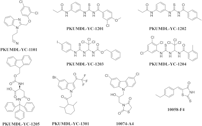
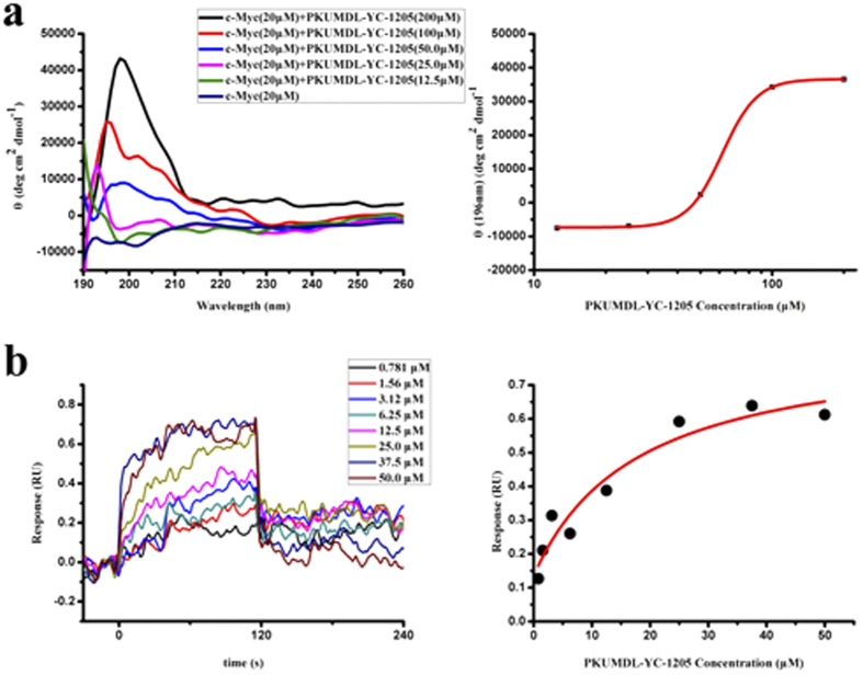
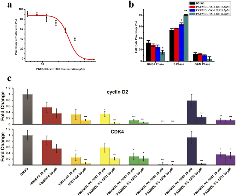
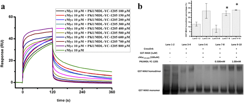
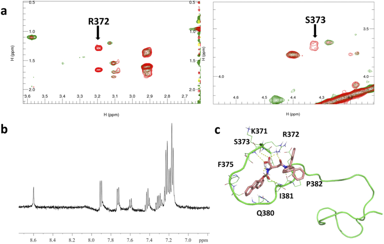
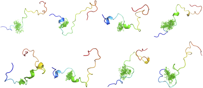
Intrinsically disordered proteins (IDPs) are associated with various diseases and have been proposed as promising drug targets. However, conventional structure-based approaches cannot be applied directly to IDPs, due to their lack of ordered structures. Here, we describe a novel computational approach to virtually screen for compounds that can simultaneously bind to different IDP conformations. The test system used c-Myc, an oncoprotein containing a disordered basic helix-loop-helix-leucine zipper (bHLH-LZ) domain that adopts a helical conformation upon binding to Myc-associated factor X (Max). For the virtual screen, we used three binding pockets in representative conformations of c-Myc370-409, which is part of the disordered bHLH-LZ domain. Seven compounds were found to directly bind c-Myc370-409 in vitro, and four inhibited the growth of the c-Myc-overexpressing cells by affecting cell cycle progression. Our approach of IDP conformation sampling, binding site identification, and virtual screening for compounds that can bind to multiple conformations provides a useful strategy for structure-based drug discovery targeting IDPs.


Figures

Figure 1
Figure 1. Chemical structures of the active compounds.
Figure 2
Figure 2. Activities of PKUMDL-YC-1205 in cell-free assays.
(a) CD spectra of c-Myc370–409 with indicated concentrations of PKUMDL-YC-1205 (left) and dose-response curve at 196 nm (right). The apparent Kd of PKUMDL-YC-1205 was 61.8 ± 0.7 μM, as predicted by the Hill equation. (b) SPR direct binding curves of the indicated concentrations of PKUMDL-YC-1205 (left). The Kd value was 18 ± 12 μM based on affinity fitting of the dose-response curve (right). Data represent the mean ± standard deviation of three independent experiments.
Figure 3
Figure 3. Activities of PKUMDL-YC-1205 in cell-based assays.
(a) Growth inhibition of HL-60 cells by PKUMDL-YC-1205 was assessed by the MTT assay after exposure to indicated concentrations of PKUMDL-YC-1205 for 72 hours. The EC50 was 40.0 ± 1.9 μM. (b) Percentage of HL-60 cells in different phases of the cell cycle after treatment with 17.8 μM, 26.7 μM, and 40.0 μM PKUMDL-YC-1205 for 24 hours. Data represent the mean ± standard deviation of three independent experiments. (c) c-Myc mediated transcriptional activity in HL-60 cells was blocked. Data represent the mean ± standard error of three independent experiments. *p < 0.05, **p < 0.01, ***p < 0.005.
Figure 4
Figure 4. PKUMDL-YC-1205 blocks the interaction between c-Myc370–409 and Max.
(a) PKUMDL-YC-1205 abolished cMyc370–409 binding to Max in the SPR competitive assay at the indicated concentrations. (b) PKUMDL-YC-1205 disrupted the Max-Max/c-Myc-Max dimerization equilibrium. Chemical cross-linking and anti-Max western blotting results are shown (left). Blackness integrals of the GST-Max homodimer percentage are shown as a histogram (right). Data represent the mean ± standard deviation of two independent experiments. *p < 0.05.
Figure 5
Figure 5. NMR study of PKUMDL-YC-1205 binding with c-Myc370–409.
(a) Partially enlarged details of overlapped 1H-1H TOCSY spectrum of c-Myc370–409 with (green) and without (red) PKUMDL-YC-1205. (b) STD NMR spectrum of PKUMDL-YC-1205 with c-Myc370–409 (molar ratio 50:1). (c) One binding model of c-Myc370–409 with PKUMDL-YC-1205. c-Myc370–409 is represented in green and PKUMDL-YC-1205 is represented in pink.
Figure 6
Figure 6. PKUMDL-YC-1205 binds to multiple conformations of c-Myc370–409 in molecular dynamics simulations.
Conformations from the five simulations of c-Myc370–409 with PKUMDL-YC-1205 were clustered. The c-Myc370–409 structures are depicted in a rainbow (from blue at the N-terminal to red at the C-terminal) and PKUMDL-YC-1205 structures are depicted at the centres of mass as green dots.

References

    1. Wright P. E. & Dyson H. J. Intrinsically unstructured proteins: re-assessing the protein structure-function paradigm. J. Mol. Biol. 293, 321–331 (1999). - PubMed
    1. Dunker A. K. et al. The unfoldomics decade: an update on intrinsically disordered proteins. BMC Genomics 9, S1 (2008). - PMC - PubMed
    1. Uversky V. N. Intrinsically disordered proteins from A to Z. Int. J. Biochem. Cell Biol. 43, 1090–1103 (2011). - PubMed
    1. Schlessinger A. et al. Protein disorder—a breakthrough invention of evolution? Curr. Opin. Struct. Biol. 21, 412–418 (2011). - PubMed
    1. Huang Y. & Liu Z. Kinetic advantage of intrinsically disordered proteins in coupled folding-binding process: a critical assessment of the “fly-casting” mechanism. J. Mol. Biol. 393, 1143–1159 (2009). - PubMed

Publication types

MeSH terms

Substances