. 2016 May;100(5):720-6.
doi: 10.1136/bjophthalmol-2015-308290. Epub 2016 Mar 1.

Increased choroidal mast cells and their degranulation in age-related macular degeneration

Affiliations

Increased choroidal mast cells and their degranulation in age-related macular degeneration

Imran A Bhutto et al. Br J Ophthalmol. 2016 May.

Abstract

Background/aims: Inflammation has been implicated in age-related macular degeneration (AMD). This study investigates the association of mast cells (MCs), a resident choroidal inflammatory cell, with pathological changes in AMD.

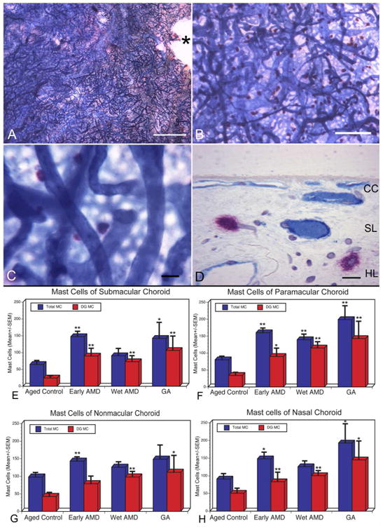
Methods: Human donor eyes included aged controls (n=10), clinically diagnosed with early AMD (n=8), geographic atrophy (GA, n=4) and exudative AMD (n=11). The choroids were excised and incubated for alkaline phosphatase (APase; blood vessels) and non-specific esterase activities (MCs). Degranulated (DG) and non-degranulated MCs in four areas of posterior choroid (nasal, non-macular, paramacular and submacular) were counted in flat mounts (4-6 fields/area). Choroids were subsequently embedded in JB-4 and sectioned for histological analyses.

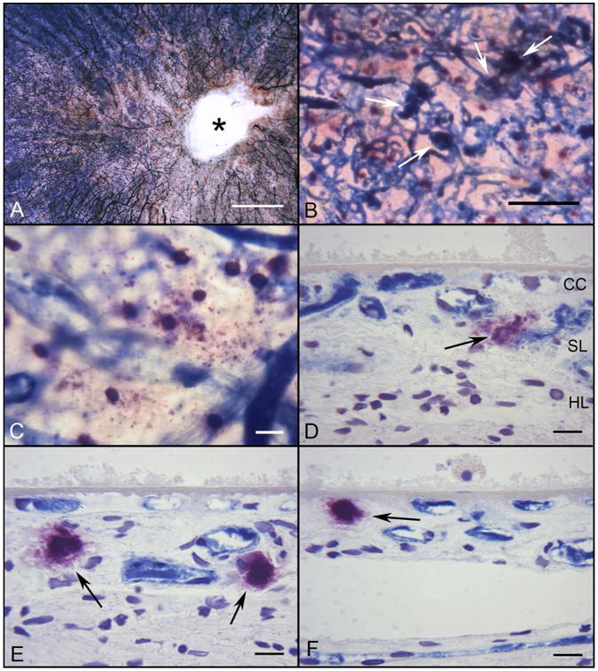
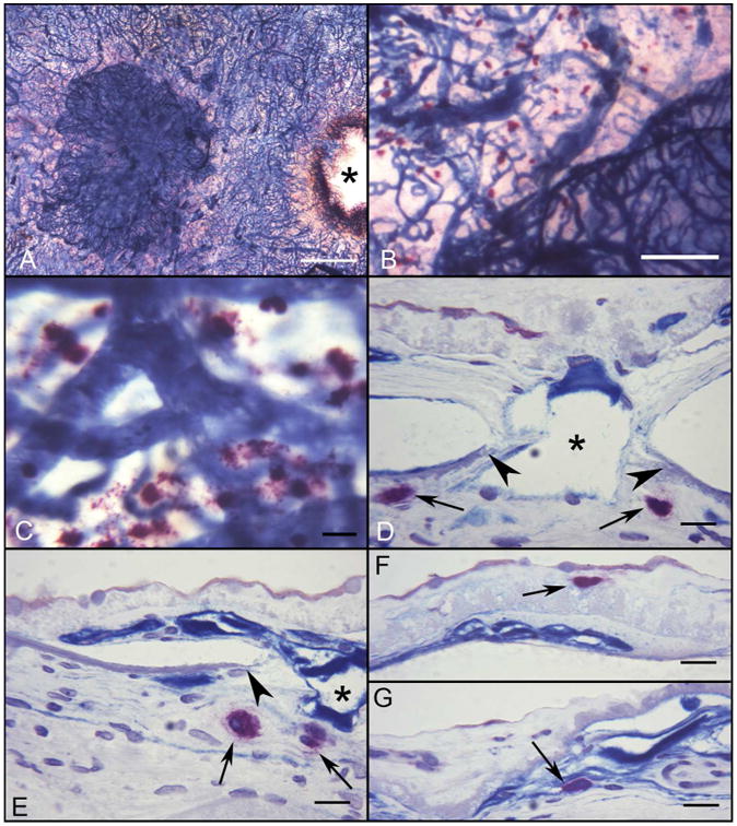
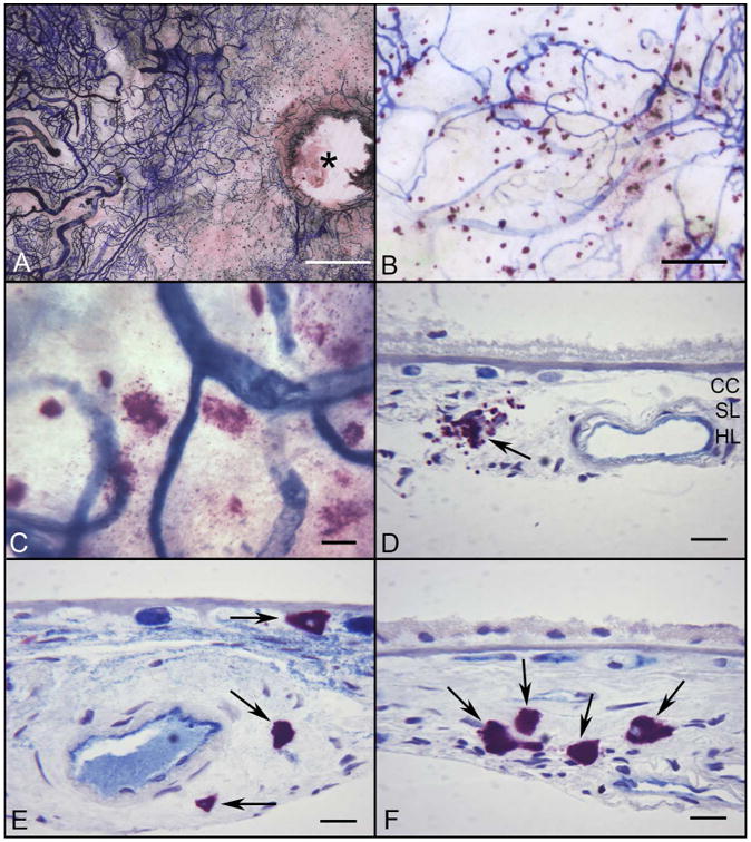
Results: The number of MCs was significantly increased in all choroidal areas in early AMD (p=0.0006) and in paramacular area in exudative AMD (139.44±55.3 cells/mm(2); p=0.0091) and GA (199.08±82.0 cells/mm(2); p=0.0019) compared with the aged controls. DG MCs were also increased in paramacular (p=0.001) and submacular choroid (p=0.02) in all forms of AMD. Areas with the greatest numbers of DG MCs had loss of choriocapillaris (CC). Sections revealed that the MCs were widely distributed in Sattler's and Haller's layer in the choroidal stroma in aged controls, whereas MCs were frequently found in close proximity with CC in GA and exudative AMD and in choroidal neovascularisation (CNV).

Conclusion: Increased MC numbers and degranulation were observed in all AMD choroids. These results suggest that MC degranulation may contribute to the pathogenesis of AMD: death of CC and retinal pigment epithelial and CNV formation. The proteolytic enzymes released from MC granules may result in thinning of AMD choroid.

Keywords: Choroid; Inflammation; Macula; Neovascularisation; Pathology.


Figures

Figure 1
Figure 1
Flat mount of submacular choroid from an aged control subject stained with alkaline phosphatase and nonspecific esterase (APase/NSE) (Subject#4). (A and B) APase+ choroidal vessels are stained blue and NSE-positive MCs are stained red with this preparation. (C) MCs are largely distributed in the intermediate and deep choroid so the choriocapillaris (CC) is out of focus. (D) Histological section of the choroid shown in (A) demonstrates red MCs are associated with blood vessels in Sattler's layer (SL) and Haller's layer (HL), which are APase+ and therefore have viable endothelial cells.(*, optic nerve; Bar=1mm in A, 200μm in B, 50μm in C, and 20mm in D). (E-H) Comparison of the number of MCs (total MCs, blue bar; degranulated or DG MCs, red bar) present per mm2 of choroid counted in the flat perspective in aged control and AMD subjects. MC numbers/mm2 are presented from four areas of posterior choroid [submacular (E), paramacular (F), nonmacular (G), and nasal (H)] and compared to the aged control in those regions. Total numbers of MCs as well as degranulated MCs were statistically significantly increased in almost all areas examined in AMD choroids compared to the aged control. The significance of the difference between aged control and AMD (p< 0.05) is indicated using the Students t-test (*) and Wilcoxon rank-sum test (**).
Figure 2
Figure 2
Flat mount of APase/NSE stained choroid from a subject with early AMD (Subject#15). A succinct area of CC degeneration (lacking APase staining) is evident in the submacular region and around the optic disc(*)(A). Several neovascular buds have dark blue APase activity (B, white arrows). (C) At higher magnification, some degranulated MCs are apparent at the level of CC, which has reduced APase activity in this area. (D, E, F) Histological sections from same choroid demonstrate the DG MCs (black arrows) present near degenerative CC channels, which are APase and around the dark blue APase+ neovascular buds. A thin basal laminar deposit is present internal to Bruchs membrane.(Bar=1mm in A, 200μm in B, 50μm in C, and 20μm in D-F)
Figure 3
Figure 3
Flat mount of APase/NSE choroid from a subject with wet AMD (Subject#20). A large APase stained choroidal neovascular formation (CNV) is present in submacular region of the choroid (A)(*, optic nerve). (B) Numerous degranulated MCs are adjacent to the edge of CNV, where the CC is greatly attenuated. (C) At higher magnification, degranulated MCs are present at the edge of CNV. (D-G) Histological sections from same choroid demonstrate the degranulated MCs (black arrows) adjacent to the CNV feeder vessel. An MC is present in a basal laminar deposit (F) and in the CNV itself (G). Breaks in Bruch's membrane are indicated by arrowheads. (*, CNV breaching Bruchs membrane; Bar=1mm in A, 200μm in B, 50μm in C, and 20μm in D-G)
Figure 4
Figure 4
Flat mount preparation of APase/NSE incubated choroid from a subject with GA (Subject#30) (A) There is a large area of CC loss (APase) around the optic disc (*) and in the submacular region of the choroid. (B) Many degranulated MCs are associated with areas of RPE atrophy and CC degeneration. (C) At higher magnification, most MCs are degranulated. (D-F) Histological sections from same choroid demonstrate the degranulated MCs (black arrows) aggregating in choroidal stroma in a region of RPE atrophy with a thin basal laminar deposit (D) and in the wall of an artery with atherosclerotic changes (E). MCs are increased in number but in their normal position in Sattler's and Haller's layers in a region without RPE atrophy (F). (Bar=1mm in A, 200μm in B, 50μm in C, and 20μm in D-F)

References

    1. Bhutto I, Lutty G. Understanding age-related macular degeneration (AMD): relationships between the photoreceptor/retinal pigment epithelium/Bruch's membrane/choriocapillaris complex. Molecular aspects of medicine. 2012;33(4):295–317. doi: 10.1016/j.mam.2012.04.005. published Online First: Epub Date. - DOI - PMC - PubMed
    1. Schramm EC, Clark SJ, Triebwasser MP, Raychaudhuri S, Seddon JM, Atkinson JP. Genetic variants in the complement system predisposing to age-related macular degeneration: a review. Molecular immunology. 2014;61(2):118–25. doi: 10.1016/j.molimm.2014.06.032. Online First: Epub Date. - DOI - PMC - PubMed
    1. Costa JJ, Weller PF, Galli SJ. The cells of the allergic response: mast cells, basophils, and eosinophils. JAMA. 1997;278(22):1815–22. - PubMed
    1. Galli SJ. Mast cells and basophils. Current opinion in hematology. 2000;7(1):32–9. - PubMed
    1. Xu JM, Shi GP. Emerging role of mast cells and macrophages in cardiovascular and metabolic diseases. Endocr Rev. 2012;33(1):71–108. doi:er.2011-0013[pii]10.1210/er.2011-0013. Online First: Epub Date. - PMC - PubMed

Publication types