. 2016 Mar 2:6:22508.
doi: 10.1038/srep22508.

Frog Virus 3 dissemination in the brain of tadpoles, but not in adult Xenopus, involves blood brain barrier dysfunction

Affiliations

Frog Virus 3 dissemination in the brain of tadpoles, but not in adult Xenopus, involves blood brain barrier dysfunction

Francisco De Jesús Andino et al. Sci Rep. .

Abstract

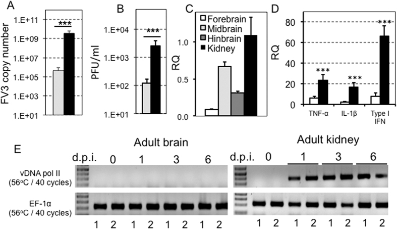
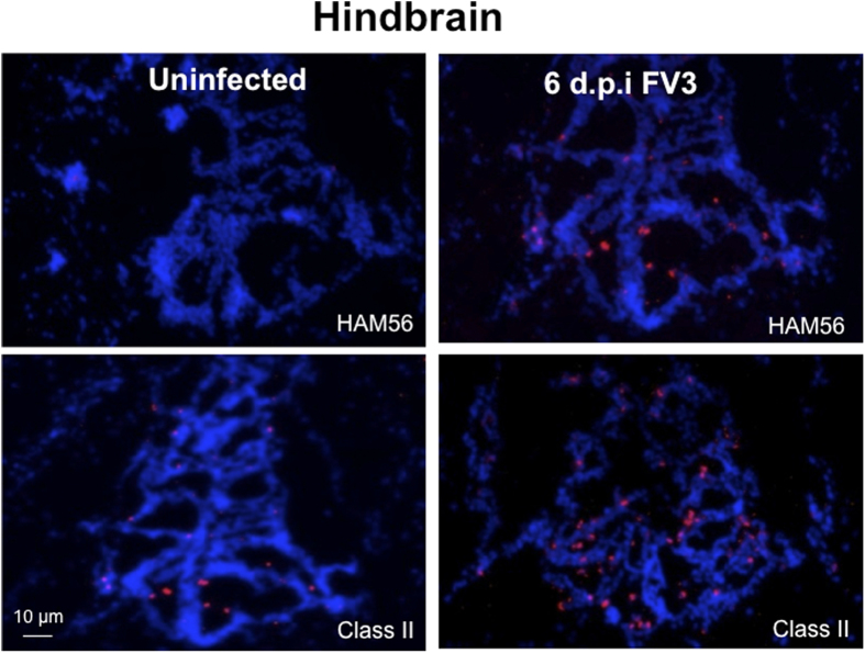
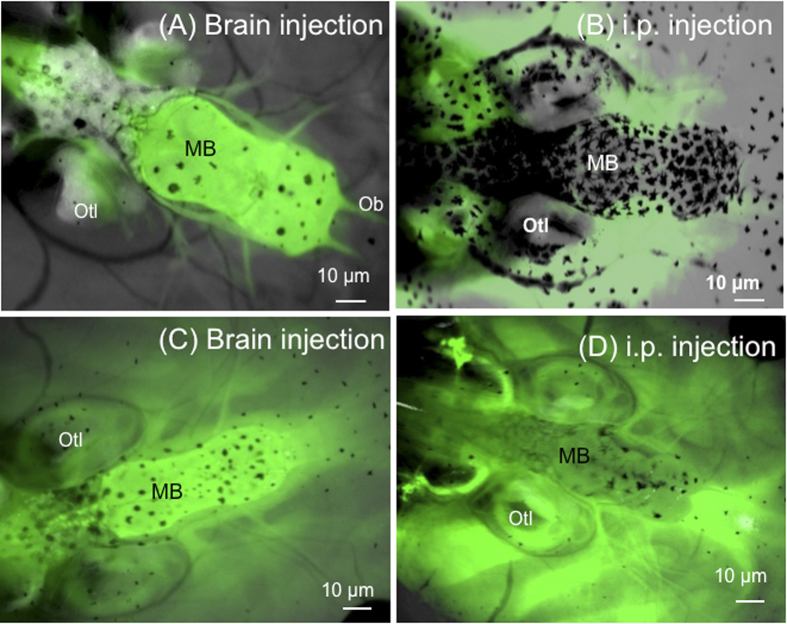
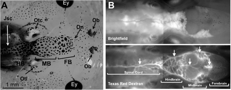
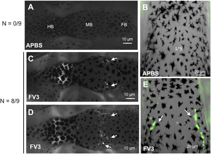
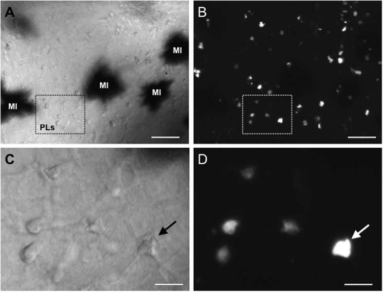
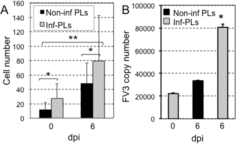
While increasing evidence points to a key role of monocytes in amphibian host defenses, monocytes are also thought to be important in the dissemination and persistent infection caused by ranavirus. However, little is known about the fate of infected macrophages or if ranavirus exploits immune privileged organs, such as the brain, in order to establish a reservoir. The amphibian Xenopus laevis and Frog Virus 3 (FV3) were established as an experimental platform for investigating in vivo whether ranavirus could disseminate to the brain. Our data show that the FV3 infection alters the BBB integrity, possibly mediated by an inflammatory response, which leads to viral dissemination into the central nervous system in X. laevis tadpole but not adult. Furthermore, our data suggest that the macrophages play a major role in viral dissemination by carrying the virus into the neural tissues.


Figures

Figure 1
Figure 1. FV3 dissemination to the brain of tadpole but not adult X. laevis.
Outbred pre-metamorphic tadpoles or adults were infected by i.p. injection of 1 × 104 PFU or 1 × 106 PFU of FV3, respectively. (A) FV3 genome copy number of tadpole brains (gray bars) and kidneys (black bars) at 6 d.p.i. (N = 6), determined by absolute qPCR using primers specific for FV3 vDNA Pol II. Results are means ± SE of the FV3 genome copy number per 50 ng of total DNA from 6 animals. ***P < 0.004 significant differences relative to tadpole brains by T-test. (B) Viral loads in tadpole brains (gray bars) and kidneys (black bars) at 6 d.p.i. (N = 6), determined by plaque assay. Results are representative of 3 replicates and displayed as means ± SE of the PFU/mL from 10 animals. ***P < 0.001 significant differences relative to tadpole’s brain by T-test. (C) Viral transcription in tadpole forebrain (white bars), midbrain (clear gray bar), hindbrain (dark gray bar) and kidneys (black bar) at 6 dpi animals, determined by qRT-PCR using primers specific for FV3 vDNA Pol II. P < 0.001 significant differences of each brain section relative to kidney using one-way ANOVA test and Tukey as post hoc test. There was no significant difference among the brain sections. (D) Change in the expression of the pro-inflammatory genes TNF-α and IL-1β as well as the antiviral gene type I IFN, in tadpole brains of 6 days post-FV3 (black bars) or sham-infected (white bars) animals, by qRT-PCR. Primers specific for Xenopus GAPDH (glyceraldehyde-3-phosphate dehydrogenase) were used as an endogenous control, and the expression of these genes were normalized to GAPDH. Results are means ± SE of gene expression from 6 animals. ***P < 0.001 significant differences relative to uninfected tadpole’s brain by T-test. (E) Detection of FV3 infection in two year-old X. laevis adult frog brains and kidneys (2 individuals per group) at 0, 1, 3 and 6 days post-infection. The presence of FV3 was detected in extracted DNA by PCR using primers specific for FV3 vDNA Pol II. EF-1α was use as a housekeeping gene control.
Figure 2
Figure 2. Accumulation of macrophage-like cells in FV3-infected tadpoles’ brain tissues.
Cryosections (8 μm) of tadpole hindbrains 6 days post-FV3 infection were fixed with 4% cold paraformaldehyde then stained with X. laevis specific anti-class II AM20 or mouse anti-HAM56 mAbs followed by Dylight 594-conjugated F (ab’)2 donkey anti-mouse IgG (H + L) (Jackson Immuno Research, PA). Cellular nuclei were stained with the DNA intercalator Hoechst-33258. Sections were mounted in anti-fade medium (Molecular Probes, Oregon) and visualized with a fluorescence microscope using an Axiovert 200 inverted fluorescence microscope and Infinity 2 digital camera (objectives x5/x10/x20; Zeiss). Digital images were analyzed and processed by ImageJ software from NIH.
Figure 3
Figure 3. Functional measures of the BBB integrity in X. laevis tadpoles.
Outbred pre-metamorphic uninfected tadpoles were injected in the brain with (A) 1 μg/mL (10 μL) NaF only or (C) NaF plus 1.36 M (5 μL) mannitol. Alternatively, tadpoles were injected in the intraperitoneal cavity with (D) NaF only or (D) NaF plus mannitol. Data shown are representative of 15–20 individuals. Diffusion of the tracer NaF was visualized by fluorescence microscopy as described in Fig. 2. MB, midbrain; Ob, olfactory bulb; Otl, otolift.
Figure 4
Figure 4. Visualization of tadpole’s brain and blood vasculature.
(A) Dorsal view of a tadpole head at developmental stage 55 under a stereomicroscope at low magnification depicting the forebrain (FB), midbrain (MB), hindbrain (HB) and the junction with the spinal cord (JsC). Other anatomical structures visible are: Ey, eye; Ob, Olfactory bulb; On, olfactory neuron, Otc, Otocyst; Otl, Otolift. (B) Albinos outbred pre-metamorphic tadpoles were injected intracardially with Texas red dextran and 20 min later were anesthetized and observed under a fluorescent microscope with a low (5x) magnification objective. Arrows indicate major blood vessels.
Figure 5
Figure 5. Dysfunction of the BBB during FV3 infection.
Outbred pre-metamorphic tadpoles were sham-infected with APBS (A) whole brain and (B) midbrain view, or infected with 1 × 104 PFU of FV3 by i.p. injection (C,D) whole brain and (E) midbrain view. At 6 d.p.i, tadpoles were i.p injected with 1 μg/mL (10 μL) NaF and the diffusion of the green fluorescent marker was visualized by fluorescence microscopy. Data shown are representative of 20 animals. FB, forebrain; MB, midbrain; HB, hindbrain.
Figure 6
Figure 6. Peritoneal leukocyte transmigration across the BBB in tadpoles.
Whole mount brain preparation from a tadpole at 6 d.p.i adoptively transferred by intracardiac injection of infected PLs labeled with PKH26. The midbrain area was examined under phase contrast (A,C) and fluorescence microscopy (B,D) at low magnification (10x objective) and high magnification (40x objective). The black spot in A are melanophores (MI). The arrow indicates the same cell. White bar =10 µm.
Figure 7
Figure 7. Infiltration of infected leukocytes and dissemination of FV3 in the tadpole’s brain.
Mock-infected or 6 d.p.i FV3 pre-metamorphic inbred J tadpoles were adoptively transferred by intracardiac injection of 100,000 (10 μL volume) uninfected or in vitro FV3-infected (1 d.p.i) PLs previously labeled with 2 μM red fluorescent membrane PKH26. One day post-transfer, tadpole recipients were anesthetized and infiltration of leukocytes into the tadpole’s brain was visualized and quantified by fluorescence microscopy analysis of whole mount brain. (A) Results are representative of 3 replicates and displayed as means ± SE from 10 animals from uninfected tadpole recipients (0 dpi) injected with either uninfected (black bar) or infected (gray bar) PLs; or from infected tadpoles recipients at (6 dpi) injected either with uninfected (black bar) or infected (gray bar) PLs. *P < 0.05 and **P < 0.01 significant differences relative to uninfected tadpole’s brain using one-way ANOVA test and Tukey post hoc test. (B) FV3 genome copy number determined by absolute qPCR using primer specific for FV3 vDNAPol.

Similar articles

Cited by

References

    1. Chen G. & Robert J. Antiviral immunity in amphibians. Viruses 3, 2065–2086, doi: 10.3390/v3112065 (2012). - DOI - PMC - PubMed
    1. Robert J. & Gregory Chinchar V. “Ranaviruses: an emerging threat to ectothermic vertebrates” report of the First International Symposium on Ranaviruses, Minneapolis MN July 8, 2011. Dev Comp Immunol 36, 259–261, doi: 10.1016/j.dci.2011.08.008 (2012). - DOI - PubMed
    1. Gray M. J., Miller D. L. & Hoverman J. T. Ecology and pathology of amphibian ranaviruses. Dis Aquat Organ 87, 243–266 (2009). - PubMed
    1. Collins J. P. Amphibian decline and extinction: what we know and what we need to learn. Dis Aquat Organ 92, 93–99, doi: 10.3354/dao02307 (2010). - DOI - PubMed
    1. Daszak P. et al.. Emerging infectious diseases and amphibian population declines. Emerg Infect Dis 5, 735–748 (1999). - PMC - PubMed

Publication types

LinkOut - more resources