. 2016 Mar 1:16:173.
doi: 10.1186/s12885-016-2203-7.

Cathepsin K induces platelet dysfunction and affects cell signaling in breast cancer - molecularly distinct behavior of cathepsin K in breast cancer

Affiliations

Cathepsin K induces platelet dysfunction and affects cell signaling in breast cancer - molecularly distinct behavior of cathepsin K in breast cancer

Sheila Siqueira Andrade et al. BMC Cancer. .

Abstract

Background: Breast cancer comprises clinically and molecularly distinct tumor subgroups that differ in cell histology and biology and show divergent clinical phenotypes that impede phase III trials, such as those utilizing cathepsin K inhibitors. Here we correlate the epithelial-mesenchymal-like transition breast cancer cells and cathepsin K secretion with activation and aggregation of platelets. Cathepsin K is up-regulated in cancer cells that proteolyze extracellular matrix and contributes to invasiveness. Although proteolytically activated receptors (PARs) are activated by proteases, the direct interaction of cysteine cathepsins with PARs is poorly understood. In human platelets, PAR-1 and -4 are highly expressed, but PAR-3 shows low expression and unclear functions.

Methods: Platelet aggregation was monitored by measuring changes in turbidity. Platelets were immunoblotted with anti-phospho and total p38, Src-Tyr-416, FAK-Tyr-397, and TGFβ monoclonal antibody. Activation was measured in a flow cytometer and calcium mobilization in a confocal microscope. Mammary epithelial cells were prepared from the primary breast cancer samples of 15 women with Luminal-B subtype to produce primary cells.

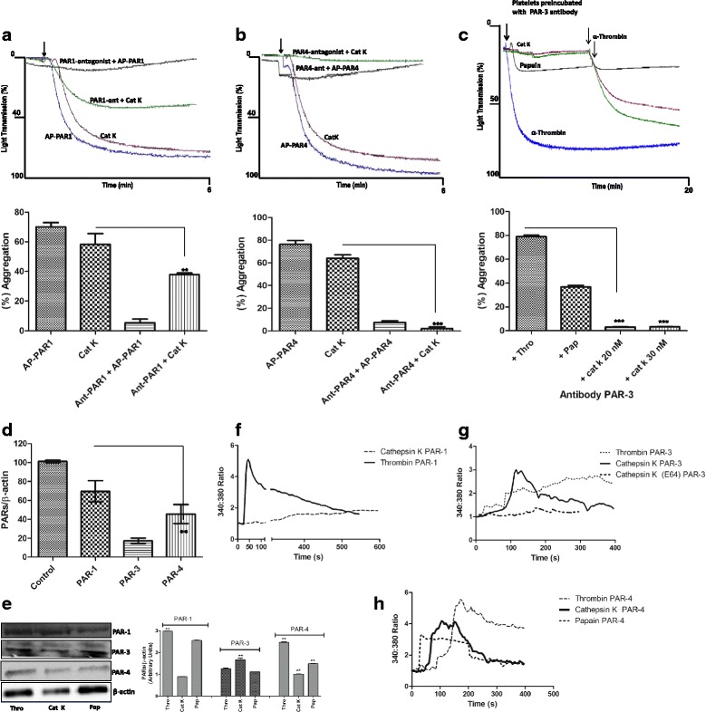
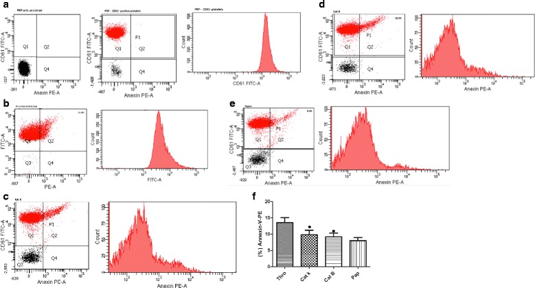
Results: We demonstrate that platelets are aggregated by cathepsin K in a dose-dependent manner, but not by other cysteine cathepsins. PARs-3 and -4 were confirmed as the cathepsin K target by immunodetection and specific antagonists using a fibroblast cell line derived from PARs deficient mice. Moreover, through co-culture experiments, we show that platelets activated by cathepsin K mediated the up-regulation of SHH, PTHrP, OPN, and TGFβ in epithelial-mesenchymal-like cells from patients with Luminal B breast cancer.

Conclusions: Cathepsin K induces platelet dysfunction and affects signaling in breast cancer cells.


Figures

Fig. 1
Fig. 1
Cat K-induced human platelet aggregation. Dose-response curve and effect of LWMK, HWMK, and E-64 under cat K activity. The washed human platelets (3.0 x 10⁵/mL) function was measured at baseline and stimulus was added after 30 s. (a) Effect of cat K (20 nM) and papain (1.6 µM) on platelet aggregation. (b) Negative effect of cathepsins L, V, S, and B (at a median effective dose - ED50 - of 0.2 µM) on platelets showing typical tracing results, α-thrombin (1.0 UNIH/500 µL) was used as the agonist control. (c) The bar graph shows percentage of aggregation. Data are presented as means ± SEM (***p<0.0001). Traces show one typical experiment out of at least six. (d) Solutions containing different concentrations of cat K (2.5 to 30 nM) were added to platelets and aggregation was measured as described. Representative traces showing the typical aggregation induced by cat K at different concentrations and α-thrombin (1.0 UNIH/500 µL). (e) Concentration-response bar graph expressed as percentage of aggregation (***p<0.001). (f) Clot aggregate analyses of human platelets treated with cat K (20 nM) and α-thrombin (1.0 UNIH/500 µL). (g) Inhibition of human cat Kactivity (black line) and papain (blue line) on human platelet aggregation by E-64 (5 µM), LWMK (1.0 µM) + HWMK (1.0 µM)
Fig. 2
Fig. 2
The influence of cat K (20 nM), papain (1.6 µM), and cat L (0.2 µM) on the effect of α-thrombin. (a) The results show platelet aggregation induced by cat K (20 nM) followed by α-thrombin stimulation (1.0 UNIH/500 µL) after 2 min. α-Thrombin stimulation was slightly prolonged (black line). Cat K does not interfer with α-thrombin activity on platelets aggregation (blue line). Data are expressed as mean ± SEM from 6 independent experiments (*p<0.05). (b) Papain (1.6 µM) followed by α-thrombin stimulation (1.0 UNIH/500 µL) (blue line). The inverse stimulation (α-thrombin followed by papain (1.6 µM) and cat L (0.2 µM)), (black and orange lines). The bar graph shows percentage of aggregation expressed as mean ± SEM from 3 independent experiments (**p<0.01, ***p<0.001). (c) Representative traces showing typical aggregation induced by cat K (20 nM), papain (1.6 µM), and α- thrombin (1.0 UNIH/500 µL). The arrows indicate when agonists were added. (d) Stimulation by papain (1.6 µM) followed by cat K (20 nM), red line. Inverse cat K stimulation (20 nM) followed by papain (1.6 µM), black line. (e) The bar graph shows percentages of aggregation. Data are expressed as mean ± SEM from 3 independent experiments (*p<0.05)
Fig. 3
Fig. 3
Effect of PAR-1 antagonist (SCH 79797), PAR-4 antagonist (transcinnamoyl- YPGKF-NH2), and PAR-3 antibody on cat K-induced platelet aggregation and intracellular calcium measurements. (a) Human platelets were preincubated with the specific PAR-1 SCH 79797 antagonist (140 nM) for 30 min at 37 °C and evaluated in the aggregometer. Representative traces show the interference of SCH 79797 on cat K-induced platelet aggregation. AP-PAR1 (0.2 µM) was completely inhibited. (b) The specific PAR-4 trans-cinnamoyl-YPGKF-NH2 antagonist (30 µM) inhibits human platelet aggregation induced by cat K (20 nM). AP-PAR-4 (60 µM) was used as control. (c) Inhibitory effect of the PAR-3 antibody (0.02 µM) on cat Kinduced human platelet aggregation. Platelets were preincubated with the PAR-3 antibody for 30 min at 37 °C and aggregation was measured. Representative traces show the inhibition of the cat K effect (red and green lines). Papain-induced human platelet aggregation inhibition, black line. The bar graph shows percentages of aggregation. Data are expressed as mean ± SEM from 3 independent experiments (**p<0.001, *** p<0.0001). (d) Reverse transcriptase-PCR using total platelet RNA. Error bars indicate S.D. from triplicate samples. **p <0.01. (e). PAR-1, -3, and -4 were detected in washed platelets treated with cat K (20 nM), papain (1.6 µM), and α-thrombin (0.001 UNIH/500 µL) for 10 min at 37 °C; lysate proteins were separated by 10% SDS-PAGE and electrotransferred to nitrocellulose membrane. Membranes were blocked and incubated with anti-PAR1, anti-PAR3, anti-PAR4 rabbit primary antibodies, and anti-ß-actin (control). Antibody binding was visualized by chemiluminescence and the relative levels of these proteins were determined by densitometric analysis. Data are expressed as mean ± SEM from 3 independent experiments (**p<0.001). (f) Intracellular calcium measurements in ThrR -/- fibroblasts (absence in PAR-1). Increases in cytosolic calcium in response to cat K (20 nM) and α-thrombin (0.001 UNIH/500 µL). (g) ThrR -/- fibroblasts (absence in PAR-3). Increases in cytosolic calcium in response to cat K (20 nM) and α-thrombin (0.001 UNIH/500 µL). Inhibition of human cat K-activity on human platelet aggregation by E-64 (5 µM). (h) ThrR -/- fibroblasts (absence in PAR-4). Increases in cytosolic calcium in response to cat K (20 nM), papain (1.6 µM), and α-thrombin (0.001 UNIH/500 µL)
Fig. 4
Fig. 4
Detection of signaling phosphoproteins by Immunoblot analysis and Ca2+ release into human platelets. (a) p-Src family, (b) p-FAK, and (c) p-p38 washed platelets were treated with cat K (20 nM), papain (1.6 µM), and α-thrombin (0.001 UNIH/500µL) for 10 min at 37 °C; lysate proteins were separated by 10 % SDS-PAGE and electrotransferred to nitrocellulose membranes. Membranes were blocked and incubated with rabbit primary antibodies, anti-phospho-Src (Tyr-416), anti-Src, anti-phospho-FAK (Tyr-397), anti-FAK, anti- phospho-p38 MAPK, anti-p38 MAPK, and anti-ß-actin. Antibody binding was visualized by chemiluminescence and the relative levels of these proteins were determined by densitometric analysis. Papain (1.6 µM) slightly induced Src- and FAK-phosphorylation (2.1 ± 0.2 and 2.5 ± 0.4, respectively). Treatment with α-thrombin resulted in increased Src phosphorylation with the pSrc/total Src ratio of 6.2 ± 0.4, and increased FAK phosphorylation with the pFAK/total FAK ratio of 6.3 ± 0.2. The use of PAR-4 antagonist and PAR-3 antibody eliminated the Src Tyr416 phosphorylation by cat K (unpublished data). The phosphorylation of p38 was required when human platelets were stimulated with cat K. Pre-treatment of platelets with PAR-3 antibody and PAR-4 antagonist abolished the Src-Tyr-416 and FAKTyr- 397 phosphorylation. Data are expressed as mean ± SEM from 3 independent experiments (*p<0.01,**p<0.001, ***p<0.0001). See also Fig. 1a. (d) The [Ca2+]i mobilization was measured as described. The arrow indicates when α-thrombin was added to platelets (around 100 s). The washed platelets responded strongly with a transient rise in [Ca2+]i. (e) Cat K (20 nM) shows a calcium spike followed by a sustained elevation in the fluorescence ratio. The presence of α-thrombin (1.0 UNIH/500µL) led to an increase in [Ca2+]i indicating that the platelets induced Ca2+ release. (f) The papain (1.6 µM) response was similar to that from cat K, however, no substantial rise in [Ca2+]i was detected with the addition of α-thrombin. (g) Cat K did not block the papain effect. Sustained elevation in the fluorescence ratio was observed. (h) Cat L (0.2 µM) does not induce a detectable effect on Ca2+ mobilization; α-thrombin (1.0 UNIH/500µL) showed a strong response with a transient rise in [Ca2+]i. Platelets were treated with indomethacin (0.1 mg/mL) to prevent thromboxane synthesis, which may also be affected by PKC [Ca2+]. The mesurements were monitored by Fluo-4/AM (4 µM) intensity in a laser scanning confocal miscroscopy; scale bar = 10 µm
Fig. 5
Fig. 5
Human platelet activation by cat K. Side and forward scatters of plasma rich in platelets (PRP) from different blood donors prepared under the same conditions. PRP were stained with lineage markers: CD61-FITC (platelets) and Annexin V-PE (phosphatidyserine exposure) or the appropriate isotype controls. PRP was analyzed by flow cytometry. Platelets were treated with apyrase (5.0 UNIH/mL). The areas Q1 and Q2 correspond to nonactivated and activated platelets, respectively. (a) Forward - by sidescatter profiles of events in PRP. Populations identified by futher gating. CD61+ nonactivated platelets. Representative histogram of CD61+ platelet resident in PRP. (b) α-thrombin (0.001UNIH/mL), (c) Platelets activated by cat K (20 nM), (d) cat B (0.2 µM), and (e) papain (1.6 µM), and their corresponding histograms. Note the increased number of events in Q2 after stimulation with cat K, papain, and α-thrombin. Flow cytometric quantification of Annexin V-PE (beads were used for size calibration). Each figure represents an analysis of 10,000 events with SSC (side scatter) on the abscissa, and FITC fluorescence intensities on the ordinate. (f) The bar graph shows percentages of Annexin-V-PE. Data are expressed as mean ± SEM from 3 independent experiments (*p<0.05). See also Fig. 1 and 3
Fig. 6
Fig. 6
Immunophenotype of breast cancer cells, from patients with Luminal B subtype, used in all experiments. Cells are shown under phase contrast microscopy and indirect immunofluorescence for vimentin, cytokeratin, Ecadherin, N-cadherin, PAI-1, claudin 1, phalloidin, and DAPI (blue, for nuclei). (a) Phase contrast–confluent culture of tumor cells with EMT (epithelialmesenchymal- transition) after 3 days. (b-e) Analysis of epithelial and mesenchymal markers by confocal microscopy; cytokeratin, vimentin, Ecadherin, and N-cadherin, respectively. (f and g) Representative flow cytometry histograms of PAI-1 and claudin 1 on tumor cells with EMT. The histogram on the left represents a control staining using an isotype-matched control antibody. (h) Zymography for MMP-9, in conditioned medium, in EMT and epithelial cells isolated from women with breast cancer. (i) Phase contrast— epithelial cells confluent culture after 2 days. (j and k) Analysis of epithelial marker cytokeratin. We observed the cytoskeletal organization pattern when using phalloidin. (l) The expression of cat K is up-regulated in co-cultured breast cancer cells and platelets. Cat K was assessed by immunoblotting. (m) Breast cancer cells in epithelial-mesenchimal transition co-cultured with platelets and activated by cat K showed up-regultation of SHH, PTHrP, OPN, and TGF-ß expression, and increased phospho-Src. The graph represents the densitometric analyses from the immunoblotting results. The results are represented as band intensities in arbitrary units relative to the respective total loading control (ß-actin). (n) The flow cytometry results are reported as percentages of CD44 human-specific antibody in breast cancer cells, and P-selectin in human platelets activated by cat K after exposure to breast cancer cells. See also Fig. 2
Fig. 7
Fig. 7
Hypothesized molecular coordination between Cat-K-induced platelet aggregation and crosstalk with epithelial-mesenchimal-like cells from patients with breast cancer. The activation and release of cat K occurs in epithelial-mesenchimal transition cells in breast cancer subtype Luminal B. (a) Breast cancer cells cultured with human platelets and activated by cat K and α-thrombin. Phase contrast images of epithelial-mesenchimal-like cells co-cultured with platelets activated by cat K for 24 h. (b) Cat K may activate PAR-3 and PAR-4 in human platelets, by receptor cleavage, and trigger platelet aggregation. Activated PAR-3 and PAR-4 can perpetuate the cohesion of tumor cells during heteroaggregation with increase in P-selectin and CD44. The crosstalk between platelets activated by cat K and epithelialmesenchimal tumor cells form microaggreates and promote up-regulation of Hedgehog ligands and growth factors such as SHH, OPN, PTHrP, and TGFß. (c) Mammary stroma (1), intravasation (2), cat K secretion by epithelialmesenchimal tumor cells (3), and activate human platelets that could directly affect the expression of the ligands of Hedgehog signaling, reported as an aberrantly activated pathway in breast cancer and related to bone metastasis markers

Similar articles

Cited by

References

    1. Mohamed MM, Sloane BF. Cysteine cathepsins: multifunctional enzymes in cancer. Nat Rev Cancer. 2006; doi:10.1038/nrc1949. - PubMed
    1. Brix K, Dunkhorst A, Mayer K, Jordans S. Cysteine cathepsins: cellular roadmap to different functions. Biochimie. 2008;90:194–207. doi: 10.1016/j.biochi.2007.07.024. - DOI - PubMed
    1. Valastyan S, Weinberg RA. Tumor metastasis: molecular insights and evolving paradigms. Cell. 2011;147:275–92. doi: 10.1016/j.cell.2011.09.024. - DOI - PMC - PubMed
    1. Turk V, Stoka V, Vasiljeva O, Renko M, Sun T, Turk B, Turk D. Cysteine cathepsins: from structure, function and regulation to new frontiers. Biochim Biophys Acta. 2012; doi: 10.1016/j.bbapap.2011.10.002. - PMC - PubMed
    1. Sanman LE, Bogyo M. Activity-based profiling of proteases. Annu Rev Biochem. 2014;83:249–73. doi: 10.1146/annurev-biochem-060713-035352. - DOI - PubMed

Publication types

MeSH terms