. 2016 May 1;25(9):1846-56.
doi: 10.1093/hmg/ddw059. Epub 2016 Feb 29.

Missense variants in the middle domain of DNM1L in cases of infantile encephalopathy alter peroxisomes and mitochondria when assayed in Drosophila

Affiliations

Missense variants in the middle domain of DNM1L in cases of infantile encephalopathy alter peroxisomes and mitochondria when assayed in Drosophila

Yu-Hsin Chao et al. Hum Mol Genet. .

Abstract

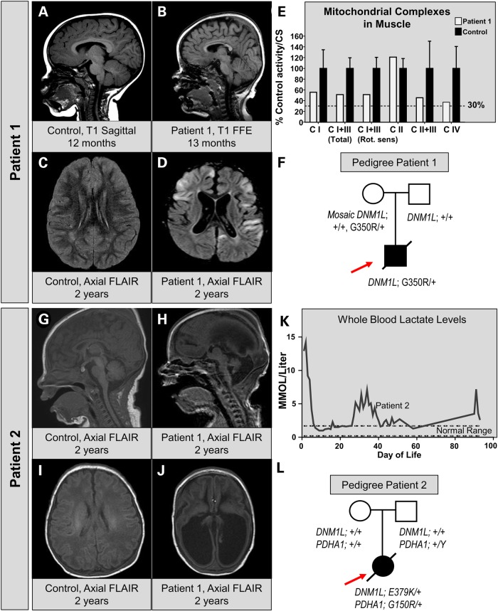
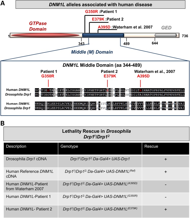
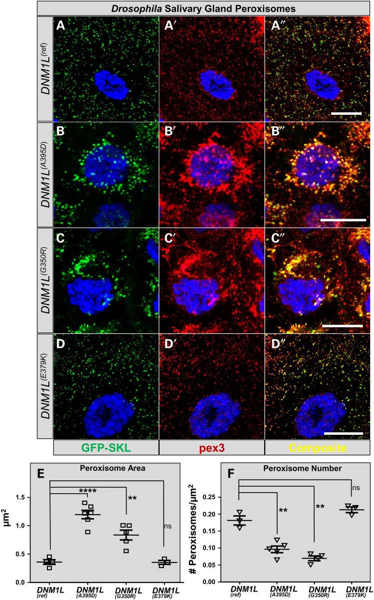
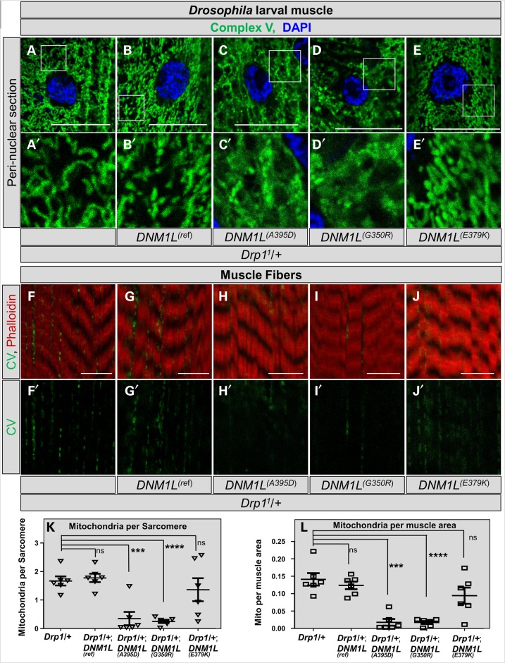
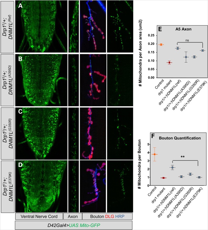
Defects in organelle dynamics underlie a number of human degenerative disorders, and whole exome sequencing (WES) is a powerful tool for studying genetic changes that affect the cellular machinery. WES may uncover variants of unknown significance (VUS) that require functional validation. Previously, a pathogenic de novo variant in the middle domain of DNM1L (p.A395D) was identified in a single patient with a lethal defect of mitochondrial and peroxisomal fission. We identified two additional patients with infantile encephalopathy and partially overlapping clinical features, each with a novel VUS in the middle domain of DNM1L (p.G350R and p.E379K). To evaluate pathogenicity, we generated transgenic Drosophila expressing wild-type or variant DNM1L. We find that human wild-type DNM1L rescues the lethality as well as specific phenotypes associated with the loss of Drp1 in Drosophila. Neither the p.A395D variant nor the novel variant p.G350R rescue lethality or other phenotypes. Moreover, overexpression of p.A395D and p.G350R in Drosophila neurons, salivary gland and muscle strikingly altered peroxisomal and mitochondrial morphology. In contrast, the other novel variant (p.E379K) rescued lethality and did not affect organelle morphology, although it was associated with a subtle mitochondrial trafficking defect in an in vivo assay. Interestingly, the patient with the p.E379K variant also has a de novo VUS in pyruvate dehydrogenase 1 (PDHA1) affecting the same amino acid (G150) as another case of PDHA1 deficiency suggesting the PDHA1 variant may be pathogenic. In summary, detailed clinical evaluation and WES with functional studies in Drosophila can distinguish different functional consequences of newly-described DNM1L alleles.


Figures

Figure 1.
Figure 1.
Clinical, neuroradiographic and molecular features of two patients with infantile encephalopathy and DNM1L variants. (A) MRI of a control patient at 12 months of age showing normal sagittal T1-weighted images. (B) MRI of Patient 1 at 13 months of age reveals hypoplasia of the corpus callosum and a simplified gyral pattern on T1 sagittal images. (C) MRI of control patient for suspected seizure showing a normal axial FLAIR sequence. (D) Axial FLAIR of Patient 1 at 2 years of age showing multi-focal areas of cortical signal abnormality with swelling and diffusion restriction. Diffuse cortical volume loss is also present. (E) Respiratory chain enzyme activities in muscle. The % control activity for Patient 1 (white bars) is shown for NADH:Ferricyanide dehydrogenase (CI), NADH:cytochrome C reductase total (CI+III), NADH:cytochrome C reductase Rotenone sensitive (CI+III), Succinate dehydrogenase (CII) and Cytochrome C oxidase (CIV). Values were normalized to citrate synthase activity. (F) Pedigree of the patient showing the WES data. Sanger verification was performed and low-level (6–8%) mosaicism was detected in the maternal sample. (G and I) A sagittal MRI in a newborn showing a normal T1 and FLAIR appearance. (H and J) MRI of Patient 2 at 4 days of age reveals ventriculomegaly, absence of the corpus callosum, volume loss and gyral simplification on T1 and FLAIR. (K) Whole blood lactate levels measured in a clinical lab over the time course of Patient 2's medical care at our institution. (L) Pedigree of Patient 2 showing the presence of two independent de novo events, one in DNM1L and one in PDHA1.
Figure 2.
Figure 2.
Genetic and domain associations for the DNM1L alleles. (A) DNM1L encodes a protein with an N-terminal GTPase domain, a middle domain and a C-terminal GTPase effector domain (GED). The three variants are missense alleles in the middle domain. Alignment shows homology between human DNM1L and Drosophila Drp1, red lines indicate the three human variants, while the A395D and G350R variants occur in highly conserved residues, the E379K is not conserved. (B) Lethality rescue experiments in Drosophila with human DNM1L alleles. The genotypes are listed as second chromosome; third chromosome. Rescue of lethality is indicated by (+).
Figure 3.
Figure 3.
Dominant effects of DNM1L expression on Drosophila salivary gland peroxisomes. (AA″) Drosophila salivary gland cells are shown with expression of the reference DNM1L with Actin-Gal4 alongside UAS-EGFP-SKL (courtesy of Hamed Jefar-Nejad) and co-stained with Drosophila anti-Pex3 antibody produces salivary gland cells that are indistinguishable from GFP-SKL expression alone (data not shown). (B–B″) The p.A395D construct (seen in Waterham et al. (23)) produces enlarged peroxisomes with abnormal distribution. Fewer peroxisomes are apparent in the cell. (CC″) The p.G350R construct (seen in Patient 1) produces enlarged peroxisomes with abnormal distribution; fewer peroxisomes are apparent in the cell, a phenotype similar to pA395D. (DD″) The p.E379K construct (Patient 2) does not appear to have a dramatic effect on peroxisomal size compared with DNM1L(Ref). (E) Quantification of the peroxisomal area per peroxisome. In cells expressing the DNM1L(Ref) peroxisomes have an average size of 0.3 µm2. (F) Quantification of peroxisomal number per µm2 of cytoplasm. Error bars = 25 µm2.
Figure 4.
Figure 4.
Drosophila larval muscle mitochondrial size and number. (AA′) Control third instar larval muscle stained with Complex V and DAPI and imaged at the level of the nucleus. Control mitochondria have clear separation and fibrillar morphology. (BB′) Mitochondria from DNM1L(Ref) expressed by MEF2-Gal4, a muscle-specific enhancer, showing normal morphology. (C–C′, D–D′) Mitochondria from DNM1L(A395D) and DNM1L(G350R) expressed by MEF2-Gal4 showing mats of inter-connected mitochondria. (EE′) Mitochondria from DNM1L(E379K) expressed by MEF2-Gal4 showing clear normal morphology. (F–F′) Control third instar larval muscle stained with Complex V and Phalloidin and imaged at the level of the muscle fibers. Control mitochondria are clearly present intercalating between the fibers. (G–G′) Mitochondria from DNM1L(Ref) intercalate between muscle fibers. (HH′, II′) DNM1L(A395D) and DNM1L(G350R) show near absence of mitochondria. (JJ′) DNM1LE379K) shows normal numbers of mitochondria. (KK′) Quantification of number of mitochondria per sarcomere from experiments shown in (F)–(J). (K–K′) Quantification of mitochondria per muscle area from experiments shown in (F)–(J).
Figure 5.
Figure 5.
Effect of DNM1L variants on mitochondrial brain trafficking. (A) Left, Drosophila larval brain ventral nerve cord on with normal mitochondrial distribution. DNM1L(Ref) was expressed by D42-Gal4 in a sensitized background (Drp11/+). Center, mitochondrial trafficking in the axon at the A5 abdominal segment. Right, mitochondria in the synaptic bouton counterstained with HRP (blue) and Discs Large (DLG, red). (B and C) Expression of DNM1L(A395D) and DNM1L(G350R) dramatically alter distribution of mitochondria with a defect in trafficking mitochondria. (D) A defect in mitochondrial trafficking observed in the synaptic boutons of DNM1L(E379K)-expressing larvae. (E) Quantification of number of mitochondria per axon area for A5 axonal segments for the experiment shown in (A)–(D). (F) Quantification of number of mitochondrial per synaptic bouton for the experiment shown in (A)–(D).

References

    1. DiMauro S., Schon E.A., Carelli V., Hirano M. (2013) The clinical maze of mitochondrial neurology. Nat. Rev. Neurol., 9, 429–444. - PMC - PubMed
    1. Leonard J.V., Schapira A.H. (2000) Mitochondrial respiratory chain disorders I: mitochondrial DNA defects. Lancet, 355, 299–304. - PubMed
    1. Leonard J.V., Schapira A.H. (2000) Mitochondrial respiratory chain disorders II: neurodegenerative disorders and nuclear gene defects. Lancet, 355, 389–394. - PubMed
    1. Lombes A., Aure K., Bellanne-Chantelot C., Gilleron M., Jardel C. (2014) Unsolved issues related to human mitochondrial diseases. Biochimie, 100, 171–176. - PubMed
    1. Tuppen H.A., Blakely E.L., Turnbull D.M., Taylor R.W. (2010) Mitochondrial DNA mutations and human disease. Biochim. Biophys. Acta, 1797, 113–128. - PubMed

MeSH terms

Supplementary concepts

LinkOut - more resources