. 2016 Mar 2:6:22311.
doi: 10.1038/srep22311.

Sequence evidence for common ancestry of eukaryotic endomembrane coatomers

Affiliations

Sequence evidence for common ancestry of eukaryotic endomembrane coatomers

Vasilis J Promponas et al. Sci Rep. .

Abstract

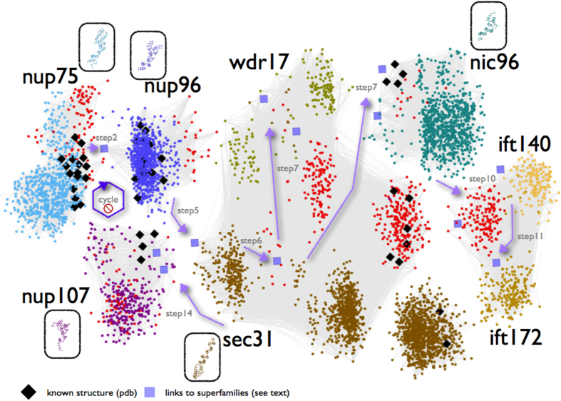
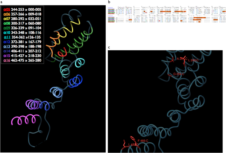
Eukaryotic cells are defined by compartments through which the trafficking of macromolecules is mediated by large complexes, such as the nuclear pore, transport vesicles and intraflagellar transport. The assembly and maintenance of these complexes is facilitated by endomembrane coatomers, long suspected to be divergently related on the basis of structural and more recently phylogenomic analysis. By performing supervised walks in sequence space across coatomer superfamilies, we uncover subtle sequence patterns that have remained elusive to date, ultimately unifying eukaryotic coatomers by divergent evolution. The conserved residues shared by 3,502 endomembrane coatomer components are mapped onto the solenoid superhelix of nucleoporin and COPII protein structures, thus determining the invariant elements of coatomer architecture. This ancient structural motif can be considered as a universal signature connecting eukaryotic coatomers involved in multiple cellular processes across cell physiology and human disease.


Figures

Figure 1
Figure 1. Pictorial representation of the sequence space walk connecting components of the nuclear pore complex, COPII and intraflagellar transport.
Members of each of the eight superfamilies are shown in distinct colors, with superfamily representatives of known structure enclosed in oval boxes following the same coloring scheme. Orientations of structural representatives are identical to the reference Nup75 structure (3F3F_C, Figure 2a). Two exceptions of the coloring scheme involve homologs of known structure (black diamonds-♦) and previously uncharacterized (unannotated) protein sequences (red dots-●). Intra-family connections detectable by pairwise sequence comparisons are represented by dense sub-networks of thin light-grey lines (see Methods). Inter-family connections revealed by iterative profile searches – otherwise undetectable, are depicted by (i) light purple arrows for the corresponding steps and (ii) squares for sequence links across superfamilies. Seven steps of the sequence space walk deemed as critical for revealing novel inter-family relationships are displayed (step 7 is shown twice, as it connects both to WDR17 and Nic96 superfamilies) – see also Table S2. The cycle with a ‘stop’ sign refers to the exclusion of Nup107 superfamily which terminates the search early (see Methods). Only a representative subset of hits is shown for clarity; this representation should only be taken as a rough sketch of the documented process (available as Data Supplement DS05). An annotated version of this two-dimensional layout is available as Data Supplement DS11.
Figure 2
Figure 2. Sequence conservation across endomembrane coatomer structure components.
(a) The Nup75 structure (3F3F_C) corresponding to the detected ACE1-like alpha-solenoid superhelix motif is shown. Individual helices α5-α16 are colored by unique colors, warm colors representing the most conserved segments (α5-α6, α15-α16). Sequence positions for each helix are shown in the legend, according to the comparative structural analysis of ACE1, followed by the corresponding positions of the alignment in Fig. 2b. This reference orientation is used throughout this work, corresponding to the crown and the second half of the ACE1 trunk – see also Fig. 5 in Ref. . (b) Sequence alignment of five alpha-solenoid superhelix motif-containing representative structures. Aligned positions are established by direct comparison of the KMAP-13 profile against the structure database, edited to match the reference Nup75 structure, using Mview. PDB codes are given, followed by the description of the corresponding protein chains. A consensus sequence and a skyline conservation plot are provided. The twelve helices listed in Fig. 2a are depicted as horizontal oval-shaped bars marked for the consensus sequence, seven at the top panel and the remaining five at the bottom panel. The total length of the alignment is 280 residues, corresponding to the KMAP-13 profile search against the common conserved elements of the available structures. Regions missing in the structure database entries are shown in orange. Structural data in PDB format are provided in Data Supplement DS09. (c) Structural context of the evolutionarily conserved positions in Nup75. N-terminal Leu-249, Gly-256 and Ala-261 (C for chain C of 3F3F) (see Fig. 2a) and C-terminal Phe-469 and Leu-473 correspond to alignment positions I1, G8, A13 and F272, L278 respectively (Fig. 2b). The C-alpha trace is shown in dark blue and the subset of conserved positions is shown in red, along with the side chain representations. Alignment position L101 is not highlighted as it is frequently substituted by a tyrosine residue (Fig. 2b).

Similar articles

Cited by

References

    1. Grossman E., Medalia O. & Zwerger M. Functional architecture of the nuclear pore complex. Annu Rev Biophys 41, 557–84 (2012). - PubMed
    1. DeGrasse J. A. et al. Evidence for a shared nuclear pore complex architecture that is conserved from the last common eukaryotic ancestor. Mol Cell Proteomics 8, 2119–30 (2009). - PMC - PubMed
    1. Devos D. et al. Simple fold composition and modular architecture of the nuclear pore complex. Proc Natl Acad Sci USA 103, 2172–7 (2006). - PMC - PubMed
    1. Stuwe T. et al. Architecture of the nuclear pore complex coat. Science 347, 1148–52 (2015). - PMC - PubMed
    1. Devos D. et al. Components of coated vesicles and nuclear pore complexes share a common molecular architecture. PLoS Biol 2, e380 (2004). - PMC - PubMed

Publication types

Substances

LinkOut - more resources