

An introduction to sample preparation and imaging by cryo-electron microscopy for structural biology
- PMID: 26931652
- PMCID: PMC4854231
- DOI: 10.1016/j.ymeth.2016.02.017
An introduction to sample preparation and imaging by cryo-electron microscopy for structural biology
Abstract
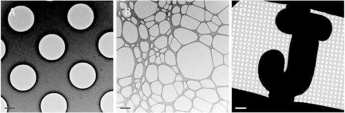
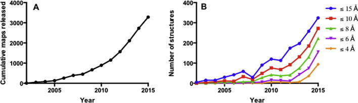
Transmission electron microscopy (EM) is a versatile technique that can be used to image biological specimens ranging from intact eukaryotic cells to individual proteins >150kDa. There are several strategies for preparing samples for imaging by EM, including negative staining and cryogenic freezing. In the last few years, cryo-EM has undergone a 'resolution revolution', owing to both advances in imaging hardware, image processing software, and improvements in sample preparation, leading to growing number of researchers using cryo-EM as a research tool. However, cryo-EM is still a rapidly growing field, with unique challenges. Here, we summarise considerations for imaging of a range of specimens from macromolecular complexes to cells using EM.
Keywords: Electron microscopy.
Copyright © 2016 The Authors. Published by Elsevier Inc. All rights reserved.
Figures
References
-
- Lau W.C.Y., Rubinstein J.L. Single particle electron microscopy. Methods Mol. Biol. 2013;955:401–426. - PubMed
-
- Briggs J.A.G. Structural biology in situ – the potential of subtomogram averaging. Curr. Opin. Struct. Biol. 2013;23:261–267. - PubMed
-
- Lucic V., Forster F., Baumeister W. Structural studies by electron tomography: from cells to molecules. Annu. Rev. Biochem. 2005;74:833–865. - PubMed
Publication types
MeSH terms
Substances
Grants and funding
LinkOut - more resources
Full Text Sources
Other Literature Sources
Medical
