

Comparative transcriptome analysis of muscular dystrophy models Large(myd), Dmd(mdx)/Large(myd) and Dmd(mdx): what makes them different?
- PMID: 26932192
- PMCID: PMC4989205
- DOI: 10.1038/ejhg.2016.16
Comparative transcriptome analysis of muscular dystrophy models Large(myd), Dmd(mdx)/Large(myd) and Dmd(mdx): what makes them different?
Abstract
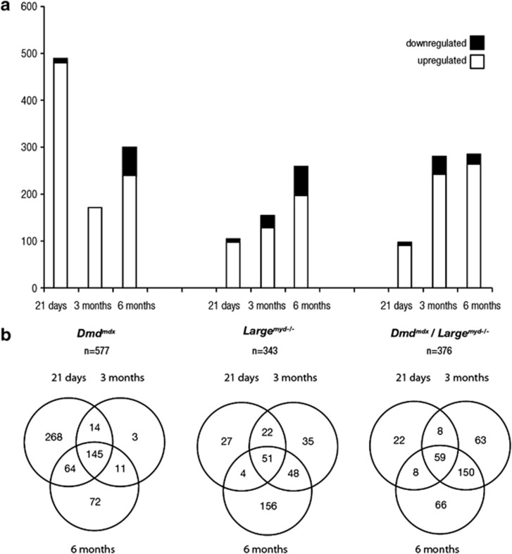
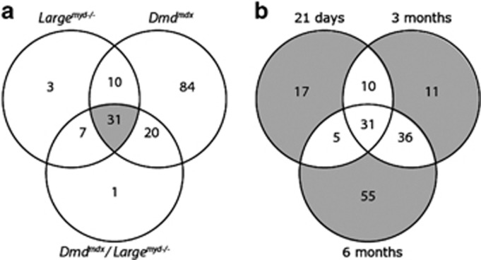
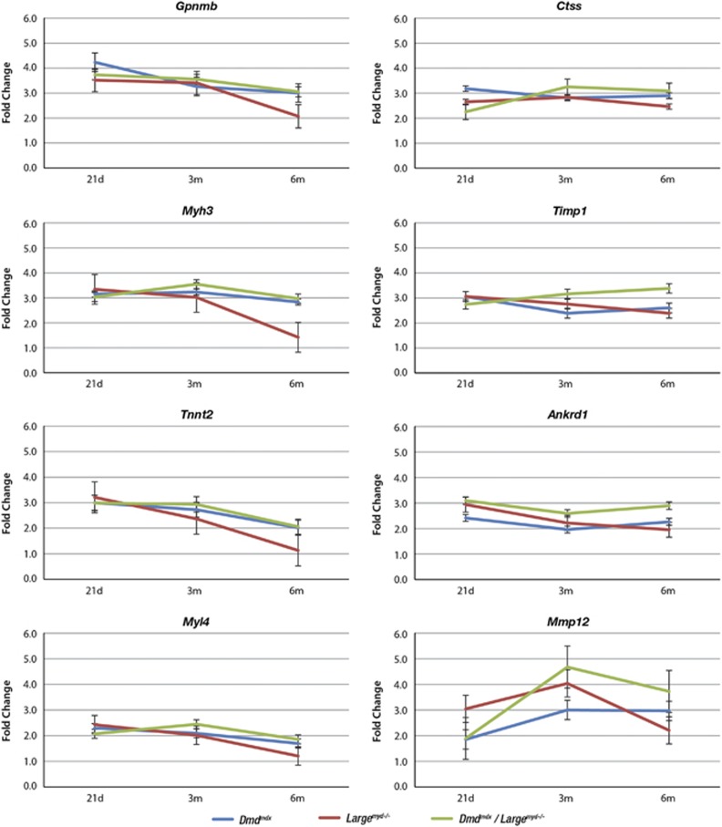
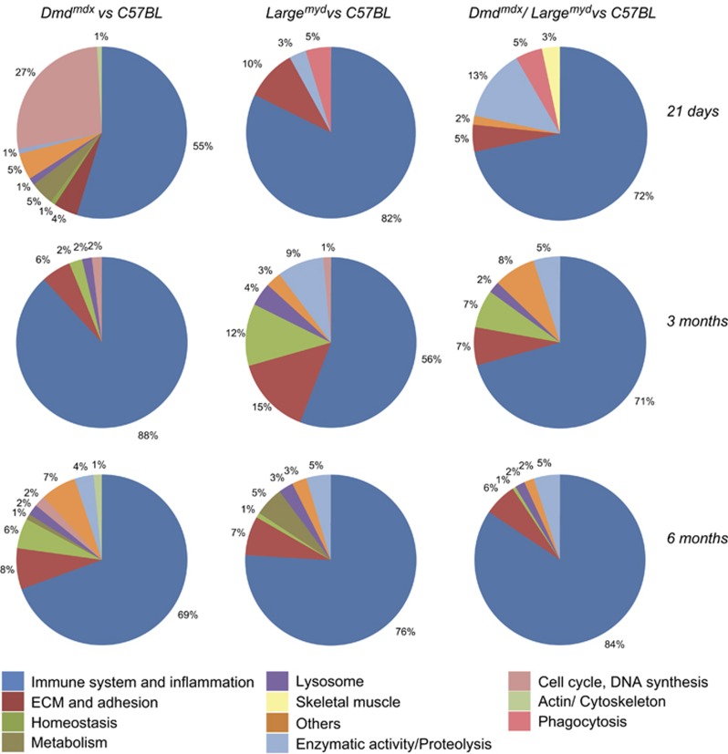
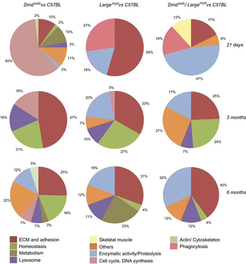
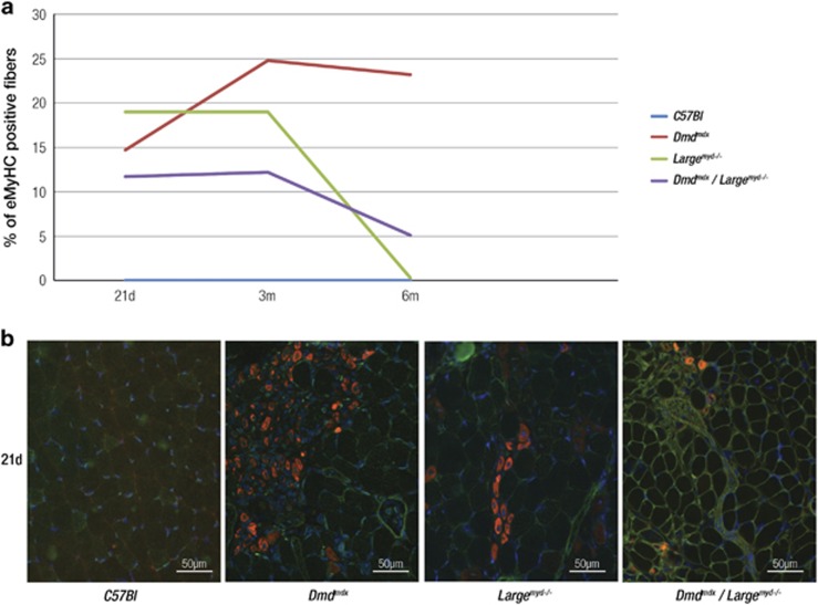
Muscular dystrophies (MD) are a clinically and genetically heterogeneous group of Mendelian diseases. The underlying pathophysiology and phenotypic variability in each form are much more complex, suggesting the involvement of many other genes. Thus, here we studied the whole genome expression profile in muscles from three mice models for MD, at different time points: Dmd(mdx) (mutation in dystrophin gene), Large(myd-/-) (mutation in Large) and Dmd(mdx)/Large(myd-/-) (both mutations). The identification of altered biological functions can contribute to understand diseases and to find prognostic biomarkers and points for therapeutic intervention. We identified a substantial number of differentially expressed genes (DEGs) in each model, reflecting diseases' complexity. The main biological process affected in the three strains was immune system, accounting for the majority of enriched functional categories, followed by degeneration/regeneration and extracellular matrix remodeling processes. The most notable differences were in 21-day-old Dmd(mdx), with a high proportion of DEGs related to its regenerative capacity. A higher number of positive embryonic myosin heavy chain (eMyHC) fibers confirmed this. The new Dmd(mdx)/Large(myd-/-) model did not show a highly different transcriptome from the parental lineages, with a profile closer to Large(myd-/-), but not bearing the same regenerative potential as Dmd(mdx). This is the first report about transcriptome profile of a mouse model for congenital MD and Dmd(mdx)/Large(myd). By comparing the studied profiles, we conclude that alterations in biological functions due to the dystrophic process are very similar, and that the intense regeneration in Dmd(mdx) involves a large number of activated genes, not differentially expressed in the other two strains.
Figures
References
-
- Flanigan KM: The muscular dystrophies. Semin Neurol 2012; 32: 255–263. - PubMed
-
- Hoffman EP, Brown RH, Kunkel LM: Dystrophin: the protein product of the Duchenne muscular dystrophy locus. Cell 1987; 51: 919–928. - PubMed
-
- Pastoret C, Sebille A: mdx mice show progressive weakness and muscle deterioration with age. J Neurol Sci 1995; 129: 97–105. - PubMed
-
- Moore CJ, Hewitt JE: Dystroglycan glycosylation and muscular dystrophy. Glycoconj J 2009; 26: 349–357. - PubMed
MeSH terms
Substances
LinkOut - more resources
Full Text Sources
Other Literature Sources
Medical
Molecular Biology Databases
