. 2016 Mar 2:7:10806.
doi: 10.1038/ncomms10806.

Biochemical reconstitution of TET1-TDG-BER-dependent active DNA demethylation reveals a highly coordinated mechanism

Affiliations

Biochemical reconstitution of TET1-TDG-BER-dependent active DNA demethylation reveals a highly coordinated mechanism

Alain R Weber et al. Nat Commun. .

Abstract

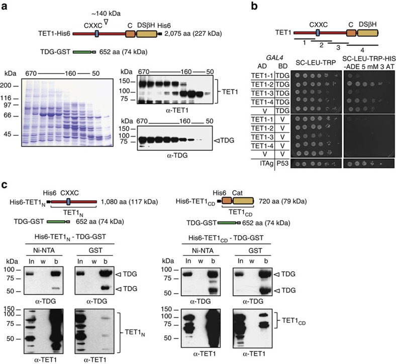
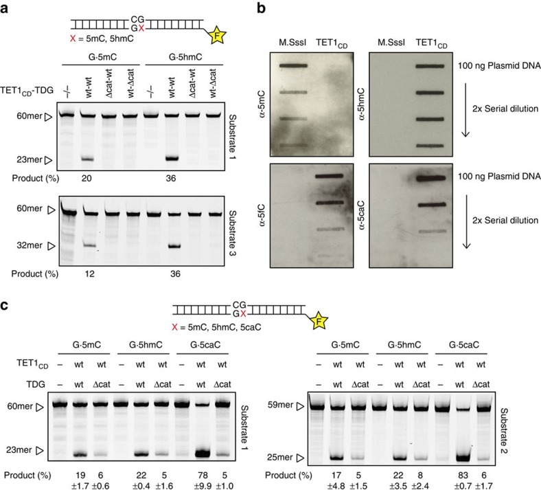
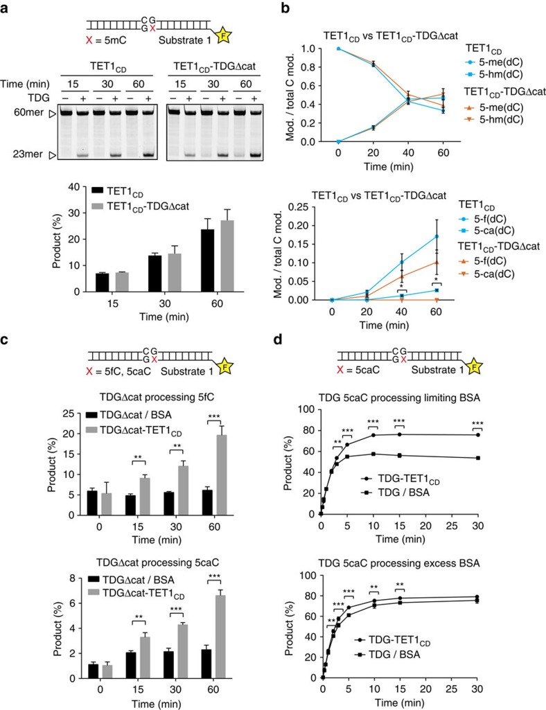
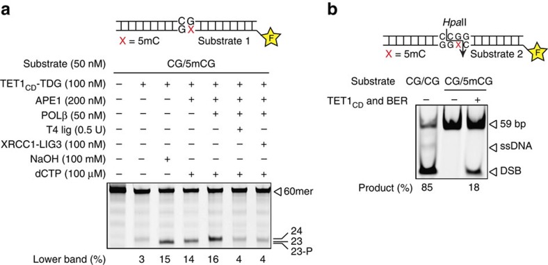
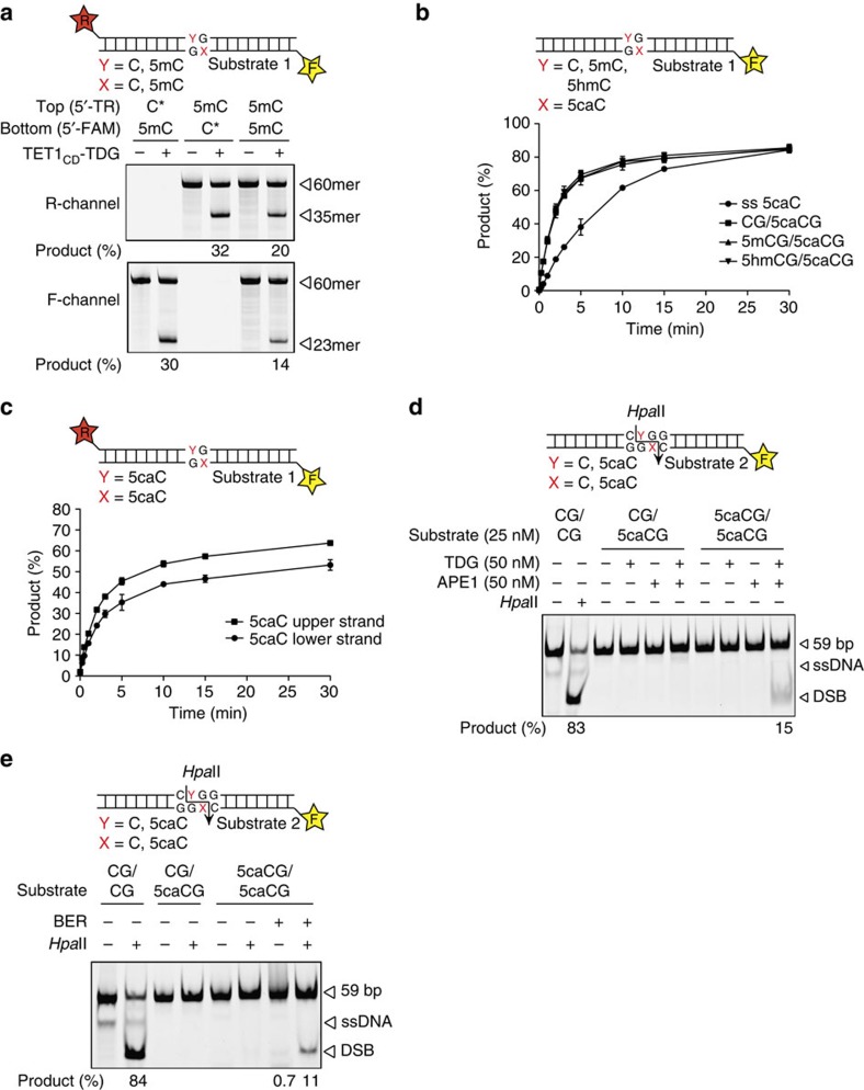
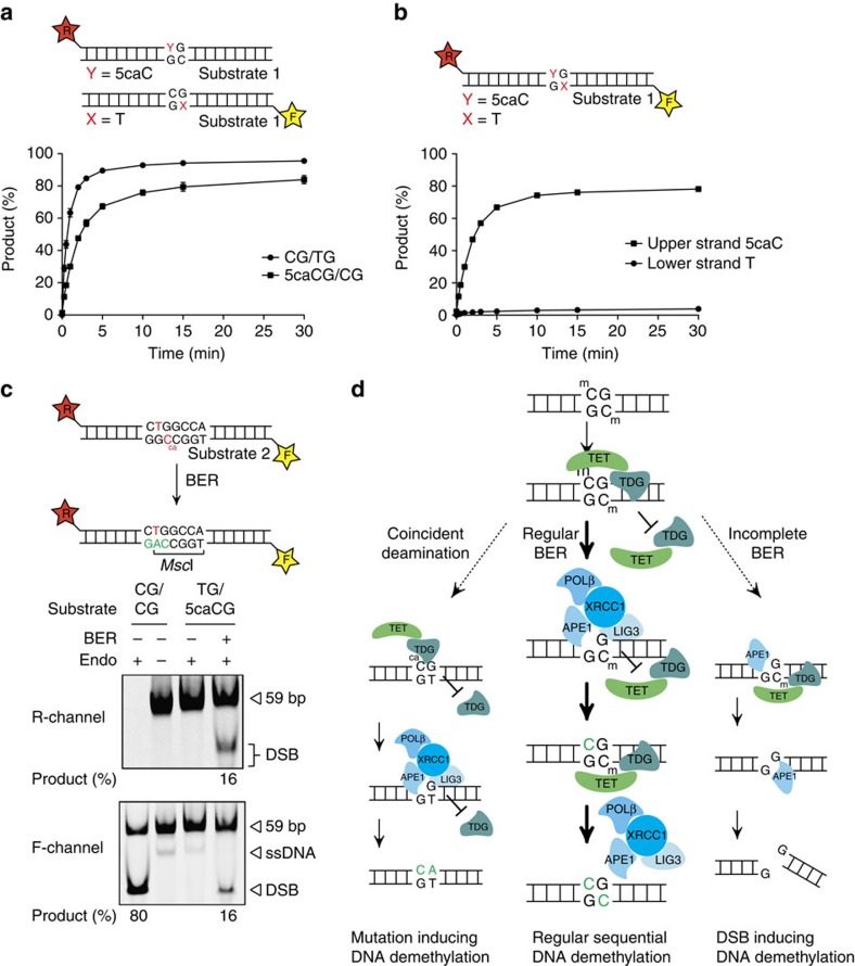
Cytosine methylation in CpG dinucleotides is an epigenetic DNA modification dynamically established and maintained by DNA methyltransferases and demethylases. Molecular mechanisms of active DNA demethylation began to surface only recently with the discovery of the 5-methylcytosine (5mC)-directed hydroxylase and base excision activities of ten-eleven translocation (TET) proteins and thymine DNA glycosylase (TDG). This implicated a pathway operating through oxidation of 5mC by TET proteins, which generates substrates for TDG-dependent base excision repair (BER) that then replaces 5mC with C. Yet, direct evidence for a productive coupling of TET with BER has never been presented. Here we show that TET1 and TDG physically interact to oxidize and excise 5mC, and proof by biochemical reconstitution that the TET-TDG-BER system is capable of productive DNA demethylation. We show that the mechanism assures a sequential demethylation of symmetrically methylated CpGs, thereby avoiding DNA double-strand break formation but contributing to the mutability of methylated CpGs.


Figures

Figure 1
Figure 1. TET1 physically interacts with TDG.
(a) Size fractionation by gel filtration at high ionic strength (500 mM NaCl) of Ni-NTA-enriched lysates of E. coli cells co-expressing TET1–His6 and TDG–GST from constructs indicated. Fractions were analysed by SDS–PAGE (left panel), and TET1 and TDG detected by immunoblotting (right panel); molecular weights of gel filtration standards are indicated. (b) Yeast two-hybrid analysis of the TET1–TDG interaction. TET1 domains cloned into the GAL4 activation domain (AD), TET1-1 (aa 1–491), TET1-2 (aa 397–931), TET1-3 (aa 870–1403) and TET1-4 (aa 1,367–2,057) are indicated at the top. Shown is the growth of serial dilutions of strains co-expressing TET1 domains fused to the AD and TDG fused to the GAL4-binding domain (BD) and respective negative controls (TET1 domains or TDG co-expressed with the vector control (V)) on permissive and selective media. The large T antigen (lTAg) and p53 fused to the AD and BD, respectively, served as a positive control. (c) Immunoblotting of fractions obtained from Ni-NTA and GST purifications using E. coli extracts co-expressing His6–TET1N and TDG–GST (left panel), or co-expressing His6–TET1CD and TDG–GST (right panel). Expression constructs used are indicated; TET1N (aa 301–1366) and TET1CD (aa 1367–2057); b, bound fraction, in, input; w, wash.
Figure 2
Figure 2. Combined TET1 and TDG activity releases 5mC through oxidized intermediates.
(a) Base-excision activity of Ni-NTA-enriched His6–TET1CD–TDG–GST on synthetic DNA substrates as indicated. The ability to generate alkaline-sensitive AP-sites in substrates containing either single G·5mC or G·5hmC base pairs was assayed with enriched His6–TET1CD–TDG–GST consisting of wild-type proteins or respective mutant variants (His6–TET1CDΔcat–TDG–GST, His6–TET1CD–TDGΔcat–GST). Products were separated by denaturing gel electrophoresis, visualized with fluorescent scanning and quantified; positions of the 60mer substrate DNA and product fragment are indicated. (b) Slot blot analysis of plasmid oxidation by purified His6–TET1CD. In-vitro-methylated pUC19 plasmid DNA (800 nM) was treated with His6–TET1CD (125 nM) and cytosine modifications were detected by immunblotting with specific antibodies against 5mC, 5hmC, 5fC and 5caC. (c) Reconstitution of 5mC/5hmC base release with purified His6–TET1CD and His6–TDG proteins. DNA substrates (25 nM) containing either G·5mC, G·5hmC or G·5caC base pairs were reacted with preassembled His6–TET1CD–His6–TDG (50 nM), reaction products separated by denaturing gel electrophoresis, visualized and quantified. Positions of the 60mer substrate DNA and product fragments are indicated. Shown are mean values with s.d. (n=3).
Figure 3
Figure 3. TET1CD stabilizes TDG activity.
(a) Stimulatory effect of His6–TDGΔcat on His6–TET1CD examined by base release assay. His6–TET1CD (50 nM) or His6–TET1CD–His6–TDGΔcat (25 nM) complex were incubated with DNA substrate (25 nM) containing a G·5mC base pair for the indicated time. Recovered DNA was then assayed by a base release assay using His6–TDG (250 nM) to monitor the presence of oxidized 5mC species. (b) LC–MS/MS analysis of plasmid oxidation assays using His6–TET1CD or TET1CD–His6–TDGΔcat. In-vitro-methylated pUC19 plasmid DNA (660 nM) was treated with either TET1CD (100 nM) or a preassembled TET1CD–TDGΔcat complex for the indicated time. DNA was analysed by LC–MS/MS; shown are normalized mean values (mod/total C mod). (c) Activity of His6–TDGΔcat on 5fC and 5caC in the presence or the absence of His6–TET1CD. His6–TDGΔcat–BSA (50 nM) or His6–TDGΔcat–His6–TET1CD (50 nM) were incubated with DNA substrate (25 nM) containing a G·5fC or G·5caC at 37 °C, reactions were stopped by the addition of NaOH at indicated time and analysed by denaturing gel electrophoresis. (d) The effect of His6–TET1CD on His6–TDG catalysis assessed in base release assays. The time-dependent generation of AP-sites was measured after reaction of a 60mer substrate containing a single G·5caC (25 nM) base pair with a preassembled His6–TDG–His6–TET1CD (25 nM) or His6–TDG–BSA (25 nM) complex in the presence (lower panel) or absence (upper panel) of a 60-fold molar excess of BSA. Reactions were stopped by the addition of NaOH after the indicated time and analysed using denaturing gel electrophoresis and fluorescent scanning. Shown are mean values with s.d. P-values were calculated by the Student's t-tests (*P<0.05, **P<0.01 and ***P<0.001).
Figure 4
Figure 4. Full reconstitution of TET–TDG–BER-mediated DNA demethylation.
(a) Intermediate steps of the oxidative DNA demethylation reaction were reconstituted and visualized by denaturing gel electrophoresis. Labelled 60mer substrate DNA containing one G·5mC base pair was incubated sequentially with TET–TDG–BER enzymes at concentrations indicated. Reaction products were separated by denaturing gel electrophoresis and visualized by fluorescent scanning; sizes of the 60mer substrate DNA and reaction products are indicated. (b) Complete DNA demethylation by the reconstituted TET–TDG–BER system analysed by the generation of a HpaII-sensitive restriction site. Reconstituted DNA demethylation was done with a 5′-labelled 59-bp substrate containing one G·5mC base pair within a HpaII recognition site (CCGG). Recovered DNA was digested with methylation-sensitive HpaII endonuclease and analysed by native PAGE; positions of the 59-bp substrate DNA and product fragment are indicated.
Figure 5
Figure 5. Processing of differentially modified CpGs by TET1CD–TDG or TDG.
(a) Base release from fully methylated CpGs by His6–TET1CD–His6–TDG. His6–TET1CD–His6–TDG (50 nM) was incubated with labelled 60mer substrates (25 nM) containing a single 5mC modification on the fluorescent labelled top (5′-Texas Red, T) or bottom strand (5′-fluorescein, F) or a fully methylated CpG with labels on both strands. Product formation was monitored and quantified by denaturing gel electrophoresis and fluorescent scanning (Texas Red, R-channel and fluorescein, F-channel); positions of the 60mer substrate DNA and the resulting base incision products of both strands are indicated. *Unlabelled DNA strand. (b) Release of 5caC from differentially modified CpGs by His6–TDG. 60mer DNA substrates (25 nM) containing 5caC opposite C, 5mC or 5hmC in a CpG dinucleotide or in single-stranded (ss) DNA were incubated with His6–TDG (25 nM) and analysed by denaturing gel electrophoresis. Shown are mean percentages of product formation with s.d. (n=3). (c) 5caC release from a symmetrically modified CpG dinucleotide by His6–TDG. A substrate (25 nM) containing 5caC on both strands within a CpG dinucleotide and labels of both strands was incubated with His6–TDG (25 nM) for indicated time, analysed by denaturing gel electrophoresis and visualized by fluorescent scanning of both labels. Shown are mean percentages of product formation with s.d. (n=3). (d) TDG and APE1 only generate DNA DSBs at symmetrically modified CpGs. Base release assay using TDG and APE1 on a labelled 59-bp substrate containing either a single 5caC or a symmetrically 5caC-modified base pair within a HpaII recognition site (CCGG). Reactions were analysed by native PAGE. Substrate DNA and product fragment are indicated. (e) Full reconstitution of TDG–BER on a symmetrically modified 5caC substrate. A labelled 59-bp substrate (25 nM) containing a symmetrically 5caC-modified base pair within a HpaII recognition site (CCGG) was incubated with TDG (40 nM) and BER factors (200 nM APE1, 40 nM POLβ and 40 nM XRCC1-LIG3). Recovered DNA was digested with HpaII endonuclease and analysed by native PAGE. Substrate DNA and product fragments are indicated; ssDNA, free single-stranded DNA.
Figure 6
Figure 6. DNA demethylation blocks G·T repair and can induce mutations.
(a) Enzymatic activity of TDG on G·5caC- and G·T-containing substrates. Release of 5caC and T by His6–TDG (25 nM) was monitored over time on 5′-labelled 60-bp substrates (25 nM) containing either a G·5caC or G·T base pair. Reactions were stopped at indicated time, separated by denaturing gel electrophoresis, visualized with fluorescent scanning and quantified. Shown are mean percentages of product formation with s.d. (n=3) (b) Base release from a substrate containing a G·5caC next to a G·T mismatch. Substrate preference of TDG (25 nM) was evaluated on a 59-bp DNA fragment (25 nM) containing 5caC on the labelled top strand (5′-Texas Red) and T on the labelled bottom strand (5′-fluorescein) within the same CpG context as illustrated. Reactions were stopped after indicated time, separated by denaturing gel electrophoresis, and both strands visualized by fluorescent scanning and quantified. Shown are mean percentages of product formation with s.d. (n=3). (c) Full reconstitution of TDG–BER on a G·5caC/G·T-containing substrate. A labelled 59-bp substrate containing a G·5caC next to a G·T mismatch was incubated with His6–TDG and BER factors. Correct repair of the 5caC and the introduction of an A opposite of T was monitored by MscI digestion and analysed by native PAGE and fluorescent scanning. Unmodified (CG/CG) substrate DNA digested with HpaII was used as size marker; positions of the substrate DNA and product fragments are indicated. ssDNA, free single-stranded DNA. (d) Mechanistic model of TET–TDG–BER-mediated DNA demethylation. In the presence of all the necessary factors, DNA demethylation at fully methylated CpGs occurs in a coordinated and sequential manner to correctly re-establish the unmodified state (regular BER). Lack of coordination, for example, in the absence of downstream BER factors, repair-mediated DNA demethylation can lead to the induction of DNA DSBs (incomplete BER). Coincident oxidation and hydrolytic deamination at fully methylated CpG sites can lead to increased C to T transitions caused by the sequential repair mechanism (coincident deamination).

References

    1. Bird A. DNA methylation patterns and epigenetic memory. Genes Dev. 16, 6–21 (2002). - PubMed
    1. Jones P. A. Functions of DNA methylation: islands, start sites, gene bodies and beyond. Nat. Rev. Genet. 13, 484–492 (2012). - PubMed
    1. Guo H. et al.. The DNA methylation landscape of human early embryos. Nature 511, 606–610 (2014). - PubMed
    1. Smith Z. D. et al.. DNA methylation dynamics of the human preimplantation embryo. Nature 511, 611–615 (2014). - PMC - PubMed
    1. Oswald J. et al.. Active demethylation of the paternal genome in the mouse zygote. Curr. Biol. 10, 475–478 (2000). - PubMed

Publication types

MeSH terms