. 2014 Dec;3(1):30.
doi: 10.1186/s40169-014-0030-5. Epub 2014 Sep 26.

Targeting met mediated epithelial-mesenchymal transition in the treatment of breast cancer

Affiliations

Targeting met mediated epithelial-mesenchymal transition in the treatment of breast cancer

Paul W Sylvester. Clin Transl Med. 2014 Dec.

Abstract

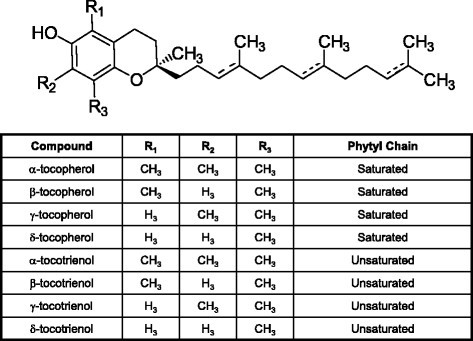
Mesenchymal epithelial transition factor receptor (Met) is a receptor tyrosine kinase that plays a critical role in promoting cancer cell malignant progression. Met is activated by its ligand hepatocyte growth factor (HGF). HGF-dependent Met activation plays an important role in stimulating epithelial-mesenchymal transition (EMT) in tumor cells, resulting in increased tumor cell proliferation, survival, motility, angiogenesis, invasion, and metastasis. The HGF/Met axis has thus attracted great interest as a potential target in the development of novel cancer therapies. In an effort to suppress tumor cell malignant progression, efforts have been made to develop agents capable of inhibiting inhibit Met-induced EMT, including specific Met tyrosine kinase inhibitors, HGF antagonists that interfere with HGF binding to Met, and antibodies that prevent Met activation and/or dimerization. Tocotrienols, a subgroup within the vitamin E family of compounds, display potent anticancer activity that results, at least in part, from inhibition of HGF-dependent Met activation and signaling. The present review will provide a brief summary of the increasing importance of the HGF/Met axis as an attractive target for cancer chemotherapy and the role of tocotrienols in suppressing Met activation, signaling and HGF-induced EMT in breast cancer cells. Evidence provided suggests that γ-tocotrienol therapy may afford significant benefit in the treatment of breast cancers characterized by Met dysregulation.

Keywords: Epithelial mesenchymal transition; HGF; Met; Targeted therapy, cancer, tocotrienols.


Figures

Figure 1
Figure 1
Schematic representation of the Met receptor and therapeutic strategies currently being developed to inhibit HGF-dependent Met activation and signaling in cancer. The Met receptor has an extracellular α-chain that binds HGF and a transmembrane β-chain that contains the tyrosine kinase domain and autophosphorylation sites that are essential for interacting with substrates. Activation of Met by HGF leads to receptor dimerization and recruitment of adaptor (GAB1, Grb2, Shc) and signaling (Ras/MAPK, PI3K/Akt, Src, STAT, Shp2) proteins. Downstream signaling promotes cell proliferation, altered cytoskeletal function, decreased cellular adhesion, increased cellular invasion, decreased apoptosis and enhanced DNA transcription. Anti-HGF approaches to inhibit Met signaling include anti-HGF antibodies that neutralize HGF and antagonists that block HGF binding to the Met receptor. A second approach includes the use of anti-Met antibodies that prevent HGF binding to Met or Met dimerization. Another approach is the use of specific Met tyrosine kinase inhibitors that prevent receptor second messenger signaling. Tocotrienols have also been found to be potent inhibitors of Met activation and signaling, but the exact mechanism mediating these effects are not completely understood at present. Targeting aberrant Met signaling in cancer cells can inhibit of downstream signaling pathways involved with tumor cell proliferation, motility, viability, morphology and epithelial-to-mesenchymal transition.
Figure 2
Figure 2
Generalized chemical structure of natural tocopherols and tocotrienols that make up the vitamin E family of compounds.
Figure 3
Figure 3
Schematic representation of HGF/Met-mediated epithelial mesenchymal transition (EMT) and promotion of malignant progression. HGF-mediated Met activation and signaling can induced multiple pathways that are involved in stimulating cancer cell proliferation, survival, motility, angiogenesis, invasion and metastasis. Normal epithelial cells display a highly differentiated morphology characterized by a single layer of cells anchored by their basal lamina to the extracellular matrix. Aberrant Met activity will stimulate cell proliferation and EMT that ultimately results in changes in morphology and behavior, characteristic of a mesenchymal-like phenotype. EMT allows cancerous epithelial cells to become more mobile, invasive and metastatic in nature.
Figure 4
Figure 4
Effects of γ-tocotrienol (γT3) and/or the Met tyrosine kinase inhibitor, SU11274 (SU), on the expression of major epithelial and mesenchymal cellular protein markers in + SA mammary tumor cells. Cells were incubated with control or treatment media containing subeffective doses of γ-tocotrienol (2 μM) and SU11274 (3 μM) either alone or in combination containing 10 ng/ml HGF as a mitogen for 3-days. (A) Afterwards, whole cell lysates were prepared and subjected to polyacrylamide gel electrophoresis and Western blot analysis for E-cadherin, β-catenin, cytokeratin-8, cytokeratin-18, and vimentin. α-Tubulin was visualized to ensure equal sample loading in each lane. Scanning densitometric analysis was performed for each blot to visualize the relative levels of proteins. Integrated optical density of each band was normalized with their corresponding α-tubulin and control treatment bands and shown in bar graphs. Vertical bars indicate the fold-change in protein levels in various treatment groups ± SEM as compared with their respective vehicle-treated control group. *P < 0.05 as compared to their respective vehicle-treated control group. (B) Treatment effects on immunocytochemical fluorescence staining of epithelial and mesenchymal markers in + SA mammary tumor cells after a 3-day culture period. +SA cells were seeded on 4-chamber culture slides at a density of 1x10⁵ cells/chamber (3 replicates/group) and allowed to attach overnight. Cells were then washed with PBS and incubated with vehicle control or treatment defined media containing 10 ng/ml HGF for 3 days in culture. At the end of treatments, cells were fixed with 4% formaldehyde/PBS and permeabilized with 0.2% triton X-100. Fixed cells were blocked and incubated with specific primary antibodies followed by incubation with Alexa Fluor 594- or 488-conjugated secondary antibodies. Red or green color indicates positive fluorescence staining for target proteins, while blue color represents nuclear counter staining with DAPI. Magnification is 200X. Figure obtained from reference 25 with permission.
Figure 5
Figure 5
γ-Tocotrienol and its oxazine derivative on CoCl2-induced hypoxia and HIF-1α expression in mammary tumor cells. HIF-1α is a hypoxia-inducible transcription factor that subsequently acts to stimulate blood vessel formation and promote survival of cancer cells during hypoxic conditions. (A) Effects of 150 μM CoCl2 (hypoxic, but not cytotoxic dose) alone and in combination with non-growth inhibitory doses (2 μM) of δ-tocotrienol or the δ-tocotrienol oxazine derivative, 12-((R)-6,8-dimethyl-8-((3E,7E)-4,8,12-trimethyltrideca-3,7,11-trienyl)-9,10-dihydrochromeno[5,6-e] [1],[3]oxazin-2(1H,3H,8H)-yl)dodecan-1-ol), on HIF-1α levels in + SA mammary tumor cells. Non-growth inhibitory doses of α-tocotrienol and its oxazine derivative were selected because high doses of these agents were found to initiate apoptosis and induce cancer cell death, which would confound the interpretation of the results regarding the effects of these agents on tumor cell compensatory response to CoCl2 hypoxia. +SA cells were seeded at concentration of 1.5X10⁶ in 100 mm culture dishes and allowed to attach overnight. The following day, cells were divided into groups and exposed to their respective treatments for a 24 hr incubation period. Afterwards, whole cell lysates were prepared for Western blot analysis. (B) Scanning densitometric analysis was performed on all blots done in triplicate and the integrated optical density of each band was normalized with corresponding α-tubulin, as shown in the bar graphs below their respective Western blot image. Vertical bars indicate the normalized integrated optical density of bands visualized in each lane ± SEM. #P < 0.05 compared to the vehicle-treated control group. *P < 0.05 as compared to the hypoxic group treated with CoCl2 alone. This figure was obtained from reference 65 with permission.

Similar articles

Cited by

References

    1. Fischgrabe J, Wulfing P. Targeted therapies in breast cancer: established drugs and recent developments. Curr Clin Pharmacol. 2009;3:85. doi: 10.2174/157488408784293732. - DOI - PubMed
    1. Prat M, Narsimhan RP, Crepaldi T, Nicotra MR, Natali PG, Comoglio PM. The receptor encoded by the human c-MET oncogene is expressed in hepatocytes, epithelial cells and solid tumors. Int JCancer. 1991;49:323. doi: 10.1002/ijc.2910490302. - DOI - PubMed
    1. Naran S, Zhang X, Hughes SJ. Inhibiton of HGF/Met as therapy for malignancy. Expert Opin Ther Targets. 2009;13:569. doi: 10.1517/14728220902853917. - DOI - PubMed
    1. Gentile A, Trusolino L, Comoglio PM. The Met tyrosine kinase receptor in development and cancer. Cancer Metastasis Rev. 2008;27:85. doi: 10.1007/s10555-007-9107-6. - DOI - PubMed
    1. Gastaldi S, Comoglio PM, Trusolino L. The Met oncogene and basal-like breast cancer: another culprit to watch out for? Breast Cancer Res. 2010;12:208. doi: 10.1186/bcr2617. - DOI - PMC - PubMed

LinkOut - more resources