

GATA4 and GATA6 regulate pancreatic endoderm identity through inhibition of hedgehog signaling
- PMID: 26932670
- PMCID: PMC4813334
- DOI: 10.1242/dev.127217
GATA4 and GATA6 regulate pancreatic endoderm identity through inhibition of hedgehog signaling
Abstract
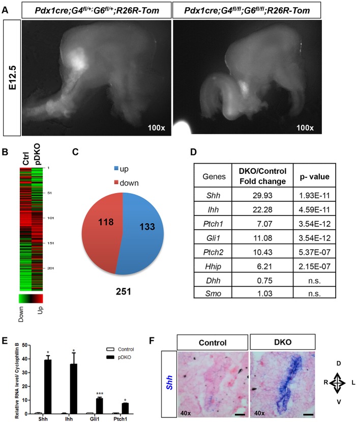
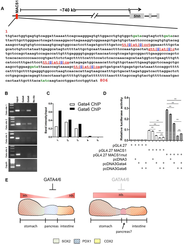
GATA4 and GATA6 are zinc finger transcription factors that have important functions in several mesodermal and endodermal organs, including heart, liver and pancreas. In humans, heterozygous mutations of either factor are associated with pancreatic agenesis; however, homozygous deletion of both Gata4 and Gata6 is necessary to disrupt pancreas development in mice. In this study, we demonstrate that arrested pancreatic development in Gata4(fl/fl); Gata6(fl/fl); Pdx1:Cre (pDKO) embryos is accompanied by the transition of ventral and dorsal pancreatic fates into intestinal or stomach lineages, respectively. These results indicate that GATA4 and GATA6 play essential roles in maintaining pancreas identity by regulating foregut endodermal fates. Remarkably, pancreatic anlagen derived from pDKO embryos also display a dramatic upregulation of hedgehog pathway components, which are normally absent from the presumptive pancreatic endoderm. Consistent with the erroneous activation of hedgehog signaling, we demonstrate that GATA4 and GATA6 are able to repress transcription through the sonic hedgehog (Shh) endoderm-specific enhancer MACS1 and that GATA-binding sites within this enhancer are necessary for this repressive activity. These studies establish the importance of GATA4/6-mediated inhibition of hedgehog signaling as a major mechanism regulating pancreatic endoderm specification during patterning of the gut tube.
Keywords: Foregut endoderm; GATA4; GATA6; Hedgehog; Mouse; Pancreas.
© 2016. Published by The Company of Biologists Ltd.
Conflict of interest statement
The authors declare no competing or financial interests.
Figures
Similar articles
-
Pancreas-specific deletion of mouse Gata4 and Gata6 causes pancreatic agenesis.J Clin Invest. 2012 Oct;122(10):3516-28. doi: 10.1172/JCI63352. Epub 2012 Sep 24. J Clin Invest. 2012. PMID: 23006325 Free PMC article.
-
GATA4 and GATA6 control mouse pancreas organogenesis.J Clin Invest. 2012 Oct;122(10):3504-15. doi: 10.1172/JCI63240. Epub 2012 Sep 24. J Clin Invest. 2012. PMID: 23006330 Free PMC article.
-
A threshold of GATA4 and GATA6 expression is required for cardiovascular development.Proc Natl Acad Sci U S A. 2006 Jul 25;103(30):11189-94. doi: 10.1073/pnas.0604604103. Epub 2006 Jul 17. Proc Natl Acad Sci U S A. 2006. PMID: 16847256 Free PMC article.
-
Progress of GATA6 in liver development.Yi Chuan. 2018 Jan 20;40(1):22-32. doi: 10.16288/j.yczz.17-163. Yi Chuan. 2018. PMID: 29367190 Review.
-
Role of GATA factors in development, differentiation, and homeostasis of the small intestinal epithelium.Am J Physiol Gastrointest Liver Physiol. 2014 Mar;306(6):G474-90. doi: 10.1152/ajpgi.00119.2013. Epub 2014 Jan 16. Am J Physiol Gastrointest Liver Physiol. 2014. PMID: 24436352 Free PMC article. Review.
Cited by
-
Control of murine brown adipocyte development by GATA6.Dev Cell. 2023 Nov 6;58(21):2195-2205.e5. doi: 10.1016/j.devcel.2023.08.003. Epub 2023 Aug 29. Dev Cell. 2023. PMID: 37647897 Free PMC article.
-
Loss of Pancreas upon Activated Wnt Signaling Is Concomitant with Emergence of Gastrointestinal Identity.PLoS One. 2016 Oct 13;11(10):e0164714. doi: 10.1371/journal.pone.0164714. eCollection 2016. PLoS One. 2016. PMID: 27736991 Free PMC article.
-
Insight into Nephrocan Function in Mouse Endoderm Patterning.Int J Mol Sci. 2019 Dec 18;21(1):8. doi: 10.3390/ijms21010008. Int J Mol Sci. 2019. PMID: 31861348 Free PMC article.
-
The Relationship between the Expression of GATA4 and GATA6 with the Clinical Characteristics and Prognosis of Resectable Pancreatic Adenocarcinoma.Biomedicines. 2023 Jan 18;11(2):252. doi: 10.3390/biomedicines11020252. Biomedicines. 2023. PMID: 36830789 Free PMC article.
-
Development of the Pancreatic Ducts and Their Contribution to Organogenesis.Adv Anat Embryol Cell Biol. 2024;239:31-55. doi: 10.1007/978-3-031-62232-8_2. Adv Anat Embryol Cell Biol. 2024. PMID: 39283481 Free PMC article. Review.
References
Publication types
MeSH terms
Substances
Grants and funding
LinkOut - more resources
Full Text Sources
Other Literature Sources
Molecular Biology Databases
