. 2016 Mar 1;143(5):841-50.
doi: 10.1242/dev.129320.

Selection of cell fate in the organ of Corti involves the integration of Hes/Hey signaling at the Atoh1 promoter

Affiliations

Selection of cell fate in the organ of Corti involves the integration of Hes/Hey signaling at the Atoh1 promoter

Yassan Abdolazimi et al. Development. .

Abstract

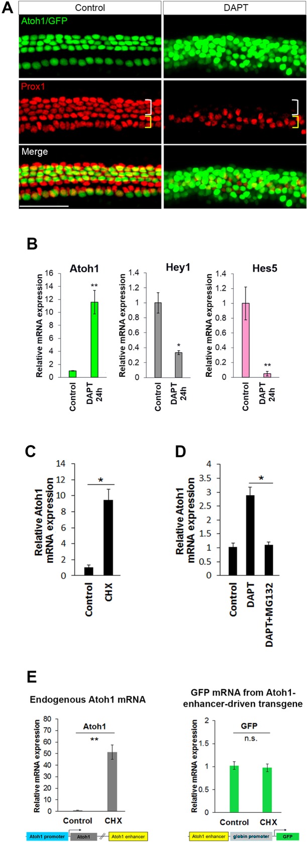
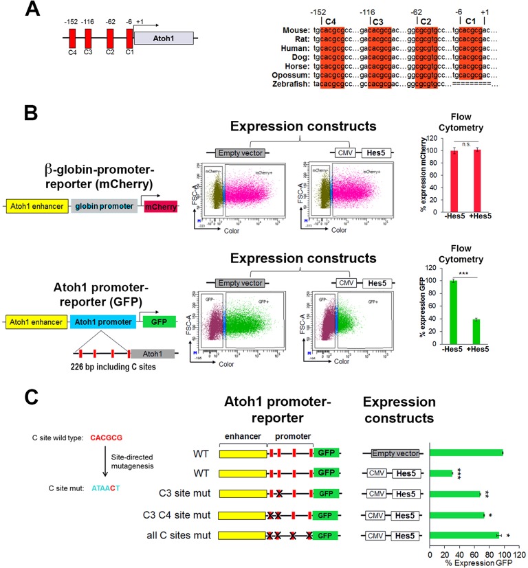
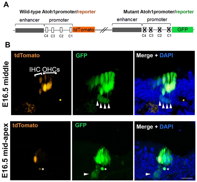
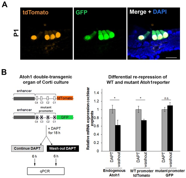
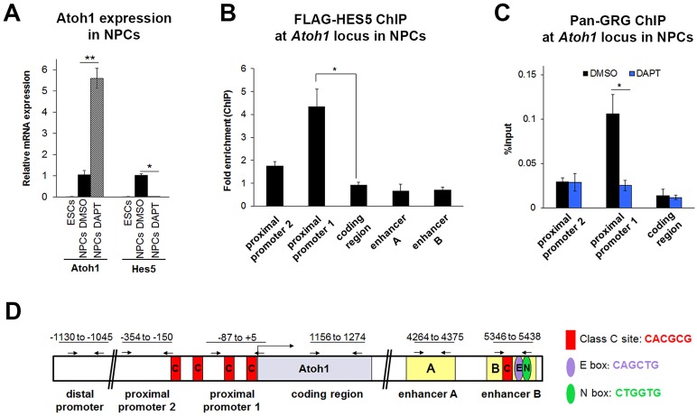
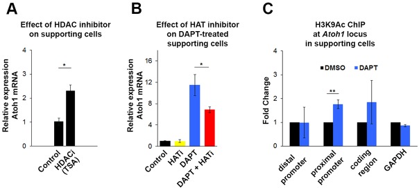
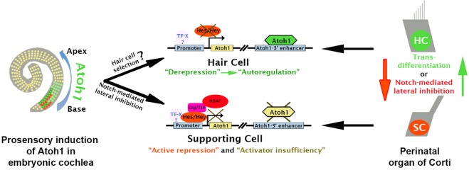
Determination of cell fate within the prosensory domain of the developing cochlear duct relies on the temporal and spatial regulation of the bHLH transcription factor Atoh1. Auditory hair cells and supporting cells arise in a wave of differentiation that patterns them into discrete rows mediated by Notch-dependent lateral inhibition. However, the mechanism responsible for selecting sensory cells from within the prosensory competence domain remains poorly understood. We show in mice that rather than being upregulated in rows of cells, Atoh1 is subject to transcriptional activation in groups of prosensory cells, and that highly conserved sites for Hes/Hey repressor binding in the Atoh1 promoter are needed to select the hair cell and supporting cell fate. During perinatal supporting cell transdifferentiation, which is a model of hair cell regeneration, we show that derepression is sufficient to induce Atoh1 expression, suggesting a mechanism for priming the 3' Atoh1 autoregulatory enhancer needed for hair cell expression.

Keywords: Atoh1; Cochlear development; Hair cell regeneration; Hes5; Mouse; Organ of Corti; Transdifferentiation.


Conflict of interest statement

Competing interests

The authors declare no competing or financial interests.

Figures

Fig. 1.
Fig. 1.
Derepression of Atoh1 in supporting cells is sufficient to drive expression and occurs through promoter elements, not through the 3′ autoregulatory enhancer. (A) P1 Atoh1 enhancer/β-globin promoter/GFP transgenic organ of Corti was cultured for 72 h in DMSO (control) or DAPT (γ-secretase inhibitor) and immunostained with anti-PROX1 antibody to label supporting cells (Deiters' and pillar cells). The appearance of ectopic GFP+ hair cell-like cells in DAPT is accompanied by loss of PROX1+ supporting cells (white bracket). (B-E) Changes in mRNA expression level were examined by real-time quantitative PCR (qPCR). (B) The observed transdifferentiation of supporting cells in A correlates with upregulation of Atoh1 and downregulation of Hes/Hey factors mRNA in FACS-purified supporting cells (p27/GFP+) after 24 h treatment with DAPT. n=4. (C) Inhibition of protein synthesis with cycloheximide (CHX) for 6 h induces Atoh1 expression in FACS-purified supporting cells (p27/GFP+). n=3. (D) Inhibition of protein degradation by MG132 prevents DAPT-induced upregulation of Atoh1 in FACS-purified supporting cells (p27/GFP+) treated for 12 h. n=3. (E) CHX induces endogenous Atoh1, but not the Atoh1 enhancer/β-globin promoter/GFP transgene. P1 cochlear organ cultures from Atoh1 enhancer/β-globin promoter/GFP mice were incubated without and with CHX for 6 h, after which organ cultures were dissociated and FACS sorted to eliminate hair cells. The GFP population from these transgenic organs includes supporting cells, cells from the greater epithelial ridge and lesser epithelial ridge. Atoh1 and GFP levels were measured in GFP cells (non-hair cells of the cochlear epithelia). n=4. All values are mean±s.e.m. *P<0.05, **P<0.005; n.s., not statistically significant. Scale bar: 100 µm. See also Fig. S1.
Fig. 2.
Fig. 2.
HES5 functions to repress Atoh1 through highly conserved binding sites in the promoter region. (A) Schematic of the murine Atoh1 promoter indicating the C-sites and their conservation among vertebrates. Numbers are relative to the transcription start site (+1) in mouse. (B) Ectopic Hes5 expression requires the Atoh1 promoter to repress Atoh1 expression in heterologous HEK 293 cells [compare β-globin promoter (top) with Atoh1 promoter (bottom)]. Reporters and expression constructs were transiently transfected and expression was quantified by flow cytometry. (Top) Reporter contains the Atoh1 enhancer and β-globin basal promoter driving the expression of mCherry. (Bottom) Reporter contains the Atoh1 enhancer and 226 bp of the Atoh1 promoter (containing the predicted C-sites) driving the expression of GFP. Reporters were cotransfected with a control (empty vector) or a plasmid expressing Hes5. The number of cells expressing GFP or mCherry was determined by flow cytometry after 48 h. Values are mean±s.e.m.; n=6. ***P<1×10⁻⁷. (C) Mutational analysis of C-site function. Five out of six nucleotides of each C-site were mutated. Quantification was performed as in B with the indicated plasmids reported as percentage of cells expressing GFP relative to the CMV-RFP co-transfected control (not shown) set to 100% in the absence of Hes5 expression (empty vector). Values are mean±s.e.m.; n=4. *P<0.0003, **P<2×10⁻⁵, ***P<4×10⁻⁷. See also Fig. S2.
Fig. 3.
Fig. 3.
The misexpression of a mutant promoter transgene in vivo shows that Hes/Hey binding sites in the Atoh1 promoter are required for the proper silencing of Atoh1 in supporting cells. (A) Schematics showing wild-type and mutant constructs used to generate the double-transgenic mouse (see Materials and Methods). (B) The expression pattern of the wild-type and mutant transgenes at E16.5 is shown in transverse section at two positions (middle and mid-apex) of the cochlear duct. The GFP from the mutant promoter transgene is misexpressed in the supporting cell layer (white arrowheads; asterisk indicates debris), in addition to the expected expression in the hair cells, indicating the need for active repression for supporting cell silencing. tdTomato from the wild-type transgene is absent from the supporting cell layer. IHC, inner hair cell; OHCs, outer hair cells. Scale bar: 20 µm.
Fig. 4.
Fig. 4.
Hes/Hey binding sites are required to rerepress Atoh1 in supporting cells after Notch signaling is first inhibited, allowing Atoh1 levels to rise, and then restored. (A) At P1, the expression of both wild-type and mutant promoters is limited to hair cells, indicating that low levels of ATOH1 (activator insufficiency) are sufficient to maintain silencing from the mutant transgene. Scale bar: 20 µm. (B) Schematic shows experimental time course: P1 cochlear cultures from the double-transgenic mouse line were treated with DAPT for 18 h, after which DAPT was washed out from half of the explants, and the explants were then collected after an additional 6 h in culture. Expression analysis of endogenous Atoh1, GFP and tdTomato by qPCR shows that endogenous Atoh1 and the wild-type (WT) transgene are actively repressed by returning Notch signaling, but the mutant transgene fails to be rerepressed in the same time period (6 h). Values are mean±s.e.m.; n=3. *P<0.05.
Fig. 5.
Fig. 5.
GRG localizes to the Atoh1 locus in a Notch-dependent manner in neural progenitor cells. Neural progenitor cells (NPCs) were differentiated from ESCs and FACS purified (Sox1-GFP+). (A) Atoh1 and Hes5 mRNA expression levels in 46C ESCs and NPCs that were treated with control (DMSO) or γ-secretase inhibitor (DAPT) for 48 h. Similar to supporting cells, inhibition of Notch signaling by DAPT induces the expression of Atoh1 and represses the expression of Hes5 in NPCs. n=4. (B) HES5 localizes to the proximal promoter and not the enhancer by ChIP-qPCR of NPCs transfected with CMV-FLAG-Hes5 plasmid (or CMV-RFP control). Results are reported as fold enrichment (FLAG-Hes5 transfected percentage input/RFP transfected percentage input). n=3. (C) GRG localizes to the Atoh1 promoter in a Notch-dependent manner. ChIP-qPCR with anti-pan-GRG antibody and primers scanning the Atoh1 locus in NPCs treated with control (DMSO) or γ-secretase inhibitor (DAPT) for 24 h after 7 days of differentiation. n=3. (D) Schematic of the Atoh1 locus in mouse showing the promoter and the autoregulatory enhancer regions. Numbers refer to the regions amplified in ChIP-qPCR. The primers used to measure the enrichment after ChIP are shown by arrows. All values are mean±s.e.m. *P<0.05, **P<0.005. See also Figs S3-S5.
Fig. 6.
Fig. 6.
Inhibition of Notch leads to increased histone acetylation at the Atoh1 promoter in supporting cells. (A) Inhibition of HDAC activity by trichostatin A (TSA) for 6 h induces Atoh1 expression in FACS-purified supporting cells (p27/GFP+). Values are mean±s.e.m.; n=3. (B) Inhibition of HAT activity by curcumin (Balasubramanyam et al., 2004) for 24 h blocks DAPT-induced Atoh1 activation in FACS-purified supporting cells (Lfng/GFP+). n=3. (C) Inhibition of Notch signaling (DAPT for 24 h) increases H3K9ac, as analyzed by ChIP-qPCR at the Atoh1 promoter in FACS-purified supporting cells (p27/GFP+). Values are mean±s.e.m.; n=3. *P<0.05, **P<0.005. See also Fig. S6.
Fig. 7.
Fig. 7.
Model of Atoh1 regulation during organ of Corti development. Upregulation of Atoh1 in groups of cells within the embryonic prosensory domain occurs in a basal-to-apical wave. Once the Atoh1 autoregulatory threshold is achieved in selected nascent hair cells, Notch-mediated active repression is triggered in the surrounding prosensory cells to stimulate the silencing of Atoh1 through Hes/Hey binding, leading to the onset of supporting cell differentiation and the patterning of the cellular mosaic of the organ of Corti. At P1, cell fate is maintained by continuing Notch-mediated repression through recruitment of GRG/HDACs. Loss of Notch activity leads to derepression of the Atoh1 promoter and increased Atoh1 expression through hypothetical positive transcriptional activity (TF-X), leading to autoregulation and transdifferentiation of supporting cells (SC) to a hair cell-like state (HC).

Similar articles

Cited by

References

    1. Balasubramanyam K., Varier R. A., Altaf M., Swaminathan V., Siddappa N. B., Ranga U. and Kundu T. K. (2004). Curcumin, a novel p300/CREB-binding protein-specific inhibitor of acetyltransferase, represses the acetylation of histone/nonhistone proteins and histone acetyltransferase-dependent chromatin transcription. J. Biol. Chem. 279, 51163-51171. 10.1074/jbc.M409024200 - DOI - PubMed
    1. Barolo S. and Posakony J. W. (2002). Three habits of highly effective signaling pathways: principles of transcriptional control by developmental cell signaling. Genes Dev. 16, 1167-1181. 10.1101/gad.976502 - DOI - PubMed
    1. Basch M. L., Ohyama T., Segil N. and Groves A. K. (2011). Canonical Notch signaling is not necessary for prosensory induction in the mouse cochlea: insights from a conditional mutant of RBPjkappa. J. Neurosci. 31, 8046-8058. 10.1523/JNEUROSCI.6671-10.2011 - DOI - PMC - PubMed
    1. Benito-Gonzalez A. and Doetzlhofer A. (2014). Hey1 and Hey2 control the spatial and temporal pattern of mammalian auditory hair cell differentiation downstream of Hedgehog signaling. J. Neurosci. 34, 12865-12876. 10.1523/JNEUROSCI.1494-14.2014 - DOI - PMC - PubMed
    1. Bermingham N. A., Hassan B. A., Price S. D., Vollrath M. A., Ben-Arie N., Eatock R. A., Bellen H. J., Lysakowski A. and Zoghbi H. Y. (1999). Math1: an essential gene for the generation of inner ear hair cells. Science 284, 1837-1841. 10.1126/science.284.5421.1837 - DOI - PubMed

Publication types

MeSH terms

LinkOut - more resources