. 2016 May 6;291(19):10104-18.
doi: 10.1074/jbc.M115.712448. Epub 2016 Mar 1.

The Combining Sites of Anti-lipid A Antibodies Reveal a Widely Utilized Motif Specific for Negatively Charged Groups

Affiliations

The Combining Sites of Anti-lipid A Antibodies Reveal a Widely Utilized Motif Specific for Negatively Charged Groups

Omid Haji-Ghassemi et al. J Biol Chem. .

Abstract

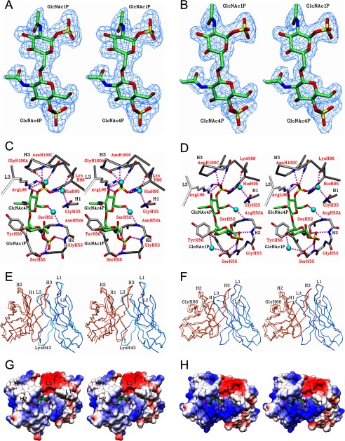
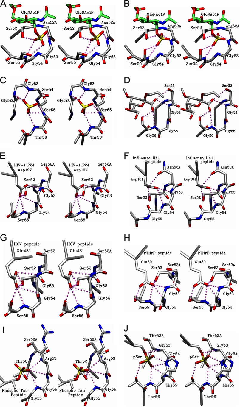
Lipopolysaccharide dispersed in the blood by Gram-negative bacteria can be a potent inducer of septic shock. One research focus has been based on antibody sequestration of lipid A (the endotoxic principle of LPS); however, none have been successfully developed into a clinical treatment. Comparison of a panel of anti-lipid A antibodies reveals highly specific antibodies produced through distinct germ line precursors. The structures of antigen-binding fragments for two homologous mAbs specific for lipid A, S55-3 and S55-5, have been determined both in complex with lipid A disaccharide backbone and unliganded. These high resolution structures reveal a conserved positively charged pocket formed within the complementarity determining region H2 loops that binds the terminal phosphates of lipid A. Significantly, this motif occurs in unrelated antibodies where it mediates binding to negatively charged moieties through a range of epitopes, including phosphorylated peptides used in diagnostics and therapeutics. S55-3 and S55-5 have combining sites distinct from anti-lipid A antibodies previously described (as a result of their separate germ line origin), which are nevertheless complementary both in shape and charge to the antigen. S55-3 and S55-5 display similar avidity toward lipid A despite possessing a number of different amino acid residues in their combining sites. Binding of lipid A occurs independent of the acyl chains, although the GlcN-O6 attachment point for the core oligosaccharide is buried in the combining site, which explains their inability to recognize LPS. Despite their lack of therapeutic potential, the observed motif may have significant immunological implications as a tool for engineering recombinant antibodies.

Keywords: X-ray crystallography; lipid A; lipopolysaccharide (LPS); monoclonal antibody; protein structure; recognition pocket.


Figures

FIGURE 1.
FIGURE 1.
Chemical structure of lipid A analogues used for ELISA and crystallization trials. BBP, B4P, B1P, and dephosphorylated lipid A backbone (BB) are shown. GlcN, glucosamine; in the deacylated lipid A backbone R1 and R2 = H; after N-acetylation R1 and R2 = Ac, and after spacered biotinylation R1 = LC-Bio and R2 = H. Shown is the structure designated LC-bio, which is attached to the free amine by reaction with sulfo-NHS-LC-biotin. The labels a–n indicate the location of protons assigned by NMR (Figs. 2 and 3).
FIGURE 2.
FIGURE 2.
1H NMR spectra and proton assignments of BBP, B4P, B1P, and BB after reaction with sulfo-NHS-LC-biotin and purification by HPAEC. For structures see Fig. 1.
FIGURE 3.
FIGURE 3.
1H,13C NMR spectrum and assignments of BBP after reaction with sulfo-NHS-LC-biotin and purification by HPAEC showing that under the conditions chosen only the proton of A2 (GlcN A) is shifted to higher frequency due to biotinylation. For structure see Fig. 1.
FIGURE 4.
FIGURE 4.
Quantitative ELISA coated with graded concentrations of biotinylated neoglycoconjugates corresponding to 2 pmol (left panel) and 20 pmol (right panel) of ligand per well and reacted with mAbs S55-3 (black squares) and S55-5 (open triangles) at concentrations indicated on the x axis. Measurements were performed as duplicates, and binding curves were generated by fitting the data to a logistic function. Ligands used were BBP (A and B), B4P (C and D), B1P (E and F), and BB (G and H). For structures see Fig. 1.
FIGURE 5.
FIGURE 5.
Stereo diagram of 2FoFc electron density map (blue) contoured at 1.0 σ for BBP-NAc lipid A analogue observed in the combining site of S55-3 (A) and S55-5 (B) mAbs post-refinement is shown. Stereo view of S55-3 (C) and S55-5 (D) in complex with BBP-NAc shows hydrogen bonds (purple dashed spheres) and water (blue spheres) bridges between the antigen and mAbs. CDR loops of the light and heavy chain are colored white and gray, respectively. Strong hydrophobic contact are shown as dashed lines (black). C-6 hydroxyl group of second glucosamine is the attachment point to inner core residues normally found on LPS. Stereo view of Fv structure alignments between liganded and unliganded structures of S55-3 (E) and S55-5 (F). Alignments were carried out using the α-carbon trace of the liganded variable light as the reference structure for each antibody. Displacement of CDR L1 and H3 is highlighted. Dark blue, liganded light chain. Cyan, unliganded light chains. Orange, unliganded heavy chains. Red, liganded heavy chain. The stereo image of the electrostatic surface potentials for Fv structures of S55-3 (G) and S55-5 (H) bound to BBP-NAc.
FIGURE 6.
FIGURE 6.
Stereo diagram of CDR H2 loop of antibodies possessing the Xaa-Xaa-Gly-Gly-(Ser/Thr/Gly) motif. Hydrogen bonds are formed via CDR H2 loop of S55-3 (A) and S55-5 Fab (B) (this study) to GlcNAc1P residue of lipid A (this study). C, N. meningitides LPT-3 Fab showing a sulfate group near the binding site and hydrogen bonds formed via the CDR H2 residues. D, CDR H2 loop of anti-chlamydial S25-26 Fab (PDB code 4M7J, Haji-Ghassemi et al. (49) showing hydrogen bonds formed via the CDR H2 residues to 3-deoxy-d-manno-oct-2-ulosonic acid carboxyl group as part of the antigen. E, hydrogen bonds between Asp-197 residue of p24 viral capsid protein and CDR H2 residues of A10F9 Fab (PDB code 3VRL (see Footnote 6)). F, residue contacts between Asp-101 residue of influenza virus hemagglutinin (HA-1) peptide and CDR H2 loop of 17/9 Fab (PDB code 1HIM, Rini et al. (70)). G, contacts between Glu-431 residue of hepatitis C virus envelope peptide and CDR H2 residues of mAb8 Fab (PDB code 4HZL, Deng et al. (57)). H, contacts between Glu-30 residue of a peptide from parathyroid hormone-related protein and CDR H2 residues of anti-PTHrP Fab (PDB code 3FFD, McKinstry et al. (59)). I, contacts between phosphorylated Thr belonging to the Tau peptide and CDR H2 residues of anti-Tau antibody Thr(P)-231/Ser(P)-235 (PDB code 4GLR, Shih et al. (60)). J, contacts between phosphorylated Ser peptide and CDR H2 residues of pSAb (PDB code 4JFZ, Koerber et al. (61)).

Similar articles

Cited by

References

    1. Engel C., Brunkhorst F. M., Bone H. G., Brunkhorst R., Gerlach H., Grond S., Gruendling M., Huhle G., Jaschinski U., John S., Mayer K., Oppert M., Olthoff D., Quintel M., Ragaller M., et al. (2007) Epidemiology of sepsis in Germany: results from a national prospective multicenter study. Intensive Care Med. 33, 606–618 - PubMed
    1. Martin G. S. (2012) Sepsis, severe sepsis and septic shock: changes in incidence, pathogens and outcomes. Expert Rev. Anti Infect. Ther. 10, 701–706 - PMC - PubMed
    1. Angus D. C., Linde-Zwirble W. T., Lidicker J., Clermont G., Carcillo J., and Pinsky M. R. (2001) Epidemiology of severe sepsis in the United States: analysis of incidence, outcome, and associated costs of care. Crit. Care Med. 29, 1303–1310 - PubMed
    1. Buttenschoen K., Radermacher P., and Bracht H. (2010) Endotoxin elimination in sepsis: physiology and therapeutic application. Langenbeck Arch. Surg. 395, 597–605 - PubMed
    1. Rietschel E. T., Brade H., Brade L., Brandenburg K., Schade U., Seydel U., Zähringer U., Galanos C., Lüderitz O., and Westphal O. (1987) Lipid A, the endotoxic center of bacterial lipopolysaccharides: relation of chemical structure to biological activity. Prog. Clin. Biol. Res. 231, 25–53 - PubMed

Publication types

MeSH terms

LinkOut - more resources