. 2016 Apr 22;291(17):9000-13.
doi: 10.1074/jbc.M116.718973. Epub 2016 Mar 1.

Family-wide Characterization of Histone Binding Abilities of Human CW Domain-containing Proteins

Affiliations

Family-wide Characterization of Histone Binding Abilities of Human CW Domain-containing Proteins

Yanli Liu et al. J Biol Chem. .

Abstract

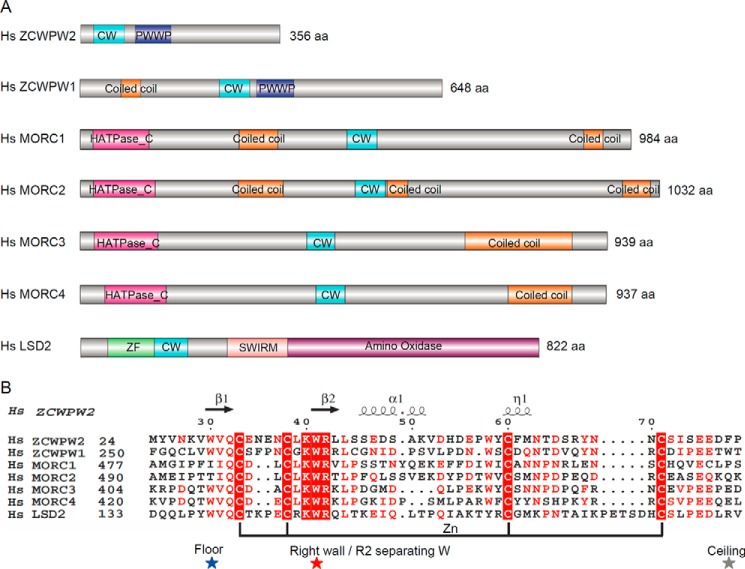
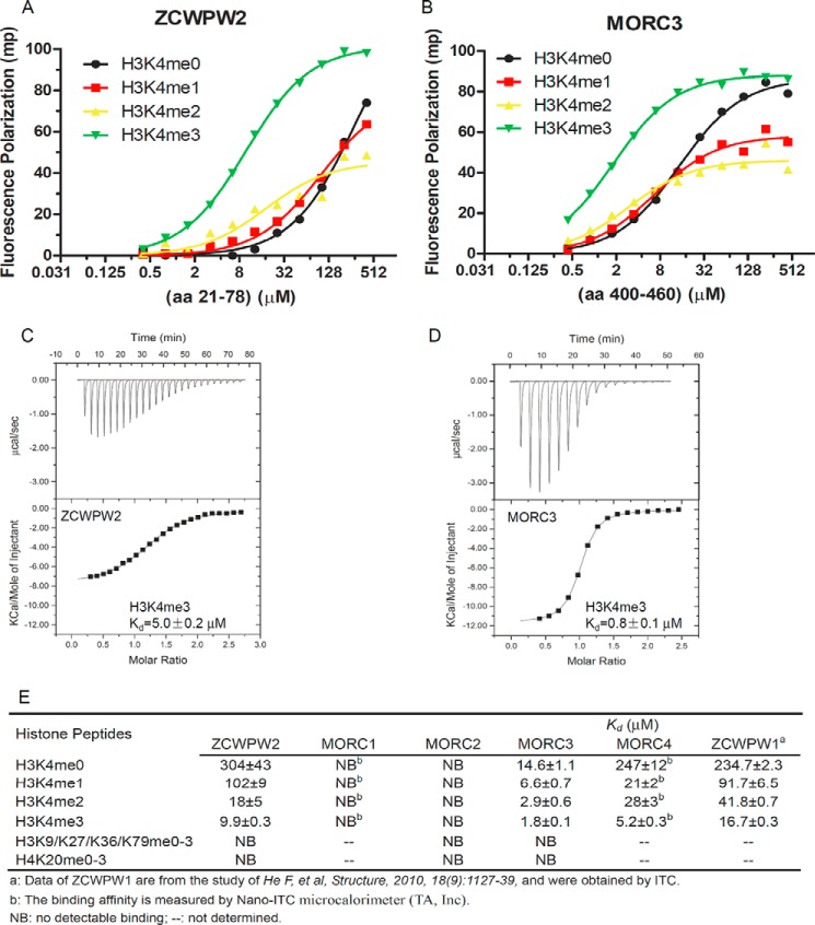
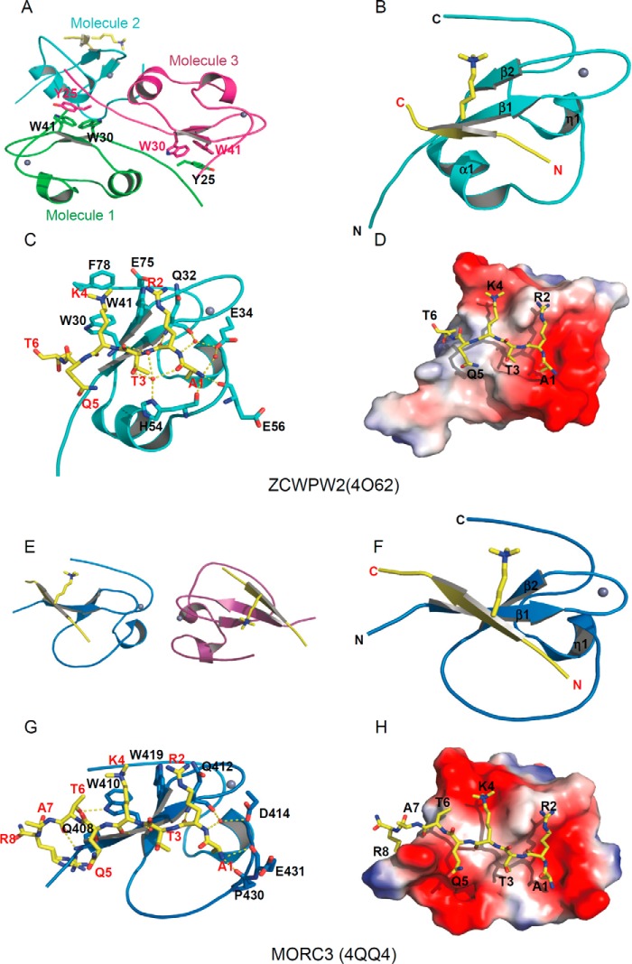
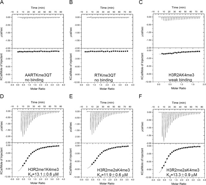
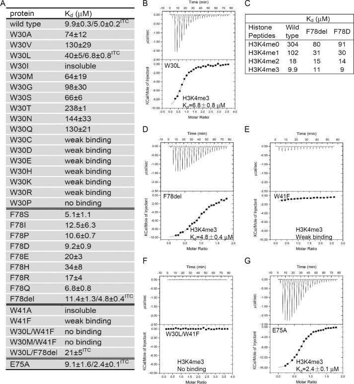
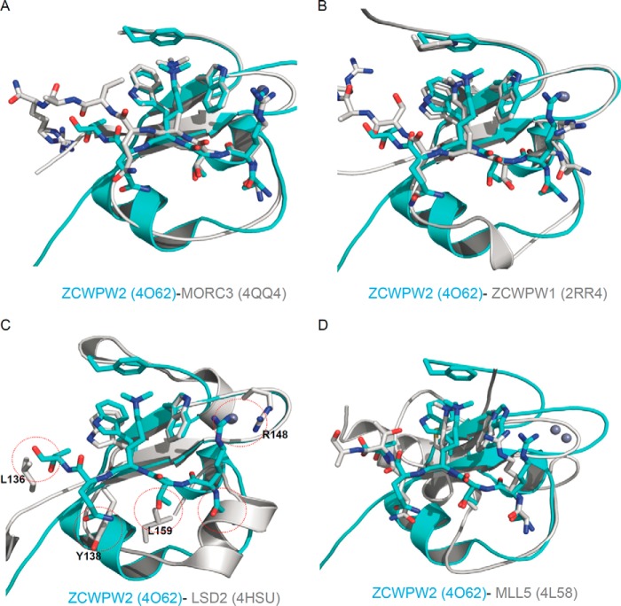
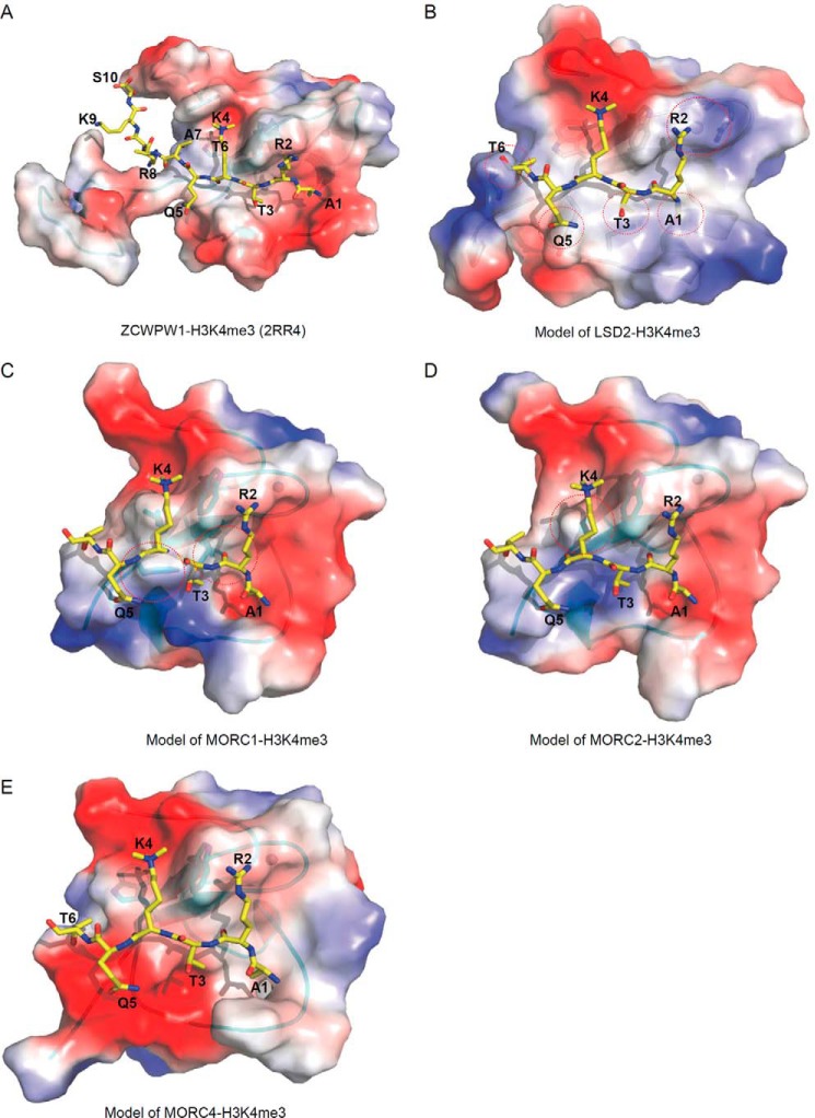
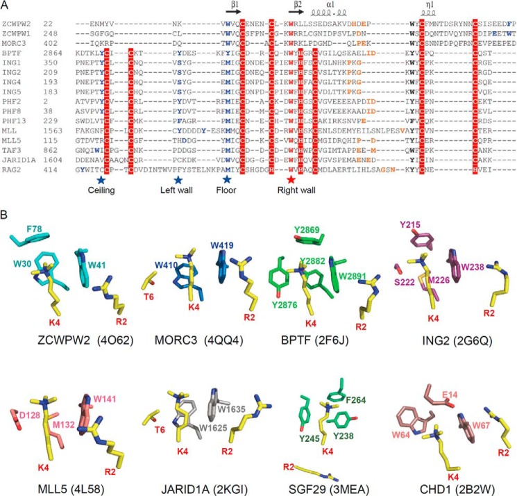
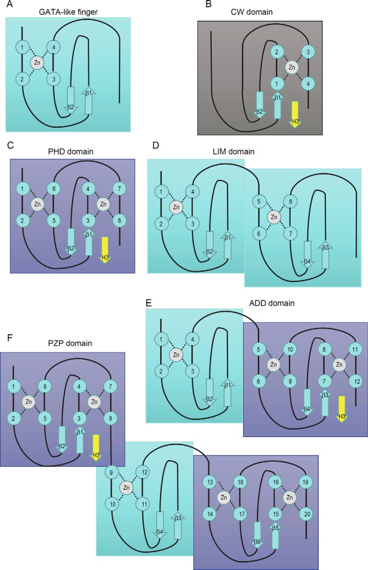
Covalent modifications of histone N-terminal tails play a critical role in regulating chromatin structure and controlling gene expression. These modifications are controlled by histone-modifying enzymes and read out by histone-binding proteins. Numerous proteins have been identified as histone modification readers. Here we report the family-wide characterization of histone binding abilities of human CW domain-containing proteins. We demonstrate that the CW domains in ZCWPW2 and MORC3/4 selectively recognize histone H3 trimethylated at Lys-4, similar to ZCWPW1 reported previously, while the MORC1/2 and LSD2 lack histone H3 Lys-4 binding ability. Our crystal structures of the CW domains of ZCWPW2 and MORC3 in complex with the histone H3 trimethylated at Lys-4 peptide reveal the molecular basis of this interaction. In each complex, two tryptophan residues in the CW domain form the "floor" and "right wall," respectively, of the methyllysine recognition cage. Our mutation results based on ZCWPW2 reveal that the right wall tryptophan residue is essential for binding, and the floor tryptophan residue enhances binding affinity. Our structural and mutational analysis highlights the conserved roles of the cage residues of CW domain across the histone methyllysine binders but also suggests why some CW domains lack histone binding ability.

Keywords: CW domain; H3K4 methylation; MORC3; ZCWPW2; chromatin structure; histone methylation; histone modification; post-translational modification (PTM); protein-protein interaction.


Figures

FIGURE 1.
FIGURE 1.
Comparison of human zinc finger CW domain-containing proteins. A, schematic representation of the domain structure of these proteins. B, structure-based sequence alignment of these CW domains. Secondary structure elements and residue numbers of the ZCWPW2 CW domain are indicated above the sequence alignment. The highly conserved “right wall” residues are marked by a red star; the partly conserved “floor” residues are marked by a blue star; the non-conserved “ceiling” residues are marked by a gray star. The alignments were constructed with ClustalW (45) and refined with ESPript (46).
FIGURE 2.
FIGURE 2.
ZCWPW2 and MORC3 are H3K4me3 binders. A and B, the binding curves of FP measurements of different H3K4 peptides to CW domains of ZCWPW2 (A) and MORC3 (B). C and D, ITC binding curves of H3K4me3 peptides to CW domains of ZCWPW2 (C) and MORC3 (D). E, binding affinities of selected CW domains to different histone peptides determined by FP or ITC. FP Kd values were calculated from duplicate measurements and fitted to ligand binding function using GraphPad Prism version 5 software. ITC Kd values were calculated from single measurement, and errors were estimated by a fitting curve.
FIGURE 3.
FIGURE 3.
Structural basis of selective binding of ZCWPW2 and MORC3 to H3K4me3. A and E, model of the crystal asymmetric unit of the CW domain and H3K4me3 peptide complexes of ZCWPW2 (A) and MORC3 (E). B and F, overall structure of the CW domain and H3K4me3 peptide complexes of ZCWPW2 (B) and MORC3 (F). The CW domains are shown in a schematic representation and colored in cyan and blue, respectively. C and G, detailed interactions of the H3K4me3 peptide with ZCWPW2 CW domain (C) and MORC3 CW domain (G). H3K4me3 peptides are shown in stick mode, and hydrogen bonds are colored in yellow. D and H, electrostatic potential surface representation of CW domain and H3K4me3 peptide complexes of ZCWPW2 (D) and MORC3 (H) (47). Structure figures were generated by using PyMOL. Surface representations were calculated with the built-in protein contact potential function of PyMOL.
FIGURE 4.
FIGURE 4.
Modification of the H3K4me3 peptide affects binding. A, ITC curve of CW domain of ZCWPW2 binding after N-terminal alanine addition of H3K4me3 peptide. B, ITC curve of CW domain of ZCWPW2 binding after N-terminal alanine deletion from H3K4me3 peptide. C, ITC curve of CW domain of ZCWPW2 binding to histone H3 R2A H3K4me3 peptide. D–F, ITC curves of CW domain of ZCWPW2 binding after H3R2 methylation of H3K4me3 peptide, monomethylation (D), symmetric methylation (E), and asymmetric methylation (F).
FIGURE 5.
FIGURE 5.
Mutation of the binding cage affects H3K4me3 binding. A, binding affinities of different ZCWPW2 CW mutants to H3K4me3 peptide determined by FP or ITC. F78del, deletion of Phe-78. B and D–G, ITC curves of selected mutants to H3K4me3 peptide. C, binding affinity of selected mutants to H3K4 peptides tested by FP, showing modest methylation selectivity after Phe-78 deletion or mutation. FP Kd values were calculated from duplicate measurements and fitted to a ligand binding curve using GraphPad Prism version 5 software. ITC Kd values were calculated from a single measurement, and errors were estimated by fit to a curve.
FIGURE 6.
FIGURE 6.
Structural comparison with other members of the CW domain family or the PHD domain of MLL5. A, superposition of the ZCWPW2 CW-H3K4me3 complex (PDB code 4O62; this work) on MORC3 CW-H3K4me3 complex (PDB code 4QQ4; this work). B, superposition of the ZCWPW2 CW-H3K4me3 complex (PDB code 4O62) on ZCWPW1 CW-H3K4me3 complex (PDB code 2RR4). C, superposition of the ZCWPW2 CW-H3K4me3 complex (PDB code 4O62) on LSD2 CW domain (PDB code 4HSU). D, superposition of the ZCWPW2 CW-H3K4me3 complex (PDB code 4O62) on MLL5 PHD-H3K4me3 complex (PDB code 4L58).
FIGURE 7.
FIGURE 7.
Electrostatic potential surface representation of other human CW-H3K4me3 complex models. A, ZCWPW1-H3K4me3 complex (PDB code 2RR4). B, model of LSD2-H3K4me3 complex. C, model of MORC1-H3K4me3 complex. D, model of MORC2-H3K4me3 complex. E, model of MORC4-H3K4me3 complex. Theoretical models were built on ZCWPW2 template by SWISS-MODEL (48).
FIGURE 8.
FIGURE 8.
Structural comparison with other H3K4me3 readers. A, sequence alignment of selected CW and PHD domains. Secondary structure elements of the ZCWPW2 CW domain are indicated above the sequence alignment. The highly conserved cage “right wall” residues are marked by a red star; the other cage-forming residues are marked by blue stars or highlighted in blue. The residues recognizing H3A1 are highlighted in orange. The alignments were constructed with ClustalW (45). B, comparison of H3K4me3 binding cage in ZCWPW2 and other H3K4me3 readers' cages.
FIGURE 9.
FIGURE 9.
Schematic representation showing the zinc-binding topology of several CW-related domains. A, GATA-like finger; B, CW domain; C, PHD domain; D, LIM domain; E, ADD domain; F, PZP domain. For the CW domain, PHD domain, ADD domain, and PZP domain, a yellow β-strand is shown to represent the binding N terminus of histone H3.

References

    1. Vaquero A., Loyola A., and Reinberg D. (2003) The constantly changing face of chromatin. Sci. Aging Knowledge Environ. 2003, RE4. - PubMed
    1. Strahl B. D., and Allis C. D. (2000) The language of covalent histone modifications. Nature 403, 41–45 - PubMed
    1. Hake S. B., Xiao A., and Allis C. D. (2004) Linking the epigenetic “language” of covalent histone modifications to cancer. Br. J. Cancer 90, 761–769 - PMC - PubMed
    1. Adams-Cioaba M. A., and Min J. (2009) Structure and function of histone methylation binding proteins. Biochem. Cell Biol. 87, 93–105 - PubMed
    1. Zhang Y., and Reinberg D. (2001) Transcription regulation by histone methylation: interplay between different covalent modifications of the core histone tails. Genes Dev. 15, 2343–2360 - PubMed

Publication types

LinkOut - more resources