

Adipose-Derived Stem Cells Induce Angiogenesis via Microvesicle Transport of miRNA-31
- PMID: 26933040
- PMCID: PMC4798737
- DOI: 10.5966/sctm.2015-0177
Adipose-Derived Stem Cells Induce Angiogenesis via Microvesicle Transport of miRNA-31
Abstract
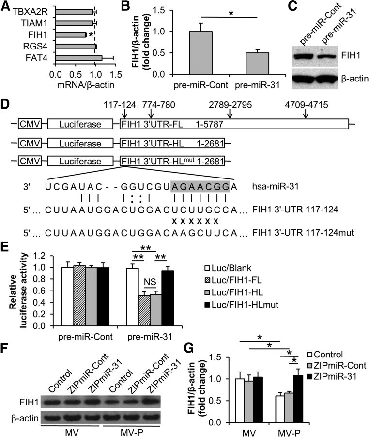
Cell secretion is an important mechanism for stem cell-based therapeutic angiogenesis, along with cell differentiation to vascular endothelial cells or smooth muscle cells. Cell-released microvesicles (MVs) have been recently implicated to play an essential role in intercellular communication. The purpose of this study was to explore the potential effects of stem cell-released MVs in proangiogenic therapy. We observed for the first time that MVs were released from adipose-derived stem cells (ASCs) and were able to increase the migration and tube formation of human umbilical vein endothelial cells (HUVECs). Endothelial differentiation medium (EDM) preconditioning of ASCs upregulated the release of MVs and enhanced the angiogenic effect of the released MVs in vitro. RNA analysis revealed that microRNA was enriched in ASC-released MVs and that the level of microRNA-31 (miR-31) in MVs was notably elevated upon EDM-preconditioning of MV-donor ASCs. Further studies exhibited that miR-31 in MVs contributed to the migration and tube formation of HUVECs, microvessel outgrowth of mouse aortic rings, and vascular formation of mouse Matrigel plugs. Moreover, factor-inhibiting HIF-1, an antiangiogenic gene, was identified as the target of miR-31 in HUVECs. Our findings provide the first evidence that MVs from ASCs, particularly from EDM-preconditioned ASCs, promote angiogenesis and the delivery of miR-31 may contribute the proangiogenic effect.
Significance: This study provides the evidence that microvesicles (MVs) from adipose-derived stem cells (ASCs), particularly from endothelial differentiation medium (EDM)-preconditioned ASCs, promote angiogenesis. An underlying mechanism of the proangiogenesis may be the delivery of microRNA-31 via MVs from ASCs to vascular endothelial cells in which factor-inhibiting HIF-1 is targeted and suppressed. The study findings reveal the role of MVs in mediating ASC-induced angiogenesis and suggest a potential MV-based angiogenic therapy for ischemic diseases.
Keywords: Adipose stem cell; Angiogenesis; Endothelial cell; Microvesicle; miRNA.
©AlphaMed Press.
Figures

Similar articles
-
Adipose-derived stem cell-derived microvesicle-released miR-210 promoted proliferation, migration and invasion of endothelial cells by regulating RUNX3.Cell Cycle. 2018;17(8):1026-1033. doi: 10.1080/15384101.2018.1480207. Epub 2018 Jul 5. Cell Cycle. 2018. PMID: 29912616 Free PMC article.
-
MicroRNA-145 Regulates the Differentiation of Adipose Stem Cells Toward Microvascular Endothelial Cells and Promotes Angiogenesis.Circ Res. 2019 Jun 21;125(1):74-89. doi: 10.1161/CIRCRESAHA.118.314290. Epub 2019 May 6. Circ Res. 2019. PMID: 31219744
-
The endothelial cell secretome as a novel treatment to prime adipose-derived stem cells for improved wound healing in diabetes.J Vasc Surg. 2018 Jul;68(1):234-244. doi: 10.1016/j.jvs.2017.05.094. Epub 2017 Jul 29. J Vasc Surg. 2018. PMID: 28760584
-
Generation of reactive oxygen species in adipose-derived stem cells: friend or foe?Expert Opin Ther Targets. 2011 Nov;15(11):1297-306. doi: 10.1517/14728222.2011.628315. Epub 2011 Oct 10. Expert Opin Ther Targets. 2011. PMID: 21981031 Free PMC article. Review.
-
Horizontal MicroRNA Transfer by Platelets - Evidence and Implications.Front Physiol. 2021 Jun 3;12:678362. doi: 10.3389/fphys.2021.678362. eCollection 2021. Front Physiol. 2021. PMID: 34149456 Free PMC article. Review.
Cited by
-
Microvesicles from Human Immortalized Cell Lines of Endothelial Progenitor Cells and Mesenchymal Stem/Stromal Cells of Adipose Tissue Origin as Carriers of Bioactive Factors Facilitating Angiogenesis.Stem Cells Int. 2020 Jun 15;2020:1289380. doi: 10.1155/2020/1289380. eCollection 2020. Stem Cells Int. 2020. PMID: 32612661 Free PMC article.
-
Research progress on the role of adipocyte exosomes in cancer progression.Oncol Res. 2024 Sep 18;32(10):1649-1660. doi: 10.32604/or.2024.043482. eCollection 2024. Oncol Res. 2024. PMID: 39308520 Free PMC article. Review.
-
Healing the Ischaemic Heart: A Critical Review of Stem Cell Therapies.Rev Cardiovasc Med. 2023 Apr 19;24(4):122. doi: 10.31083/j.rcm2404122. eCollection 2023 Apr. Rev Cardiovasc Med. 2023. PMID: 39076280 Free PMC article. Review.
-
The microenvironment-a general hypothesis on the homeostatic function of extracellular vesicles.FASEB Bioadv. 2022 Mar 12;4(5):284-297. doi: 10.1096/fba.2021-00155. eCollection 2022 May. FASEB Bioadv. 2022. PMID: 35520390 Free PMC article.
-
Extracellular vesicles in autoimmune vasculitis - Little dirts light the fire in blood vessels.Autoimmun Rev. 2019 Jun;18(6):593-606. doi: 10.1016/j.autrev.2018.12.007. Epub 2019 Apr 5. Autoimmun Rev. 2019. PMID: 30959208 Free PMC article. Review.
References
-
- Madonna R, Geng YJ, De Caterina R. Adipose tissue-derived stem cells: Characterization and potential for cardiovascular repair. Arterioscler Thromb Vasc Biol. 2009;29:1723–1729. - PubMed
-
- Cai X, Lin Y, Hauschka PV, et al. Adipose stem cells originate from perivascular cells. Biol Cell. 2011;103:435–447. - PubMed
Publication types
MeSH terms
Substances
Grants and funding
LinkOut - more resources
Full Text Sources
Other Literature Sources
Molecular Biology Databases
Miscellaneous
