

Adult Presentation of Dyke-Davidoff-Masson Syndrome: A Case Report
- PMID: 26933427
- PMCID: PMC4772644
- DOI: 10.1159/000443521
Adult Presentation of Dyke-Davidoff-Masson Syndrome: A Case Report
Abstract
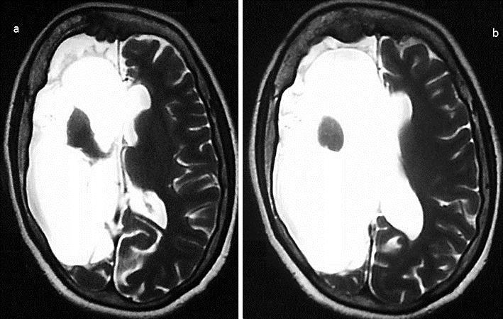
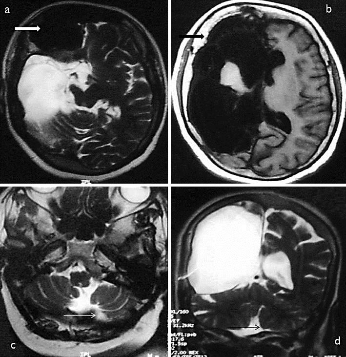
Dyke-Davidoff-Masson syndrome (DDMS) is a rare disease which is clinically characterized by hemiparesis, seizures, facial asymmetry, and mental retardation. The classical radiological findings are cerebral hemiatrophy, calvarial thickening, and hyperpneumatization of the frontal sinuses. This disease is a rare entity, and it mainly presents in childhood. Adult presentation of DDMS is unusual and has been rarely reported in the medical literature.
Key messages: DDMS is a rare disease of childhood. However, it should be kept in mind as a diagnostic possibility in an adult who presents with a long duration of progressive hemiparesis with seizures and mental retardation. Cerebral hemiatrophy, calvarial thickening, and hyperpneumatization of the frontal sinuses are diagnostic for this illness on brain imaging.
Keywords: Calvarial thickening; Cerebral hemiatrophy; Dyke-Davidoff-Masson syndrome.
Figures
References
-
- Dyke CG, Davidoff LM, Masson CB. Cerebral hemiatrophy and homolateral hypertrophy of the skull and sinuses. Surg Gynecol Obstet. 1933;57:588–600.
-
- Unal O, Tombul T, Cirak B, Anlar O, Incesu L, Kayan M. Left hemisphere and male sex dominance of cerebral hemiatrophy (DDMS) Clin Imaging. 2004;28:163–165. - PubMed
-
- Afifi AK, Godersky JC, Menezes A, et al. Cerebral hemiatrophy, hypoplasia of internal carotid artery and intracranial aneurysm. Arch Neurol. 1987;44:232–235. - PubMed
-
- Sener RN, Jinkins JR. MR of craniocerebral hemiatrophy. Clin Imaging. 1992;16:93–97. - PubMed
-
- Stred SE, Byrum CJ, Bove EL, Oliphant M. Coarctation of midaortic arch presenting with monoparesis. Ann Thorac Surg. 1986;42:210–212. - PubMed
Publication types
LinkOut - more resources
Full Text Sources
Other Literature Sources
