. 2016 Feb 22;5(1):9.
doi: 10.1167/tvst.5.1.9. eCollection 2016 Feb.

A Pilot Study of Perceptual-Motor Training for Peripheral Prisms

Affiliations

A Pilot Study of Perceptual-Motor Training for Peripheral Prisms

Kevin E Houston et al. Transl Vis Sci Technol. .

Abstract

Purpose: Peripheral prisms (p-prisms) shift peripheral portions of the visual field of one eye, providing visual field expansion for patients with hemianopia. However, patients rarely show adaption to the shift, incorrectly localizing objects viewed within the p-prisms. A pilot evaluation of a novel computerized perceptual-motor training program aiming to promote p-prism adaption was conducted.

Methods: Thirteen patients with hemianopia fitted with 57Δ oblique p-prisms completed the training protocol. They attended six 1-hour visits reaching and touching peripheral checkerboard stimuli presented over videos of driving scenes while fixating a central target. Performance was measured at each visit and after 3 months.

Results: There was a significant reduction in touch error (P = 0.01) for p-prism zone stimuli from pretraining median of 16.6° (IQR 12.1°-19.6°) to 2.7° ( IQR 1.0°-8.5°) at the end of training. P-prism zone reaction times did not change significantly with training (P > 0.05). P-prism zone detection improved significantly (P = 0.01) from a pretraining median 70% (IQR 50%-88%) to 95% at the end of training (IQR 73%-98%). Three months after training improvements had regressed but performance was still better than pretraining.

Conclusions: Improved pointing accuracy for stimuli detected in prism-expanded vision of patients with hemianopia wearing 57Δ oblique p-prisms is possible and training appears to further improve detection.

Translational relevance: This is the first use of this novel software to train adaptation of visual direction in patients with hemianopia wearing peripheral prisms.

Keywords: Peli Lens; Peripheral Prisms; brain injury; hemianopia or hemianopsia; prism adaptation; stroke.


Figures

Figure 1
Figure 1
(a) The binocular visual field of a patient with left HH as measured by Goldmann perimetry with V4e stimulus. (b) The binocular field of the same patient wearing oblique 57Δ p-prisms. (c) The oblique design in the permanent p-prism fitted unilaterally over the left eye as for the patient in 1b.
Figure 2
Figure 2
(a) Point of view of a driver with normal vision entering a driveway with a hazard approaching from the left (pedestrian in a red jacket). The cross signifies the point of fixation. (b) Illustration of the same view as seen by a driver with left HH. In complete left HH, anything left of the point of fixation (cross), which includes the pedestrian, would not be seen. The HH field is shaded here for illustration purposes only; patients with dense HH do not describe seeing a black or shaded area in their vision. (c) Illustration of the left HH driver's view when wearing oblique 57Δ p-prisms fitted conventionally for left HH (unilaterally on the left lens). The areas within the solid white lines represent the physical locations of the prism segments. The dashed lines outline the areas that are imaged by the oblique p-prisms. The portion of the blind left field containing the pedestrian is visible only to the left (p-prism) eye. The right eye has no p-prisms and so sees the regular view (pedestrian not visible to right eye). This results in binocular confusion in the area of the p-prism image (illustrated as transparency), but no binocular scotomas due to the prisms, and no diplopia. The pedestrian is visible partially in both segments and may be detected, but his location is expected to be misinterpreted as being to the right of its veridical position. If the patient is asked to point at the pedestrian without looking over, they should point 20° to 25° to the right (and perhaps either up or down). (d) Illustration of the presumed result of perceptual adaptation to p-prisms: the pedestrian is perceived in the veridical position/direction left of the fixation (cross). The slight contrast reduction in (d) in the prism vision left of fixation depicts the light scattering effect of the Fresnel prisms.
Figure 3
Figure 3
Participant training station showing a person with left HH performing a pretraining trial. The checkerboard stimulus was presented over a video scene in the blind left hemifield (illustrated by shading) within an oblique prism (lower segment) expansion area (outlined by the dashed rectangle). The prism image of the checkerboard is optically shifted toward the seeing hemifield by the p-prisms so that it appears to the patient as though on the right side of the screen, at the lower position pointed to by the arrow head (note: patient's hand is shown in midreach, eventually ending at the position of the tip of the arrowhead). The prism image results in an incorrect patient response (touching of the apparent position). The participant is unaware of their error since the view of their finger (seen by the nonprism eye) is in the same direction as the prism image of the target. Even once they reach the screen and have visual feedback of their finger and the target, they are still unaware of their error because the smooth touch screen provides no tactile feedback. They continue to believe they are touching the correct location unless auditory feedback is given.
Figure 4
Figure 4
Photo illustration of the operator screen (37 × 28 cm) showing zone placement for a patient with left HH wearing oblique p-prisms. Using kinetic perimetry, the operator marked the border of the field on the prism side (white line), while the patient fixated the white cross, and then drew the borders of the prism zones (dashed rectangles). During training, targets were only presented within the shaded areas of the zones, leaving a buffer (area between the dashed red lines and solid black lines of each zone). The catch zone was set in an area of the blind hemifield where detection was not expected (upper left). There were also three seeing side zones corresponding to each prism zone.
Figure 5
Figure 5
Horizontal touch error for targets presented in prism vision for the visit one pre-session task (pretraining) and the visit six pre-session task at the final training visit. Pretraining, most patients showed error consistent in magnitude and direction with the p-prism deviation (only horizontal error is plotted here), pointing to the apparent target location (data points clustered on the right side of the plot, not circled). Two patients showed fairly good accuracy pretraining (circled). At visit six most patients showed improved accuracy (points below dashed diagonal line). Eight met the 4° success criterion (points on or below solid horizontal line). Only patient 5 showed no improvement.
Figure 6
Figure 6
Number of patients passing each training level (L1–L5; Table 2) by visit. Before moving on to the next level the prior level had to be completed. At visit six, 10 of 13 (77%) had passed all training levels. The blank box at visit one represents the seven patients who did not manage to complete the first level of training at that visit.
Figure 7
Figure 7
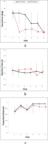
Changes on the performance task across the six training visits for (a) median horizontal touch error, (b) median reaction time, and (c) detection rates for prism zone data for all 13 patients. The pre-session task (solid lines), administered at the beginning of each visit, represents between-visit retention of training effects and the mid-session task (dashed lines) represents within-visit training effects. Black solid error bars represent the IQR (25%–75%) for the pre-session task whereas red dashed error bars are the IQR for the mid-session task. In (a), improvements in touch error were significant by visit two for the mid-session task and by visit six for the pre-session task. (b) Reaction times did not show any significant changes. (c) Pre-session task detection improved significantly by visit four and was maintained, but there was no mid-session task improvement in detection. Mid-session data points are slightly offset to make error bars more visible.
Figure 8
Figure 8
Median reaction times from the visit six pre-session task for the prism and seeing sides were highly correlated (Spearman's rho = 0.85, P < 0.001). Patients 1 and 9 had prism-side reaction times equivalent to seeing-side reaction times (on the diagonal dashed line). Solid and open data points denote successful and less successful cases (in terms of accuracy), respectively.
Figure 9
Figure 9
Changes on the performance task across the 6 training visits for successful and less successful patients: (a) median touch error, (b) median reaction time, and (c) detection rates for the prism zone. Data are shown for patients who met the training success criterion (n = 8) and those who did not (n = 5) for the pre-session task (solid lines) at the beginning of each visit, and the mid-session task (dashed lines) at the mid-way point of each visit. (a) Changes in touch error were only significant for the group of successful patients. (b) For reaction times, there were no differences between successful and less successful patients. (c) For detection, successful patients tended to have better rates. Error bars represent the IQR (25%–75%). Data points are slightly offset to make error bars more visible.

References

    1. Peli E. Field expansion for homonymous hemianopia by optically-induced peripheral exotropia. Optom Vis Sci. 2000; 77: 453–464. - PubMed
    1. Peli E. Peripheral field expansion device. United States Patent 7374,284; 2008.
    1. Jung J-H,, Peli E. Impact of high power and angle of incidence on prism corrections for visual field loss. Opt Eng. 2014; 53. - PMC - PubMed
    1. Peli E,, Bowers A,, Keeney K,, Jung J-H. High power prismatic devices for oblique peripheral prisms. Optom Vis Sci. 2016; 93 (5): e-pub ahead of print, doi:10.1097/OPX.0000000000000820. - DOI - PMC - PubMed
    1. Apfelbaum HL,, Ross NC,, Bowers AR,, Peli E. Considering apical scotomas when prescribing prisms for homonymous hemianopia. Transl Vis Sci Technol. 2013; 2: 2. - PMC - PubMed

LinkOut - more resources