. 2016 Feb 26;2(2):e1501733.
doi: 10.1126/sciadv.1501733. eCollection 2016 Feb.

Bidirectional radial Ca(2+) activity regulates neurogenesis and migration during early cortical column formation

Affiliations

Bidirectional radial Ca(2+) activity regulates neurogenesis and migration during early cortical column formation

Brian G Rash et al. Sci Adv. .

Abstract

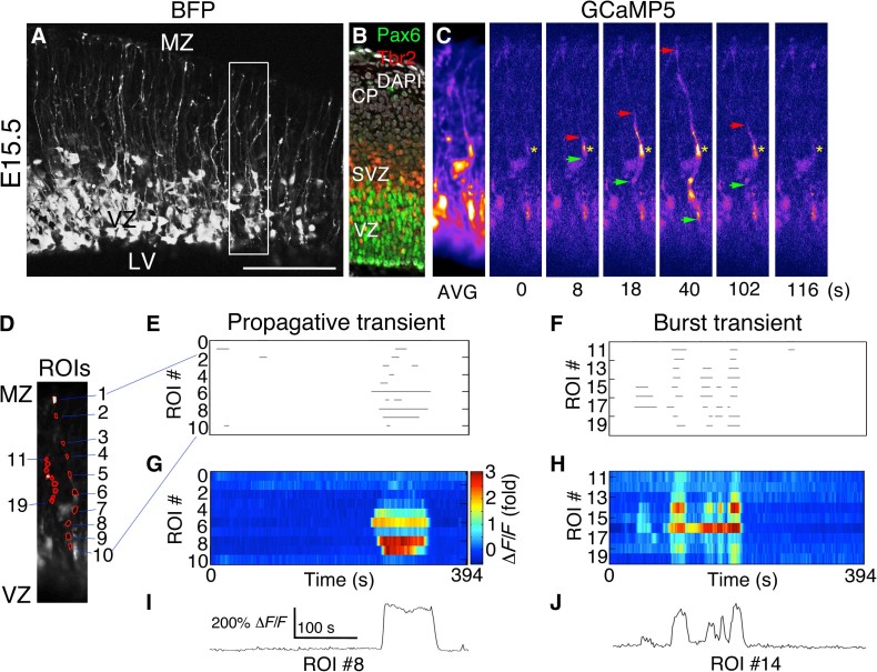
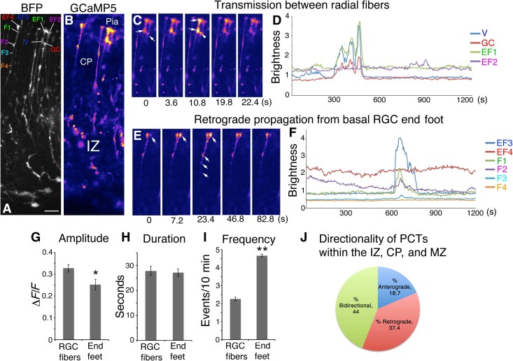
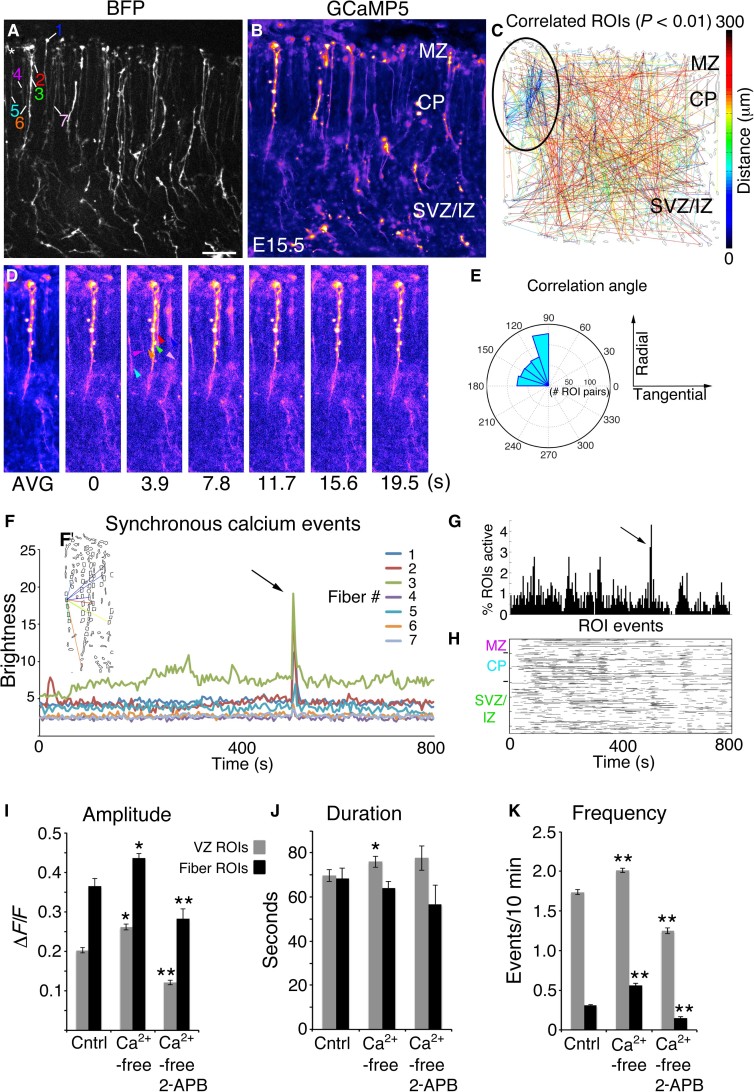
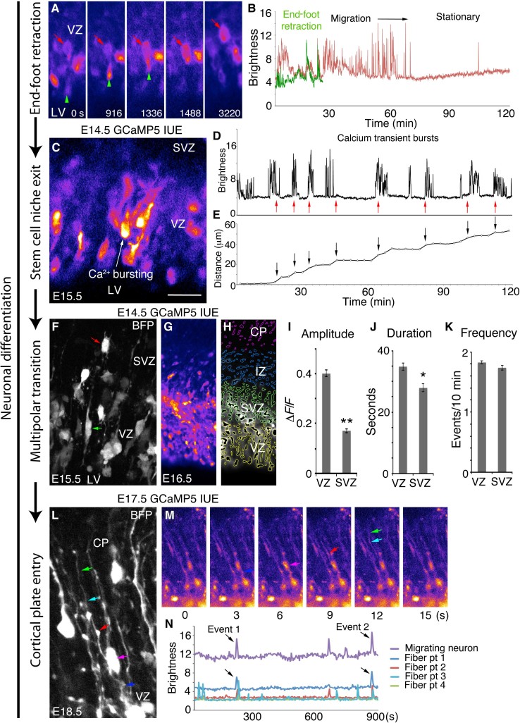
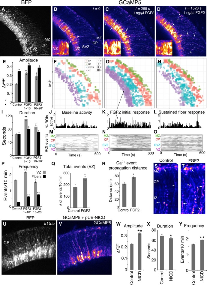
Cortical columns are basic cellular and functional units of the cerebral cortex that are malformed in many brain disorders, but how they initially develop is not well understood. Using an optogenetic sensor in the mouse embryonic forebrain, we demonstrate that Ca(2+) fluxes propagate bidirectionally within the elongated fibers of radial glial cells (RGCs), providing a novel communication mechanism linking the proliferative and postmitotic zones before the onset of synaptogenesis. Our results indicate that Ca(2+) activity along RGC fibers provides feedback information along the radial migratory pathway, influencing neurogenesis and migration during early column development. Furthermore, we find that this columnar Ca(2+) propagation is induced by Notch and fibroblast growth factor activities classically implicated in cortical expansion and patterning. Thus, cortical morphogens and growth factors may influence cortical column assembly in part by regulating long-distance Ca(2+) communication along the radial axis of cortical development.

Keywords: Calcium; GCaMP; migration; neuron; radial glial cells.


Figures

Fig. 1
Fig. 1. PCTs in RGC fibers during neurogenesis.
(A) Blue fluorescent protein (BFP) Z-stack of a live E15.5 mouse cortical slice from an embryo coelectroporated with pCAG-BFP and pCAG-GCaMP5G at E14.5. (B) Immunohistochemistry to visualize VZ and SVZ (subventricular zone) at the time of electroporation. DAPI, 4′,6-diamidino-2-phenylindole. (C) Time-lapse images corresponding to the boxed region in (A) showing a Ca2+ transient originating in an RGC fiber segment (yellow asterisk) and propagating bidirectionally (red and green arrows). AVG, average. (D) Regions of interest (ROIs) corresponding to two separate Ca2+ events in the same movie, drawn in MATLAB. (E to H) Ca2+ event properties were extracted from Ca2+ movies and rendered as rasterplots or heat map intensity plots. (I and J) Ca2+ activity traces for individual ROIs showing single or burst Ca2+ transients in RGC fibers. LV, lateral ventricle; MZ, marginal zone. Scale bars, 125 μm (A) and 100 μm (B and C).
Fig. 2
Fig. 2. Calcium transient activity and directionality in RGC fibers and pial end feet.
(A) BFP Z-stack of RGC fibers. (B) Corresponding image of GCaMP5 fluorescence. (C) Time-lapse images of coherent activity in two RGC end feet. (D) Calcium activity traces of the series in (C); note that the multiple fluorescence events of a growth cone (GC) of an adjacent RGC fiber accurately mirror activity of the adjacent end foot. (E) Time-lapse series of pial end foot initiating a Ca2+ transient, which propagates retrogradely through the CP. (F) Calcium activity traces for the series in (E) showing temporal progression of the Ca2+ wavefront in the RGC fiber. (G to I) Population comparison of Ca2+ event properties within RGC fibers and pial end feet showing respective amplitude, duration, and frequency. (J) PCTs originating in the IZ, CP, and MZ categorized by directionality. Note that MZ transients may be nonpropagative or retrograde only. EF1 to EF4, end foot 1 to end foot 4; V, varicosity; F1 to F4, fiber point 1 to fiber point 4. *P < 0.05; **P < 0.001. Error bars, mean ± SEM. Scale bar, 20 μm.
Fig. 3
Fig. 3. Radial glial fibers form synchronous activation groups.
(A) BFP Z-stack of an E15.5 cortical slice. (B) Averaged GCaMP5 movie. (C) Correlation map; clustered correlations indicated by black oval. (D) Time-lapse series; coherently active RGC fibers are indicated by colored arrowheads. (E) Correlation angles from correlated pairs in (C) binned and plotted on polar coordinates. (F) Absolute brightness traces from ROIs of coherently active RGC fibers. (F’) Correlated pairs within the synchronous cluster. (G and H) Extraction of Ca2+ event properties in MATLAB revealed the synchronous firing event in percent active and rasterplot form. (I to K) RGC cell body (gray bars) and fiber (black bars) activity in control (Cntrl) ACSF, Ca2+-free ACSF, and Ca2+-free ACSF plus 100 μM 2-APB. *P < 0.05; **P < 0.001, t test. Error bars, mean ± SEM. Scale bar, 40 μm.
Fig. 4
Fig. 4. Bursting calcium activity in newborn migrating neurons and communication with RGC fibers.
(A) Retracting apical end foot (green arrowhead) and its cell body (red arrow). (B) Calcium activity traces of the cell in (A). (C) Putative migrating neuron exiting the VZ. (D) Calcium bursting activity (red arrows) of cell indicated in (C). (E) Distance traveled by cell in (C) (arrows) corresponding to Ca2+ bursts in (D). (F) BFP Z-stack showing electroporated RGCs (green arrow) and multipolar progenitors (red arrow). (G) GCaMP5G image of an E16.5 cortical slice after E14.5 electroporation. (H) MATLAB ROIs organized in embryonic zones corresponding to the image in (G). (I to K) Ca2+ activity properties of the VZ and SVZ. (L) BFP Z-stack of an RGC fiber (dark blue, red, light blue, and green arrows) and adjacent migrating neuron (magenta arrow) at E18.5. (M) Time-lapse GCaMP5 images of the cell in (L). (N) Correlated Ca2+ activity of the RGC fiber and neuron in (L) and (M). *P < 0.05; **P < 0.001, t test. Error bars, mean ± SEM. Scale bars, 25 μm (A, C, F, and L) and 80 μm (G and H).
Fig. 5
Fig. 5. Calcium activity induced by FGF2 and Notch.
(A) BFP Z-stack of an E15.5 cortical slice. (B) Baseline calcium activity. (C and D) Calcium activity after FGF2 exposure; inset panels are high-magnification images of boxed region. (E, I, and P) ΔF/F amplitude, duration, and frequency of Ca2+ events in RGC cell bodies and fibers under control conditions, after initial FGF2 exposure, and sustained FGF2 exposure. (F to H) ΔF/F amplitude of active ROIs plotted spatially; square size represents relative amplitude. (J to L) Percentages of ROIs active. (M to O) Rasterplots of all detected Ca2+ events binned according to embryonic zone (VZ, SVZ, CP, and MZ). (Q) Total number of events in the VZ. (R) Ca2+ event propagation distance within RGC fibers. (S and T) Averaged movies show accumulated Ca2+ activity within RGC fibers and end feet in 400-s epochs. (U) BFP Z-stack of an E15.5 slice coexpressing GCaMP5G and Notch intracellular domain (NICD). (V) GCaMP5 Ca2+ signal. (W to Y) Effects of NICD overexpression on Ca2+ event properties of RGC cell bodies. *P < 0.05; **P < 0.001, t test. Error bars, mean ± SEM. Scale bar, 100 μm (A to D, U, and V) and 50 μm in insets of (B) to (D), (S), and (T).

References

    1. Mountcastle V. B., The columnar organization of the neocortex. Brain 120, 701–722 (1997). - PubMed
    1. Rakic P., Specification of cerebral cortical areas. Science 241, 170–176 (1988). - PubMed
    1. Noctor S. C., Flint A. C., Weissman T. A., Dammerman R. S., Kriegstein A. R., Neurons derived from radial glial cells establish radial units in neocortex. Nature 409, 714–720 (2001). - PubMed
    1. Rakic P., Evolution of the neocortex: A perspective from developmental biology. Nat. Rev. Neurosci. 10, 724–735 (2009). - PMC - PubMed
    1. Torii M., Hashimoto-Torii K., Levitt P., Rakic P., Integration of neuronal clones in the radial cortical columns by EphA and ephrin-A signalling. Nature 461, 524–528 (2009). - PMC - PubMed

Publication types

LinkOut - more resources