Review
. 2016 Jan 21;23(1):122-136.
doi: 10.1016/j.chembiol.2015.12.003.

A Bright Future for Precision Medicine: Advances in Fluorescent Chemical Probe Design and Their Clinical Application

Affiliations
Review

A Bright Future for Precision Medicine: Advances in Fluorescent Chemical Probe Design and Their Clinical Application

Megan Garland et al. Cell Chem Biol. .

Abstract

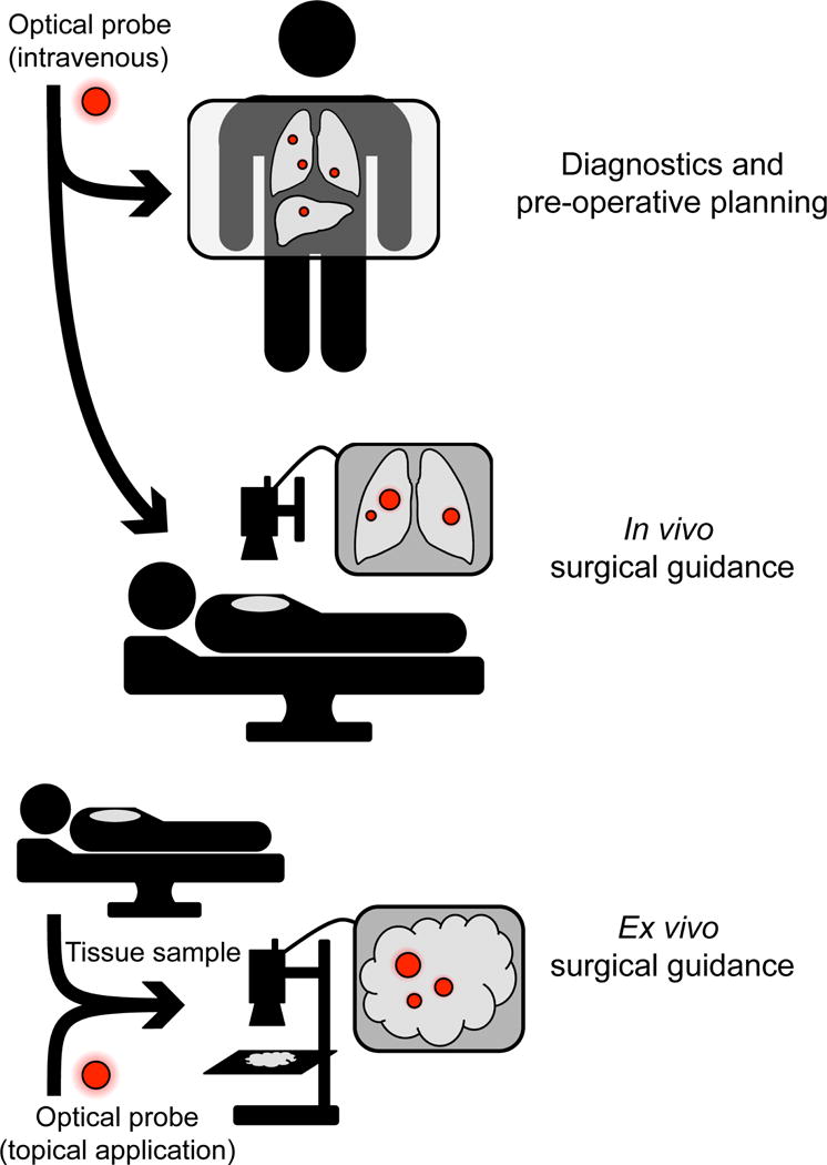
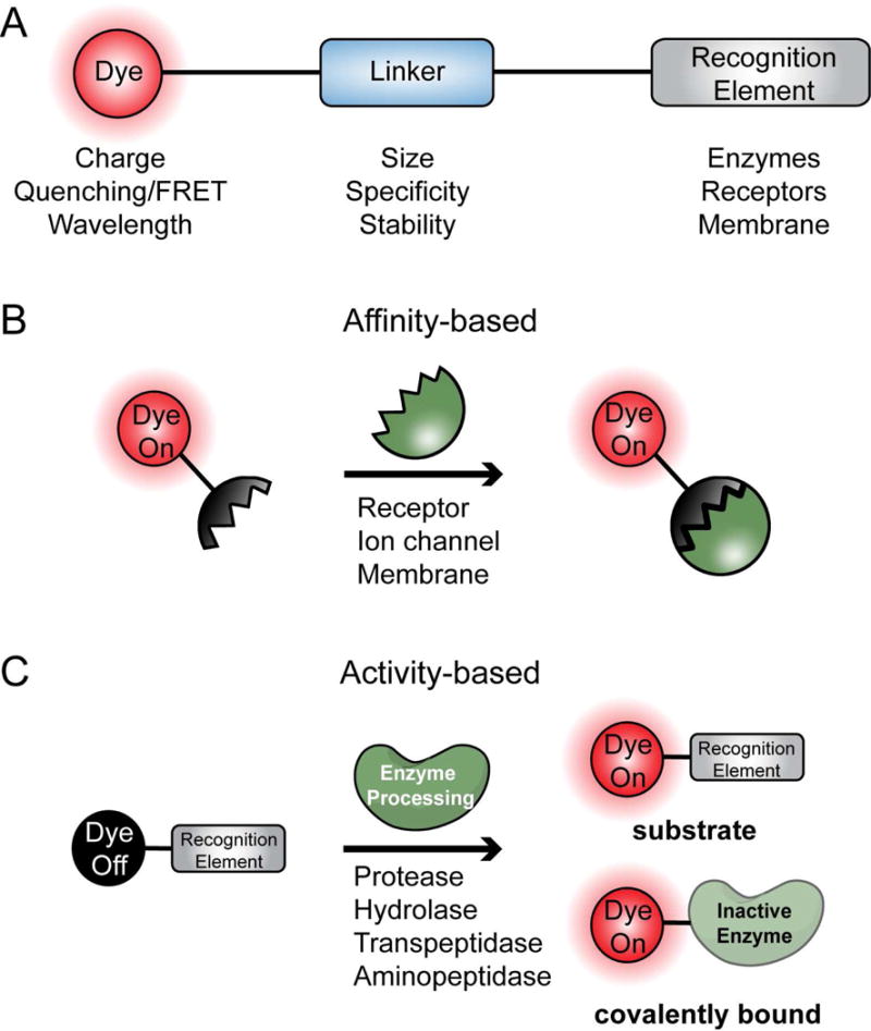
The Precision Medicine Initiative aims to use advances in basic and clinical research to develop therapeutics that selectively target and kill cancer cells. Under the same doctrine of precision medicine, there is an equally important need to visualize these diseased cells to enable diagnosis, facilitate surgical resection, and monitor therapeutic response. Therefore, there is a great opportunity for chemists to develop chemically tractable probes that can image cancer in vivo. This review focuses on recent advances in the development of optical probes, as well as their current and future applications in the clinical management of cancer. The progress in probe development described here suggests that optical imaging is an important and rapidly developing field of study that encourages continued collaboration among chemists, biologists, and clinicians to further refine these tools for interventional surgical imaging, as well as for diagnostic and therapeutic applications.


Figures

Figure 1
Figure 1. Current and Future Clinical applications for optical chemical probes
Top: future application of optical contrast agents includes intravenous administration for preoperative diagnostics and planning. Middle: current use of optical contrast agents for in vivo surgical guidance. Bottom: topical probe labeling can be used for ex vivo surgical guidance.
Figure 2
Figure 2. General considerations for probe design
A. Optical chemical probes consist of 3 main structural elements: the dye, linker, and recognition element. B. General model for affinity-based probes. The probe binds to targets such as receptors, ion channels and membranes with high affinity. C. General model for activity-based probes. These probes give off a fluorescent signal upon processing by enzymes such as proteases and peptidases.
Figure 3
Figure 3. Affinity-based Probes
A. Chlorotoxin-based probe BLZ-100. B. Alkylphosphocholine (APC) structure and APC-derived probe CLR1502. C. Folic acid structure and folate receptor α-targeting probe OTL38. D. Cyclic RGD peptides as tumor contrast agents. E. NAAG structure and PSMA targeting probes YC27 and PSMA-1-IR800. F. Structure of Indomethacin and the corresponding labeled probe Fluorocoxib A.
Figure 4
Figure 4. Substrate and activity-based probes
(ABPs) A. The cathespin targeted covalent probe BMV109 B. The cathepsin targeted substrate LUM015 C. The polyer-based protease substrate probe C-PGC probe D. A polymer MMP-2-sensitive NIRF substrate E. The turn-on fluorophore based probe for cathepsins, Z-Phe-Arg-HMRG.
Figure 5
Figure 5. Substrate and activity-based probes (ABPs) structures
A. Cathespin targeted lipidated probe 3 B. The cathepsin targeting probe, 6QCNIR C. The cell penetrating probe, RACPP D. Nanoaggregating probe for caspases, C-SNAF.
Figure 6
Figure 6. Examples of optical chemical probes being used in animal models for noninvasive cancer diagnostics, in vivo, and ex vivo surgical guidance
A. LEFT: 6QC (20 nmol IV), imaged after 4 hr in 4T1 Breast cancer mouse model, MIDDLE: Folate-Dylight680 (10 nmol IV), imaged after 4 hr in FR-expressing L1210A tumor mouse model, RIGHT: C-SNAF (5 nmol IV), imaged after 1 hr in three times DOX treated tumor-bearing mice. B. TOP: CTX:Cy5.5. Image shows white light and white light with NIR overlay on a canine tumor tissue sample. BOTTOM: 6QCNIR injected 6h prior to surgery. Image shows white light on the left and fluorescence using the da Vinci® surgical system on right. C. gGlu-HMRG (3 mL of 5 μM in 0.5% v/v DMSO in RPMI1640) topically applied to patient specimen diagnosed with invasive ductal carcinoma (papillotubular) and imaged after 5 min.

References

    1. Akcan M, Stroud MR, Hansen SJ, Clark RJ, Daly NL, Craik DJ, Olson JM. Chemical re-engineering of chlorotoxin improves bioconjugation properties for tumor imaging and targeted therapy. J Med Chem. 2011;54:782–787. - PMC - PubMed
    1. Andreesen R, Modolell M, Weltzien HU, Eibl H, Common HH, Lohr GW, Munder PG. Selective destruction of human leukemic cells by alkyl-lysophospholipids. Cancer Res. 1978;38:3894–3899. - PubMed
    1. Antaris AL, Chen H, Cheng K, Sun Y, Hong G, Qu C, Diao S, Deng Z, Hu X, Zhang B, et al. A small-molecule dye for NIR-II imaging. Nat Mater 2015 - PubMed
    1. Antony AC. Folate receptors. Annu Rev Nutr. 1996;16:501–521. - PubMed
    1. Aoki T, Murakami M, Yasuda D, Shimizu Y, Kusano T, Matsuda K, Niiya T, Kato H, Murai N, Otsuka K, et al. Intraoperative fluorescent imaging using indocyanine green for liver mapping and cholangiography. J Hepatobiliary Pancreat Sci. 2010;17:590–594. - PubMed

Publication types

MeSH terms